实验动物与比较医学 ›› 2021, Vol. 41 ›› Issue (4): 271-283.DOI: 10.12300/j.issn.1674-5817.2021.085
所属专题: 专家论坛
• 创刊40 周年专家论坛 • 下一篇
胡志斌1,2, 黄缨1, 丁玉强1,2
收稿日期:2021-04-21
修回日期:2021-05-18
出版日期:2021-08-25
发布日期:2021-08-25
作者简介:胡志斌(1997—), 男, 博士研究生, 研究方向: 疾病动物模型与神经系统疾病发生机制; E-mail: zbhu20@fudan.edu.cn
基金资助:HU Zhibin1,2, HUANG Ying1, DING Yuqiang1,2
Received:2021-04-21
Revised:2021-05-18
Published:2021-08-25
Online:2021-08-25
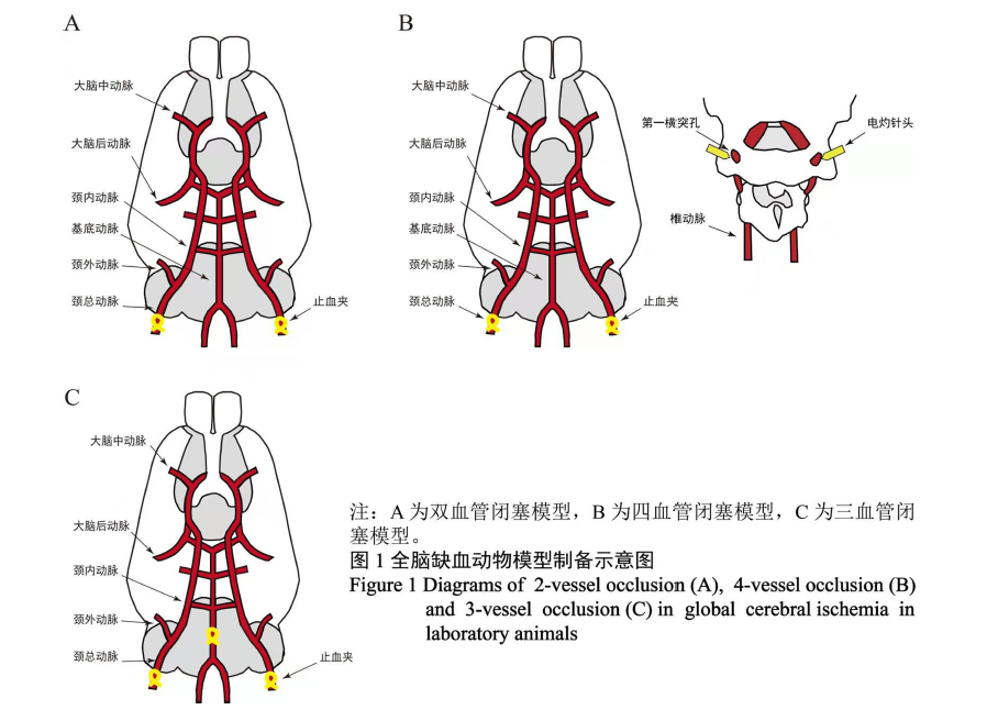
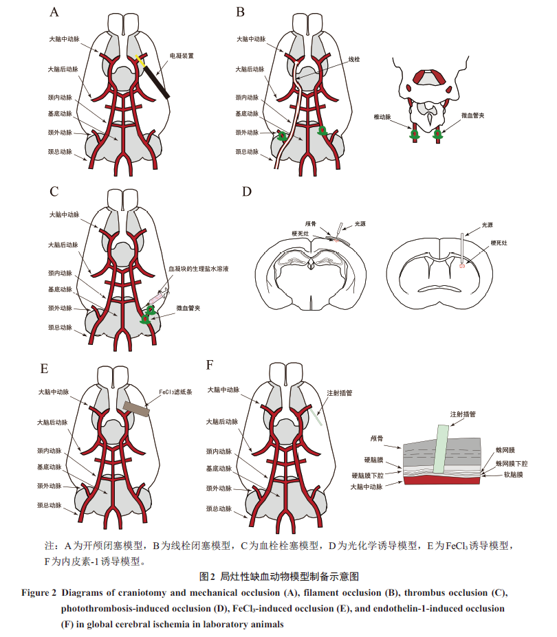
摘要: 脑缺血是临床上常见的脑血管疾病,约占所有脑血管疾病的80%,其主要是由于脑血管供血不足引起。目前已有多种动物模型被用来进行脑缺血致病机制及治疗研究。本文结合造模示意图,介绍全脑及局灶性相关脑缺血动物模型的种类及制备方法,并对其优缺点进行了概括;同时对脑缺血模型制备所用的动物种类、啮齿类动物相关感觉运动能力评估及组织学评估的研究进展进行综述,以期为脑缺血动物模型的选择和应用提供参考。
中图分类号:
胡志斌,黄缨,丁玉强. 脑缺血动物模型的制备及评估进展[J]. 实验动物与比较医学, 2021, 41(4): 271-283. DOI: 10.12300/j.issn.1674-5817.2021.085.
HU Zhibin,HUANG Ying,DING Yuqiang. Construction and Evaluation of Animal Models for Cerebral Ischemia[J]. Laboratory Animal and Comparative Medicine, 2021, 41(4): 271-283. DOI: 10.12300/j.issn.1674-5817.2021.085.
| [1] KAHL A, BLANCO I, JACKMAN K, et al.Cerebral ischemia induces the aggregation of proteins linked to neurodegenerative diseases[J]. Sci Rep, 2018, 8:2701. DOI:10.1038/s41598-018-21063-z. [2] NAGATA K, YAMAZAKI T, TAKANO D, et al.Cerebral circulation in aging[J]. Ageing Res Rev, 2016, 30:49-60. DOI:10.1016/j.arr.2016.06.001. [3] HOSSMANN K A.Pathophysiology and therapy of experimental stroke[J]. Cell Mol Neurobiol, 2006, 26(7-8):1057-1083. DOI:10.1007/s10571-006-9008-1. [4] FERRER I, VIDAL N.Neuropathology of cerebro-vascular diseases[J]. Handb Clin Neurol, 2017, 145:79-114. DOI:10.1016/b978-0-12-802395-2.00007-9. [5] DURUKAN A, TATLISUMAK T.Acute ischemic stroke: verview of major experimental rodent models, pathophysiology, and therapy of focal cerebral ischemia[J]. Pharmacol Biochem Behav, 2007, 87(1):179-197. DOI:10.1016/j.pbb.2007.04.015. [6] WANG L D, YIN L, HUA Y, et al.Fixed-dose combination treatment after stroke for secondary prevention in China: national community-based study[J]. Stroke, 2015, 46(5):1295-1300. DOI:10.1161/STROKEAHA.114.007384. [7] 孙海欣, 王文志. 中国60万人群脑血管病流行病学抽样调查报告[J]. 中国现代神经疾病杂志, 2018, 18(2):83-88. DOI:10.3969/j.issn.1672-6731.2018.02.002. [8] MCCABE C, ARROJA M M, REID E, et al.Animal models of ischaemic stroke and characterisation of the ischaemic penumbra[J]. Neuropharmacology, 2018, 134(Pt B):169-177. DOI:10.1016/j.neuropharm.2017.09.022. [9] 甘勇, 杨婷婷, 刘建新, 等. 国内外脑卒中流行趋势及影响因素研究进展[J]. 中国预防医学杂志, 2019, 20(2):139-144. DOI:10.16506/j.1009-6639.2019.02.013. [10] 王琮, 陈宇, 张阿龙, 等. 局灶性脑缺血动物模型制作方法研究进展[J]. 齐齐哈尔医学院学报, 2021, 42(6):506-509. [11] ZHOU Y X, WANG X, TANG D, et al.IL-2mAb reduces demyelination after focal cerebral ischemia by suppressing CD8+ T cells[J]. CNS Neurosci Ther, 2019, 25(4):532-543. DOI:10.1111/cns.13084. [12] LV Z M, ZHAO R J, ZHI X S, et al.Expression of DCX and transcription factor profiling in photothrombosis-induced focal ischemia in mice[J]. Front Cell Neurosci, 2018, 12:455. DOI:10.3389/fncel.2018.00455. [13] KARATAS H, ERDENER S E, GURSOY-OZDEMIR Y, et al.Thrombotic distal middle cerebral artery occlusion produced by topical FeCl3 application: novel model suitable for intravital microscopy and thrombolysis studies[J]. J Cereb Blood Flow Metab, 2011, 31(6):1452-1460. DOI:10.1038/jcbfm.2011.8. [14] YANG G, KITAGAWA K, MATSUSHITA K, et al.C57BL/6 strain is most susceptible to cerebral ischemia following bilateral common carotid occlusion among seven mouse strains: elective neuronal death in the murine transient forebrain ischemia[J]. Brain Res, 1997, 752(1-2):209-218. DOI:10.1016/s0006-8993(96)01453-9. [15] ABDEL-RAHMAN R F, ALQASOUMI S I, OGALY H A, et al. Propolis ameliorates cerebral injury in focal cerebral ischemia/reperfusion (I/R) rat model via upregulation of TGF-β1[J]. Saudi Pharm J, 2020, 28(1):116-126. DOI:10.1016/j.jsps.2019.11.013. [16] KERBRAT A, GROS C, BADJI A, et al.Multiple sclerosis lesions in motor tracts from brain to cervical cord: patial distribution and correlation with disability[J]. Brain, 2020, 143(7):2089-2105. DOI:10.1093/brain/awaa162. [17] STRÖM J O, INGBERG E, THEODORSSON A, et al. Method parameters' impact on mortality and variability in rat stroke experiments: meta-analysis[J]. BMC Neurosci, 2013, 14:41. DOI:10.1186/1471-2202-14-41. [18] KIRINO T.Delayed neuronal death in the gerbil [19] WASHIDA K, HATTORI Y, IHARA M.Animal models of chronic cerebral hypoperfusion: rom mouse to primate[J]. Int J Mol Sci, 2019, 20(24): E6176. DOI:10.3390/ijms20246176. [20] IKEDA S, HARADA K, OHWATASHI A, et al.A new non-human primate model of photochemically induced cerebral infarction[J]. PLoS One, 2013, 8(3): e60037. DOI:10.1371/journal.pone.0060037. [21] MOISENOVICH M M, SILACHEV D N, MOYSENOVICH A M, et al.Effects of recombinant spidroin rS1/9 on brain neural progenitors after photothrombosis-induced ischemia[J]. Front Cell Dev Biol, 2020, 8:823. DOI:10.3389/fcell.2020.00823. [22] DEWAR D, YAM P, MCCULLOCH J.Drug development for stroke: importance of protecting cerebral white matter[J]. Eur J Pharmacol, 1999, 375(1-3):41-50. DOI:10.1016/s0014-2999(99)00280-0. [23] MADIGAN J B, WILCOCK D M, HAINSWORTH A H.Vascular contributions to cognitive impairment and dementia: topical review of animal models[J]. Stroke, 2016, 47(7):1953-1959. DOI:10.1161/STROKEAHA.116.012066. [24] CHEN A Q, AKINYEMI R O, HASE Y, et al.Frontal white matter hyperintensities, clasmatodendrosis and gliovascular abnormalities in ageing and post-stroke dementia[J]. Brain, 2016, 139(Pt 1):242-258. DOI:10.1093/brain/awv328. [25] STEELE P R, CAVARSAN C F, DOWALIBY L, et al.Altered motoneuron properties contribute to motor deficits in a rabbit hypoxia-ischemia model of cerebral palsy[J]. Front Cell Neurosci, 2020, 14:69. DOI:10.3389/fncel.2020.00069. [26] ZHANG R, BERTELSEN L B, FLØ C, et al.Establishment and characterization of porcine focal cerebral ischemic model induced by endothelin-1[J]. Neurosci Lett, 2016, 635:1-7. DOI:10.1016/j.neulet.2016.10.036. [27] EKLÖF B, SIESJÖ B K. The effect of bilateral carotid artery ligation upon the blood flow and the energy state of the rat brain[J]. Acta Physiol Scand, 1972, 86(2):155-165. DOI:10.1111/j.1748-1716.1972.tb05322.x. [28] SANDERSON T H, WIDER J M.2-vessel occlusion/hypotension: rat model of global brain ischemia[J]. J Vis Exp, 2013(76): e50173. DOI:10.3791/50173. [29] HUANG T H, LIN Y W, HUANG C P, et al.Short-term auricular electrical stimulation rapidly elevated cortical blood flow and promoted the expression of nicotinic acetylcholine receptor α4 in the 2 vessel occlusion rats model[J]. J Biomed Sci, 2019, 26(1):36. DOI:10.1186/s12929-019-0526-9. [30] PULSINELLI W A, BRIERLEY J B.A new model of bilateral hemispheric ischemia in the unanesthetized rat[J]. Stroke, 1979, 10(3):267-272. DOI:10.1161/01.str.10.3.267. [31] SUGIO K, HORIGOME N, SAKAGUCHI T, et al.A model of bilateral hemispheric ischemia: modified four-vessel occlusion in rats[J]. Stroke, 1988, 19(7):922. DOI:10.1161/str.19.7.922a. [32] YOSHIMURA S, UCHIDA K, SAKAI N, et al.Safety of early administration of apixaban on clinical outcomes in patients with acute large vessel occlusion[J]. Transl Stroke Res, 2021, 12(2):266-274. DOI:10.1007/s12975-020-00839-4. [33] 李兵, 章翔, 蒋晓帆, 等. 改良四血管阻塞法建立大鼠全脑缺血模型[J]. 中华神经外科疾病研究杂志, 2005, 4(2):110-113. DOI:10.3969/j.issn.1671-2897.2005. 02.005. [34] ZAGHI G G, GODINHO J, FERREIRA E D, et al.Robust and enduring atorvastatin-mediated memory recovery following the 4-vessel occlusion/internal carotid artery model of chronic cerebral hypoperfusion in middle-aged rats[J]. Prog Neuropsychopharmacol Biol Psychiatry, 2016, 65:179-187. DOI:10.1016/j.pnpbp.2015.10.004. [35] KAMEYAMA M, SUZUKI J, SHIRANE R, et al.A new model of bilateral hemispheric ischemia in the rat: three vessel occlusion model[J]. Stroke, 1985, 16(3):489-493. DOI:10.1161/01.str.16.3.489. [36] PANAHIAN N, YOSHIDA T, HUANG P L, et al.Attenuated hippocampal damage after global cerebral ischemia in mice mutant in neuronal nitric oxide synthase[J]. Neuroscience, 1996, 72(2):343-354. DOI:10.1016/0306-4522(95)00563-3. [37] THAL S C, THAL S E, PLESNILA N.Characterization of a 3-vessel occlusion model for the induction of complete global cerebral ischemia in mice[J]. J Neurosci Methods, 2010, 192(2):219-227. DOI:10.1016/j.jneumeth.2010.07.032. [38] LÜ P, JIAO Q B, SHIMURA D, et al.Distinct vascular remodeling pattern of adult rats with carotid-jugular shunt[J]. Ann Vasc Surg, 2018, 49:168-178. DOI:10.1016/j.avsg.2017.12.011. [39] TAMURA A, GRAHAM D I, MCCULLOCH J, et al.Focal cerebral ischaemia in the rat: 1. escription of technique and early neuropathological consequences following middle cerebral artery occlusion[J]. J Cereb Blood Flow Metab, 1981, 1(1):53-60. DOI:10.1038/jcbfm.1981.6. [40] BEDERSON J B, PITTS L H, TSUJI M, et al.Rat middle cerebral artery occlusion: valuation of the model and development of a neurologic examination[J]. Stroke, 1986, 17(3):472-476. DOI:10.1161/01.str. 17.3.472. [41] EJAZ S, WILLIAMSON D J, AHMED T, et al.Characterizing infarction and selective neuronal loss following temporary focal cerebral ischemia in the rat: multi-modality imaging study[J]. Neurobiol Dis, 2013, 51:120-132. DOI:10.1016/j.nbd.2012.11.002. [42] SHMONIN A, MELNIKOVA E, GALAGUDZA M, et al.Characteristics of cerebral ischemia in major rat stroke models of middle cerebral artery ligation through craniectomy[J]. Int J Stroke, 2014, 9(6):793-801. DOI:10.1111/j.1747-4949.2012.00947.x. [43] KOIZUMI J I, YOSHIDA Y, NAKAZAWA T, et al.Experimental studies of ischemic brain edema. A new experimental model of cerebral embolism in rats in which recirculation can be introduced in the ischemic area[J]. Nosotchu, 1986, 8(1):1-8. [44] LONGA E Z, WEINSTEIN P R, CARLSON S, et al.Reversible middle cerebral artery occlusion without craniectomy in rats[J]. Stroke, 1989, 20(1):84-91. DOI:10.1161/01.str.20.1.84. [45] CSIBA L, BERECZKI D, SHIMA T, et al.A modified model of reversible middle cerebral artery embolization in rats without craniectomy[J]. Acta Neurochir, 1992, 114(1-2):51-58. DOI:10.1007/BF01401114. [46] ZUO X L, WU P, JI A M.Nylon filament coated with paraffin for intraluminal permanent middle cerebral artery occlusion in rats[J]. Neurosci Lett, 2012, 519(1):42-46. DOI:10.1016/j.neulet.2012.05.017. [47] 康瑜, 杨小芳. 纳米脂质体槲皮素对脑缺血再灌注损伤的神经保护作用及机制[J]. 实验动物与比较医学, 2020, 40(2):116-122. DOI:10.3969/j.issn.1674-5817.2020.02.005. [48] SVOBODA J, LITVINEC A, KALA D, et al.Strain differences in intraluminal thread model of middle cerebral artery occlusion in rats[J]. Physiol Res, 2019, 68(1):37-48. DOI:10.33549/physiolres.933958. [49] KUDO M, AOYAMA A, ICHIMORI S, et al.An animal model of cerebral infarction. Homologous blood clot emboli in rats[J]. Stroke, 1982, 13(4):505-508. DOI:10.1161/01.str.13.4.505. [50] DINAPOLI V A, ROSEN C L, NAGAMINE T, et al.Selective MCA occlusion: precise embolic stroke model[J]. J Neurosci Methods, 2006, 154(1-2):233-238. DOI:10.1016/j.jneumeth.2005.12.026. [51] ORSET C, MACREZ R, YOUNG A R, et al.Mouse model of [52] CHEN Y X, ZHU W B, ZHANG W R, et al.A novel mouse model of thromboembolic stroke[J]. J Neurosci Methods, 2015, 256:203-211. DOI:10.1016/j.jneumeth. 2015.09.013. [53] ISHRAT T, FOUDA A Y, PILLAI B, et al. Dose-response, therapeutic time-window and tPA-combina-torial efficacy of compound 21: randomized, blinded preclinical trial in a rat model of thromboembolic stroke[J]. J Cereb Blood Flow Metab, 2019, 39(8)1635-1647. DOI:10.1177/0271678X18764773. [54] VANERIO N, STIJNEN M, DE MOL B A J M, et al. Biomedical applications of photo- and sono-activated rose Bengal: a review[J]. Photobiomodul Photomed Laser Surg, 2019, 37(7):383-394. DOI:10.1089/photob.2018.4604. [55] WATSON B D, DIETRICH W D, BUSTO R, et al.Induction of reproducible brain infarction by photochemically initiated thrombosis[J]. Ann Neurol, 1985, 17(5):497-504. DOI:10.1002/ana.410170513. [56] LI H, ROY CHOUDHURY G, ZHANG N, et al.Photothrombosis-induced focal ischemia as a model of spinal cord injury in mice[J]. J Vis Exp, 2015(101): e53161. DOI:10.3791/53161. [57] XUE N Y, GE D Y, DONG R J, et al.Effect of electroacupuncture on glial fibrillary acidic protein and nerve growth factor in the [58] YU C L, ZHOU H, CHAI A P, et al.Whole-scale neurobehavioral assessments of photothrombotic ischemia in freely moving mice[J]. J Neurosci Methods, 2015, 239:100-107. DOI:10.1016/j.jneumeth. 2014.10.004. [59] OWENS A P 3rd, LU Y, WHINNA H C, et al. Towards a standardization of the murine ferric chloride-induced carotid arterial thrombosis model[J]. J Thromb Haemost, 2011, 9(9):1862-1863. DOI:10.1111/j.1538-7836.2011.04287.x. [60] SYEARA N, ALAMRI F F, JAYARAMAN S, et al.Motor deficit in the mouse ferric chloride-induced distal middle cerebral artery occlusion model of stroke[J]. Behav Brain Res, 2020, 380:112418. DOI:10.1016/j.bbr.2019.112418. [61] LI W, NIEMAN M, SEN GUPTA A.Ferric chloride-induced murine thrombosis models[J]. J Vis Exp, 2016(115): e54479. DOI:10.3791/54479. [62] BARTON M, YANAGISAWA M.Endothelin: 30 years from discovery to therapy[J]. Hypertension, 2019, 74(6):1232-1265. DOI:10.1161/HYPERTENSIONAHA. 119.12105. [63] MACRAE I M, ROBINSON M J, GRAHAM D I, et al.Endothelin-1-induced reductions in cerebral blood flow: dose dependency, time course, and neuro-pathological consequences[J]. J Cereb Blood Flow Metab, 1993, 13(2):276-284. DOI:10.1038/jcbfm. 1993.34. [64] SHARKEY J, RITCHIE I M, KELLY P A.Perivascular microapplication of endothelin-1: new model of focal cerebral ischaemia in the rat[J]. J Cereb Blood Flow Metab, 1993, 13(5):865-871. DOI:10.1038/jcbfm. 1993.108. [65] WANG N Y, LI J N, LIU W L, et al. Ferulic acid ameliorates Alzheimer's disease-like pathology and repairs cognitive decline by preventing capillary hypofunction in APP/PS1 mice[J]. Neurotherapeutics, 2021-03-30 [2021-07-18]. https://pubmed.ncbi.nlm.nih.gov/33786807/ DOI:10.1007/s13311-021-01024-7. [66] CUI L L, GOLUBCZYK D, JOLKKONEN J.Top 3 behavioral tests in cell therapy studies after stroke: difficult to stop a moving train[J]. Stroke, 2017, 48(11):3165-3167. DOI:10.1161/STROKEAHA.117.018950. [67] SCHALLERT T, UPCHURCH M, LOBAUGH N, et al.Tactile extinction: istinguishing between sensorimotor and motor asymmetries in rats with unilateral nigrostriatal damage[J]. Pharmacol Biochem Behav, 1982, 16(3):455-462. DOI:10.1016/0091-3057(82)90452-x. [68] CHEN J L, ZHANG C L, JIANG H, et al.Atorvastatin induction of VEGF and BDNF promotes brain plasticity after stroke in mice[J]. J Cereb Blood Flow Metab, 2005, 25(2):281-290. DOI:10.1038/sj.jcbfm.9600034. [69] KARHUNEN H, VIRTANEN T, SCHALLERT T, et al.Forelimb use after focal cerebral ischemia in rats treated with an alpha 2-adrenoceptor antagonist[J]. Pharmacol Biochem Behav, 2003, 74(3):663-669. DOI:10.1016/s0091-3057(02)01053-5. [70] ROGERS D C, CAMPBELL C A, STRETTON J L, et al.Correlation between motor impairment and infarct volume after permanent and transient middle cerebral artery occlusion in the rat[J]. Stroke, 1997, 28(10):2060-2065, 2066. DOI:10.1161/01.str.28.10.2060. [71] BENEDEK A, MÓRICZ K, JURÁNYI Z, et al. Use of TTC staining for the evaluation of tissue injury in the early phases of reperfusion after focal cerebral ischemia in rats[J]. Brain Res, 2006, 1116(1):159-165. DOI:10.1016/j.brainres.2006.07.123. [72] YU C L, LI J N, GAN P, et al.Developing of focal ischemia in the ENEURO.0398-ENEURO.0320.2021. DOI:10.1523/ENEURO.0398-20.2021. [73] XU S B, LU J N, SHAO A W, et al.Glial cells: role of the immune response in ischemic stroke[J]. Front Immunol, 2020, 11:294. DOI:10.3389/fimmu. 2020.00294. [74] VAN BEEK J, CHAN P, BERNAUDIN M, et al.Glial responses, clusterin, and complement in permanent focal cerebral ischemia in the mouse[J]. Glia, 2000, 31(1):39-50. DOI:10.1002/(sici)1098-1136(200007)31:1<39: aid-glia40>3.0.co;2-1. [75] LU J F, BRADLEY R A, ZHANG S C.Turning reactive [76] NAKASE T, SÖHL G, THEIS M, et al. Increased apoptosis and inflammation after focal brain ischemia in mice lacking connexin43 in astrocytes[J]. Am J Pathol, 2004, 164(6):2067-2075. DOI:10.1016/s0002-9440(10)63765-0. [77] MORIZAWA Y M, HIRAYAMA Y, OHNO N, et al.Reactive astrocytes function as phagocytes after brain ischemia via ABCA1-mediated pathway[J]. Nat Commun, 2017, 8(1):28. DOI:10.1038/s41467-017-00037-1. [78] RUPALLA K, ALLEGRINI P R, SAUER D, et al.Time course of microglia activation and apoptosis in various brain regions after permanent focal cerebral ischemia in mice[J]. Acta Neuropathol, 1998, 96(2):172-178. DOI:10.1007/s004010050878. [79] VILLA GONZÁLEZ M, VALLÉS-SAIZ L, HERNÁNDEZ I H, et al. Focal cerebral ischemia induces changes in oligodendrocytic tau isoforms in the damaged area[J]. Glia, 2020, 68(12):2471-2485. DOI:10.1002/glia.23865. [80] KASE Y, SHIMAZAKI T, OKANO H.Current understanding of adult neurogenesis in the mammalian brain: how does adult neurogenesis decrease with age?[J]. Inflamm Regen, 2020, 40:10. DOI:10.1186/s41232-020-00122-x. [81] MARQUES B L, CARVALHO G A, FREITAS E M M, et al. The role of neurogenesis in neurorepair after ischemic stroke[J]. Semin Cell Dev Biol, 2019, 95:98-110. DOI:10.1016/j.semcdb.2018.12.003. [82] ARVIDSSON A, COLLIN T, KIRIK D, et al.Neuronal replacement from endogenous precursors in the adult brain after stroke[J]. Nat Med, 2002, 8(9):963-970. DOI:10.1038/nm747. [83] MACRAE I M.Preclinical stroke research: dvantages and disadvantages of the most common rodent models of focal ischaemia[J]. Br J Pharmacol, 2011, 164(4):1062-1078. DOI:10.1111/j.1476-5381.2011.01398.x. |
| [1] | 刘亚益, 贾云凤, 左一鸣, 张军平, 吕仕超. 心气阴两虚证动物模型的构建方法与评价进展[J]. 实验动物与比较医学, 2025, 45(4): 411-421. |
| [2] | 赵鑫, 王晨曦, 石文清, 娄月芬. 斑马鱼在炎症性肠病机制及药物研究中的应用进展[J]. 实验动物与比较医学, 2025, 45(4): 422-431. |
| [3] | 李会萍, 高洪彬, 温金银, 杨锦淳. 疾病动物模型数字化图谱数据库平台的构建与初步应用[J]. 实验动物与比较医学, 2025, 45(3): 300-308. |
| [4] | 潘颐聪, 蒋汶洪, 胡明, 覃晓. 慢性肾脏病大鼠主动脉钙化模型的术式优化及效果评价[J]. 实验动物与比较医学, 2025, 45(3): 279-289. |
| [5] | 陈钰涵, 陈瑾玲, 李欣, 区燕华, 王斯, 陈镜伊, 王兴易, 袁嘉丽, 段媛媛, 羊忠山, 牛海涛. 基于中西医临床病证特点的重症肌无力动物模型分析[J]. 实验动物与比较医学, 2025, 45(2): 176-186. |
| [6] | 连辉, 姜艳玲, 刘佳, 张玉立, 谢伟, 薛晓鸥, 李健. 异常子宫出血大鼠模型的构建与评价[J]. 实验动物与比较医学, 2025, 45(2): 130-146. |
| [7] | 罗世雄, 张赛, 陈慧. 常见哮喘动物模型的建立方法与评价研究进展[J]. 实验动物与比较医学, 2025, 45(2): 167-175. |
| [8] | 王碧莹, 鲁家铄, 昝桂影, 陈若松, 柴景蕊, 刘景根, 王瑜珺. 啮齿类动物药物成瘾模型的构建方法和应用进展[J]. 实验动物与比较医学, 2025, 45(2): 158-166. |
| [9] | 费彬, 郭文科, 郭建平. 疝疾病动物模型研究及新型疝修补材料应用进展[J]. 实验动物与比较医学, 2025, 45(1): 55-66. |
| [10] | 杨家豪, 丁纯蕾, 钱风华, 孙旗, 姜旭升, 陈雯, 沈梦雯. 脓毒症相关脏器损伤动物模型研究进展[J]. 实验动物与比较医学, 2024, 44(6): 636-644. |
| [11] | 孙效容, 苏丹, 贵文娟, 陈玥. 手术诱导大鼠中重度膝骨关节炎模型的建立与评价[J]. 实验动物与比较医学, 2024, 44(6): 597-604. |
| [12] | 田芳, 潘滨, 史佳怡, 徐燕意, 李卫华. 大气细颗粒物PM2.5暴露动物模型建立方法及在生殖毒性研究中的应用进展[J]. 实验动物与比较医学, 2024, 44(6): 626-635. |
| [13] | 赵小娜, 王鹏, 叶茂青, 曲新凯. 应用Triacsin C构建新型高血糖肥胖小鼠心功能减退模型[J]. 实验动物与比较医学, 2024, 44(6): 605-612. |
| [14] | 涂颖欣, 纪依澜, 王菲, 杨东明, 王冬冬, 孙芷馨, 戴悦欣, 王言吉, 阚广捍, 吴斌, 赵德明, 杨利峰. 小型猪后肢去负荷模拟失重模型的建立与组织损伤研究[J]. 实验动物与比较医学, 2024, 44(5): 475-486. |
| [15] | 黄冬妍, 吴建辉. 生殖毒理学研究动物模型的建立方法及应用评价[J]. 实验动物与比较医学, 2024, 44(5): 550-559. |
| 阅读次数 | ||||||
|
全文 |
|
|||||
|
摘要 |
|
|||||