
News
- 2026 Online Exchange Meeting of the International Edito...
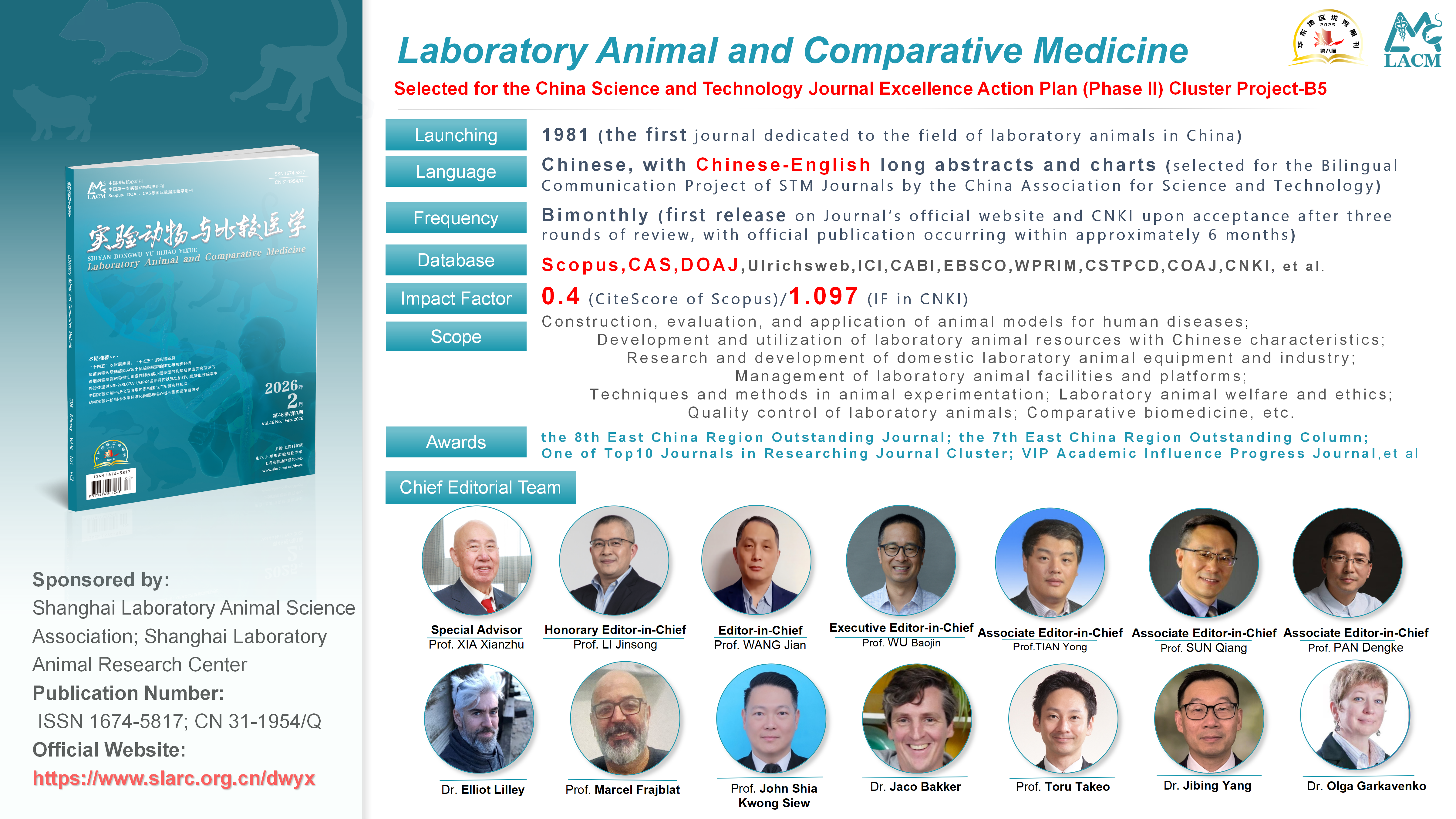
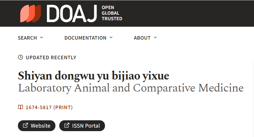
- LACM Successfully Indexed by Scopus Database
- Announcement for the 2024 Special Topic Draft of Labora...
- Honorary List of Outstanding Youth Editorial Committees...
- The journal is included in the WHO Western Pacific Regi...
- List of Thanks for the 2023 Reviewers
- List of Thanks from Supporting Units in 2023