实验动物与比较医学 ›› 2020, Vol. 40 ›› Issue (5): 367-.DOI: 10.3969/j.issn.1674-5817.2020.05.002

• 论著 • 上一篇    下一篇

不同低氧胁迫方式构建SD大鼠高原肺水肿模型的比较研究

林雪, 雷有芳, 蒲小燕   

  1. 青海大学医学院基础医学部,西宁 810016
  • 收稿日期:2020-02-26 出版日期:2020-10-15 发布日期:2020-11-19
  • 作者简介:林雪(1999—), 女, 临床医学专业在读本科生,主要研究低氧对机体的影响。E-mail: 1810845344@qq.com
  • 基金资助:

    青海省科技计划项目(2019-ZJ-7047); 

    国家级大学生创新创业训练计划(201910743003); 

    青海大学医学院中青年科技项目(2019-kyy-5)


Establishment of High Altitude Pulmonary Edema Model in SD Rats by Different Hypoxia Stress Methods

LIN Xue, LEI Youfang, PU Xiaoyan   

  1. Department of Basic Medicine, Medical College of Qinghai University, Xining 810016, China
  • Received:2020-02-26 Online:2020-10-15 Published:2020-11-19

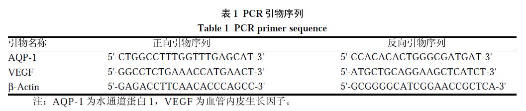
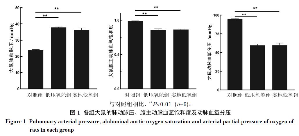
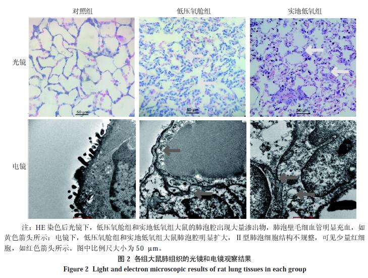
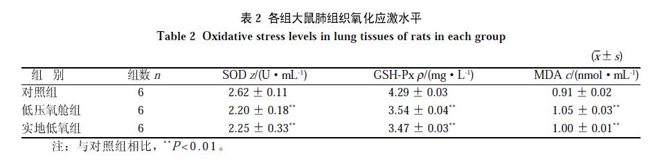
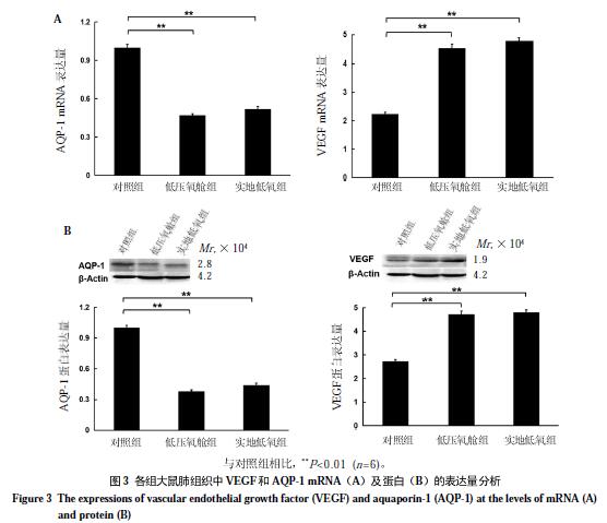
摘要: 目的    研究不同低氧胁迫方式构建高原肺水肿模型的效果。方法    将60只SD大鼠随机分为3组:对照组(海拔400 m),低压氧舱组(模拟海拔6 000 m低氧胁迫48 h),实地低氧组(海拔4 200 m低氧胁迫28 d);每组20只。通过检测实验大鼠肺组织的干湿比、形态学和病理生理学特征、关键基因水通道蛋白1(aquaporin-1,AQP-1)和血管内皮生长因子(vascular endothelial growth factor,VEGF)表达,以及氧化应激水平,比较不同低氧胁迫方式构建SD大鼠高原肺水肿模型的效果。结果    与对照组相比,低压氧舱组和实地低氧组大鼠的肺动脉压和肺组织含水量均显著升高(均P<0.01),而氧分压及氧饱和度显著下降(均P<0.01)。对照组肺组织形态在光镜和电镜下均显示结构正常;低压氧舱组和实地低氧组肺组织在光镜下均可见肺泡壁且肺泡间隔明显增宽,大量红细胞和炎性细胞溢出,且在肺泡间隔出现明显水肿。两实验组大鼠肺组织中AQP-1 mRNA和蛋白水平均较对照组明显升高(均P<0.01),VEGF mRNA和蛋白水平均较对照组明显降低(均P<0.01),而且血清中谷胱甘肽过氧化物酶(glutathione peroxidase,GSH-Px)和超氧化物歧化酶(superoxide dismutase,SOD)水平明显降低,丙二醛(malondialdehyde,MDA)水平明显升高。结论    低压氧舱模拟海拔6 000 m低氧胁迫48 h,以及海拔4 200 m实地低氧胁迫28 d,均可有效构建SD大鼠高原肺水肿模型;其中,应用低压氧舱构建SD大鼠高原肺水肿模型相对更占优势。

关键词: 高原急性肺水肿, 低氧, 低压氧舱, 实地低氧, 动物模型, SD大鼠

Abstract: Objective    To study the effects of different hypoxia stress on the construction of high altitude pulmonary edema (HAPE) model. Methods    Total of 60 SD rats were randomly divided into 3 groups: the control group (at an altitude of 400 m), the hypobaric oxygen chamber group (simulating altitude of 6 000 m, and hypoxia stress for 48 h), and the field hypoxia group (at an altitude of 4 200 m, 28 days of hypoxia stress), 20 animals in each group. By detecting lung tissue dry-wet ratio, morphological and pathophysiological characteristics, expressions of key genes including aquaporin-1 (AQP-1) and vascular endothelial growth factor (VEGF), and oxidative stress level, the effect of different hypoxia stress on the establishment of HAPE model in SD rats was compared. Results    Compared with the control group, the results of hypobaric oxygen chamber group and the field hypoxia group were as follows: the pulmonary artery pressure of the rats in the two groups increased significantly (P<0.01), while the oxygen partial pressure and oxygen saturation decreased significantly (both P<0.01); the lung tissue moisture content in both groups increased significantly (P<0.01). The lung tissue morphology of the control group was normal under a light microscope and an electron microscope, while the alveolar wall and alveolar septum of the two experimental groups were significantly widened under a light microscope, with a large number of red blood cells and inflammatory cells overflowing, and the obvious edema of alveolar septum was observed under a light microscope. The mRNA and protein levels of AQP-1 in the lung tissues of the two experimental groups were significantly higher than those of the control group, while the mRNA and protein levels of VEGF were significantly lower than those of the control group (all P<0.01). There were low GSH-Px and SOD levels and high MDA level in the serum of the two experimental groups. Conclusion     Both the hypobaric oxygen chamber simulating the hypoxia stress at an altitude of 6 000 m for 48 h and the field hypoxia stress at an altitude of 4 200 m for 28 d effectively can construct the HAPE model of SD rats, and the hypobaric oxygen chamber for constructing the SD rat model of HAPE was more dominant.


Key words: Acute pulmonary edema at high altitude, Hypoxia, Hypobaric oxygen chamber;