


实验动物与比较医学 ›› 2026, Vol. 46 ›› Issue (2): 191-204.DOI: 10.12300/j.issn.1674-5817.2025.084
宋静1,2,3, 杨宗统2( )(
)( ), 李晓晶2, 李自发1, 苏凤云3, 徐东川2, 隋在云2()()
收稿日期:2025-06-10
修回日期:2025-08-21
出版日期:2026-04-25
发布日期:2026-04-18
通讯作者:
杨宗统(1992—),男,博士,助理研究员,研究方向:中药免疫药理及药效学。E-mail: YangZT2021@163.com。ORCID:0009-0005-5401-7697;作者简介:宋 静(1987—),女,硕士研究生,研究方向:中药及其有效成分作用机制及安全性评价。E-mail:583023072@qq.com
基金资助:
SONG Jing1,2,3, YANG Zongtong2()(), LI Xiaojing2, LI Zifa1, SU Fengyun3, XU Dongchuan2, SUI Zaiyun2()()
Received:2025-06-10
Revised:2025-08-21
Published:2026-04-25
Online:2026-04-18
Correspondence to:
YANG Zongtong (ORCID: 0009-0005-5401-7697), E-mail: YangZT2021@163.com摘要:
目的 探讨泻白散对过敏性哮喘大鼠呼吸道和肠道黏膜免疫的作用机制。 方法 40只雄性SD大鼠按体重随机分为空白组、模型组、阳性药组和泻白散组,每组10只。模型组、阳性药组和泻白散组均使用卵清蛋白致敏法建立大鼠过敏性哮喘模型。自雾化激发期(第21天)开始,各组同时进行灌胃干预,即阳性药组大鼠灌胃地塞米松(0.068 mg/kg),泻白散组大鼠灌胃泻白散药液(2 g/mL,11.3 mL/kg),空白组与模型组大鼠灌胃等体积生理盐水,每日1次,连续14 d。将大鼠安乐死后,采集各组大鼠肺、肠组织,经HE染色观察组织病理学变化;透射电子显微镜观察组织超微病理结构;免疫组织化学法观察磷脂酰肌醇3-激酶(phosphatidylinositol 3-kinase,PI3K)和蛋白激酶B(protein kinase B,Akt)蛋白阳性面积。提取大鼠肺、肠组织中的总蛋白和总RNA,采用蛋白质印迹法和实时荧光定量PCR法分别检测PI3K和Akt基因的蛋白和mRNA表达水平。 结果 组织病理学结果显示,与空白组相比,模型组大鼠出现肺泡性肺气肿伴炎性细胞浸润,以及肠黏膜损伤伴炎性细胞浸润;肺组织细胞结构被破坏,细胞器数量减少,但肠超微结构病变较轻。与模型组相比,泻白散组大鼠肺组织病变程度较低,偶见肺泡壁损伤伴少量炎性细胞浸润,肠黏膜结构改善,腺体排列整齐,疏松、浸润等病理改变均见好转;肺组织细胞结构比较完整,病变程度减轻,肠组织无超微结构病理变化。免疫组织化学和蛋白质印迹结果显示,与空白组相比,模型组大鼠肺、肠组织中PI3K和Akt特异性阳性反应面积均显著增大(均P<0.001),PI3K和Akt蛋白表达量也均显著增加(均P<0.05);与模型组相比,泻白散组大鼠肺组织中Akt蛋白阳性面积显著减少(P<0.001),肠组织中PI3K阳性面积显著减少(P<0.000 1),同时肺、肠组织中PI3K和Akt蛋白表达量均显著减少(均P<0.01)。实时荧光定量PCR结果显示,与空白组相比,模型组大鼠肺、肠组织中PI3K、Akt基因的mRNA表达量均显著增加(均P<0.05);与模型组相比,泻白散组大鼠肺、肠组织中PI3K、Akt基因的mRNA表达量均显著减少(均P<0.05)。 结论 泻白散可通过抑制过敏性哮喘大鼠肺、肠组织中PI3K-Akt信号通路关键核酸和蛋白表达,改善肺组织形态结构,保护肠道黏膜完整性,并调节肠道黏膜免疫功能等机制,发挥对过敏性哮喘大鼠的保护作用。
中图分类号:
宋静,杨宗统,李晓晶,等. 泻白散对过敏性哮喘大鼠肺、肠组织形态结构及PI3K和Akt表达水平的影响[J]. 实验动物与比较医学, 2026, 46(2): 191-204. DOI: 10.12300/j.issn.1674-5817.2025.084.
SONG Jing,YANG Zongtong,LI Xiaojing,et al. Effects of Xiebai San on the Morphological Structures of Lung and Intestinal Tissues and Expression Levels of PI3K and Akt in Rats with Allergic Asthma[J]. Laboratory Animal and Comparative Medicine, 2026, 46(2): 191-204. DOI: 10.12300/j.issn.1674-5817.2025.084.
基因名称 Gene name | 引物序列(5'→3') Primer sequence (5'→3') | 产物大小/bp Product size/bp |
|---|---|---|
| PI3K | F: GCGTGACATGTAGGCTCTCG | 349 |
| R: GGGCAGTGCTGGTGGAT | ||
| Akt | F: CCGCCTGATCAAGTTCTCCT | 118 |
| R: TTCAGATGATCCATGCGGGG | ||
| β-actin | F: CGCAGCCACTGTCGAGTC | 96 |
| R: GTCATCCATGGCGAACTGGT |
表1 引物设计与合成
Table 1 Primer design and synthesis
基因名称 Gene name | 引物序列(5'→3') Primer sequence (5'→3') | 产物大小/bp Product size/bp |
|---|---|---|
| PI3K | F: GCGTGACATGTAGGCTCTCG | 349 |
| R: GGGCAGTGCTGGTGGAT | ||
| Akt | F: CCGCCTGATCAAGTTCTCCT | 118 |
| R: TTCAGATGATCCATGCGGGG | ||
| β-actin | F: CGCAGCCACTGTCGAGTC | 96 |
| R: GTCATCCATGGCGAACTGGT |
程序 Program | 温度/℃ Temperature/℃ | 时间 Time |
|---|---|---|
预变性 Initial denaturation | 95 | 3 min |
扩增 Amplification | 95 | 20 s |
45个循环 45 cycles | 61 | 15 s |
熔解曲线 Melting curve | 95 | 5 s |
| 65 | 1 min | |
| 97 | 10 s |
表2 PCR反应程序
Table 2 PCR cycling program
程序 Program | 温度/℃ Temperature/℃ | 时间 Time |
|---|---|---|
预变性 Initial denaturation | 95 | 3 min |
扩增 Amplification | 95 | 20 s |
45个循环 45 cycles | 61 | 15 s |
熔解曲线 Melting curve | 95 | 5 s |
| 65 | 1 min | |
| 97 | 10 s |
组别 Group | 肺气肿积分 Pulmonary emphysema score | 肺充/出血积分 Pulmonary congestion/bleeding score | 肺间质增厚积分 Pulmonary interstitial thickening score | 炎细胞浸润积分 Inflammatory cell infiltration score | 总积分 Total score |
|---|---|---|---|---|---|
空白组 Control group | 0.00±0.00 | 0.67±0.58 | 0.67±0.58 | 0.67±0.58 | 2.00±1.00 |
模型组 Model group | 2.56±0.53** | 1.56±0.53* | 1.89±0.60* | 1.89±0.78* | 7.89±1.05** |
阳性药组 Positive control group | 1.86±0.69## | 1.43±0.53 | 2.14±0.38 | 1.57±0.53 | 7.00±1.15 |
泻白散组 Xiebai San group | 1.46±0.88## | 1.54±0.52 | 1.46±0.52 | 1.46±0.52 | 5.92±1.26# |
表3 泻白散给药后大鼠肺组织McGuigan病理评分的比较 (n=8,
Table 3 Comparison of McGuigan pathological scores in lung tissues of rats after Xiebai San administration
组别 Group | 肺气肿积分 Pulmonary emphysema score | 肺充/出血积分 Pulmonary congestion/bleeding score | 肺间质增厚积分 Pulmonary interstitial thickening score | 炎细胞浸润积分 Inflammatory cell infiltration score | 总积分 Total score |
|---|---|---|---|---|---|
空白组 Control group | 0.00±0.00 | 0.67±0.58 | 0.67±0.58 | 0.67±0.58 | 2.00±1.00 |
模型组 Model group | 2.56±0.53** | 1.56±0.53* | 1.89±0.60* | 1.89±0.78* | 7.89±1.05** |
阳性药组 Positive control group | 1.86±0.69## | 1.43±0.53 | 2.14±0.38 | 1.57±0.53 | 7.00±1.15 |
泻白散组 Xiebai San group | 1.46±0.88## | 1.54±0.52 | 1.46±0.52 | 1.46±0.52 | 5.92±1.26# |
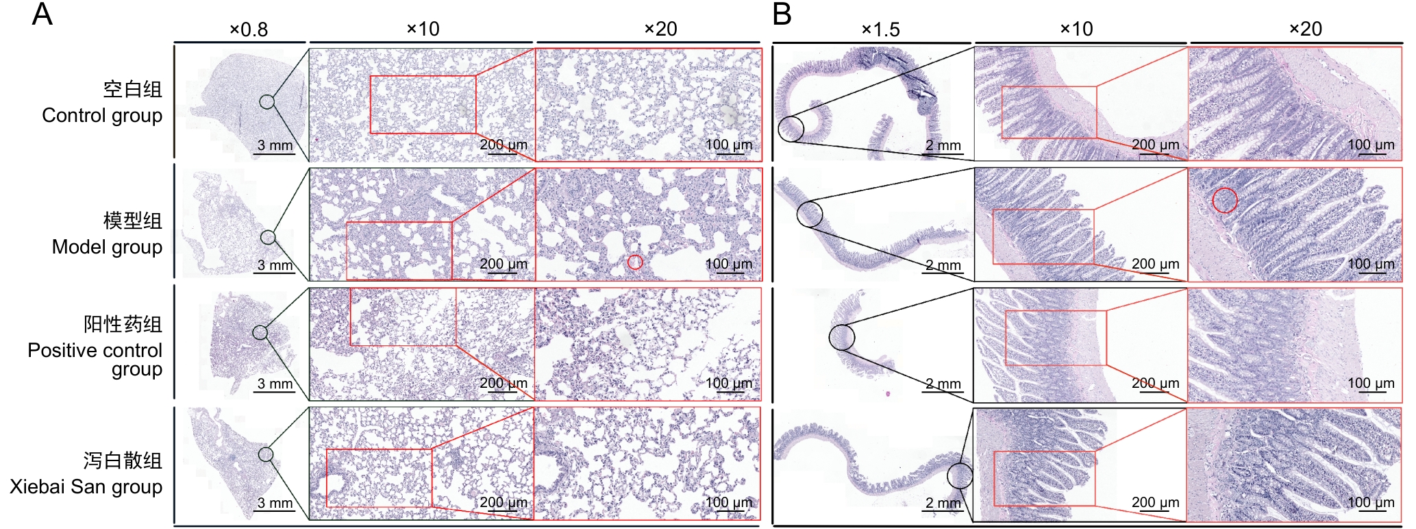
图1 泻白散给药对哮喘大鼠肺、肠组织病理变化的影响
Figure 1 Effects of Xiebai San on histopathological changes in lung and intestinal tissues of asthmatic rats
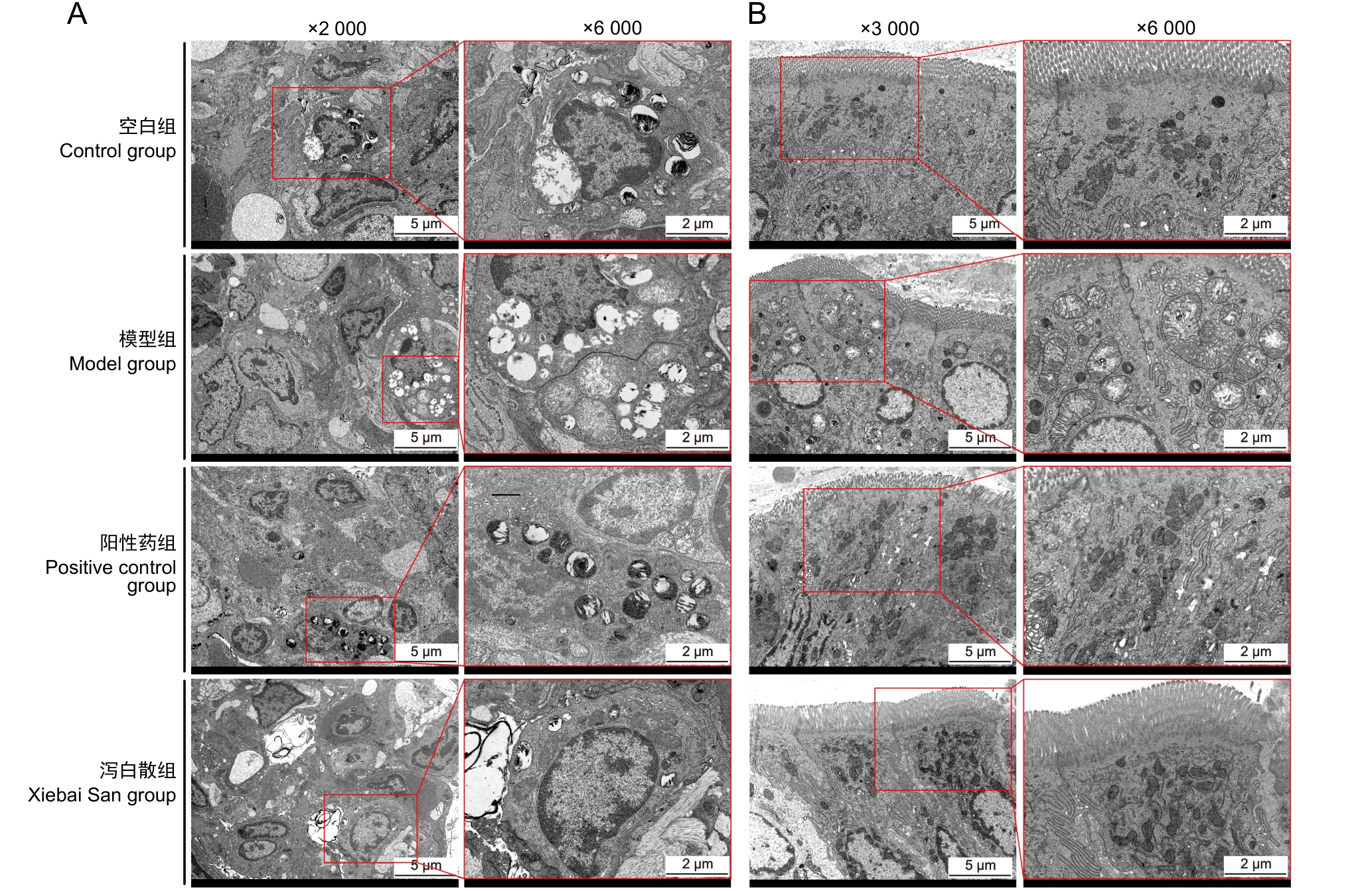
图2 泻白散给药对哮喘大鼠肺、肠组织超微病理结构的影响
Figure 2 Effects of Xiebai San administration on ultrastructural pathology of lung and intestinal tissues in asthmatic rats
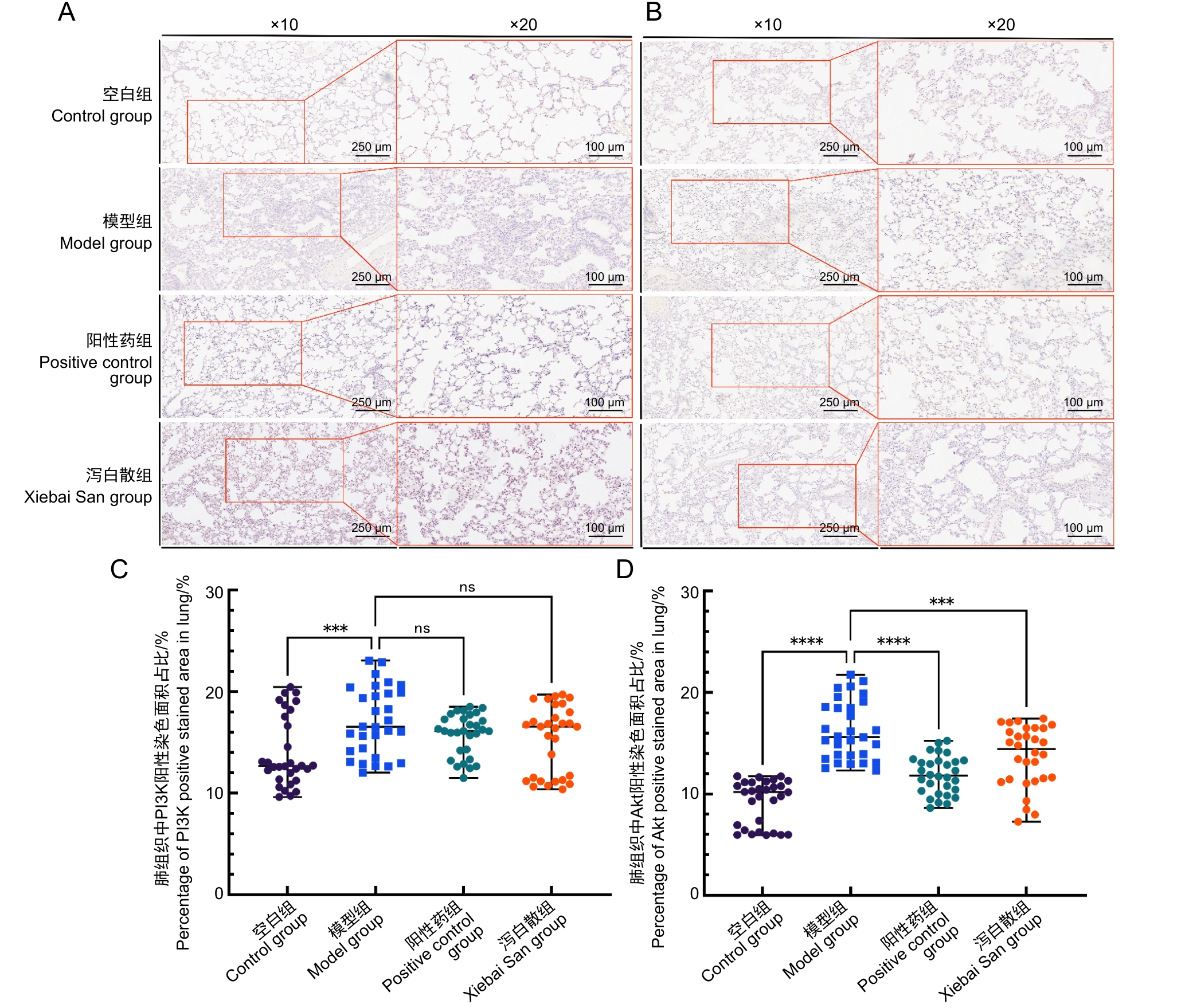
图3 泻白散给药对哮喘大鼠肺组织中PI3K、Akt蛋白分布的影响
Figure 3 Effects of Xiebai San administration on PI3K and Akt protein distribution in lung tissues of asthmatic rats
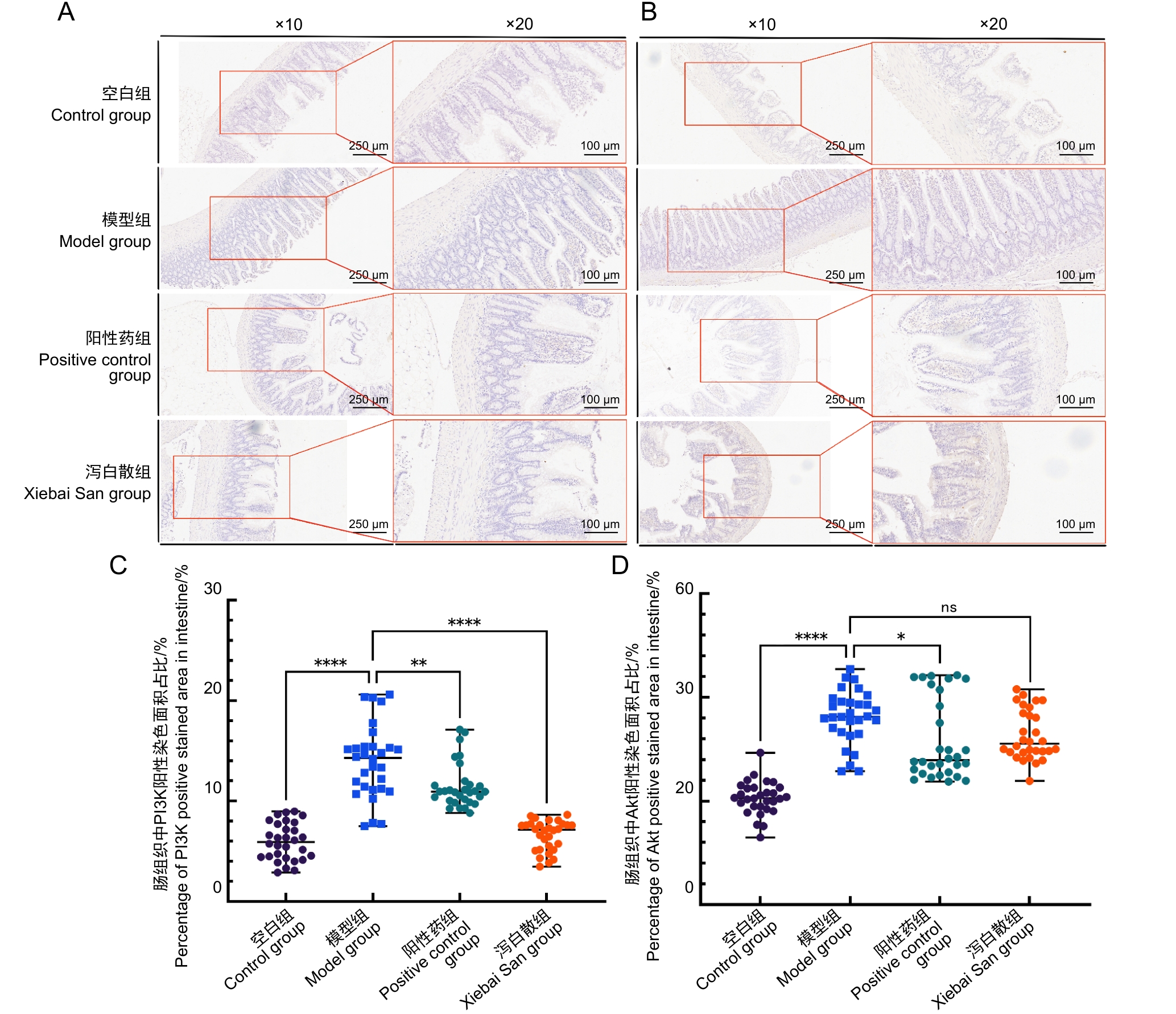
图4 泻白散给药对哮喘大鼠肠组织中PI3K、Akt蛋白分布的影响
Figure 4 Effects of Xiebai San administration on PI3K and Akt protein distribution in intestinal tissues of asthmatic rats
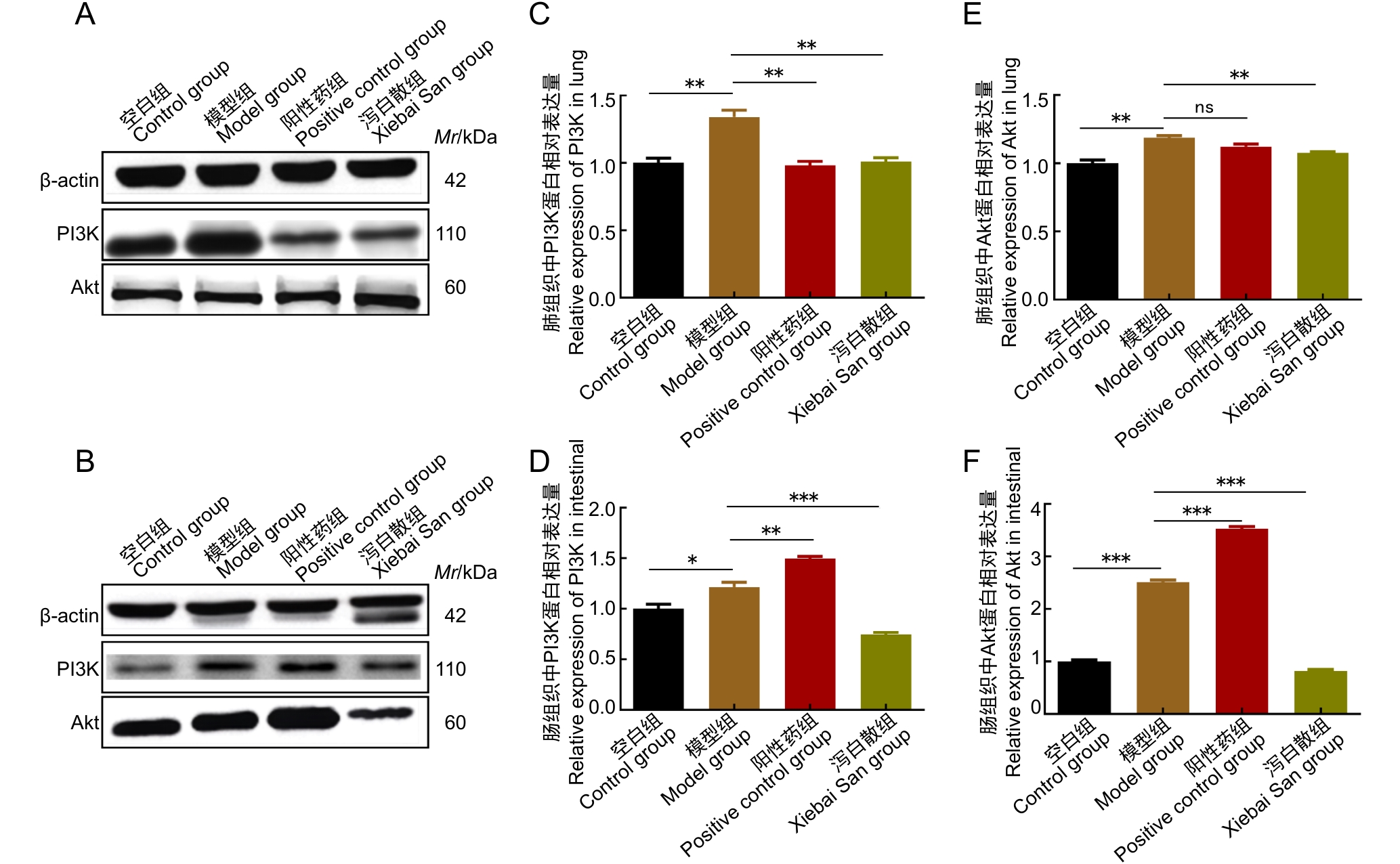
图5 泻白散给药对哮喘大鼠肺、肠组织PI3K、Akt蛋白表达的影响
Figure 5 Effects of Xiebai San administration on PI3K and Akt protein expression in lung and intestinal tissues of asthmatic rats
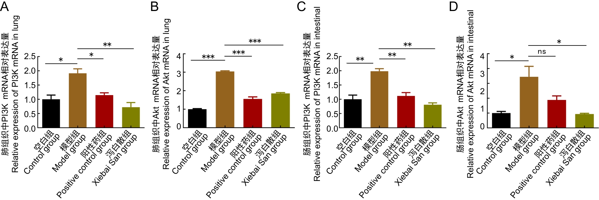
图6 泻白散给药对哮喘大鼠肺、肠组织PI3K、Akt基因表达的影响
Figure 6 Effects of Xiebai San administration on PI3K and Akt gene expression in lung and intestinal tissues of asthmatic rats
| [1] | 任雪交, 黄巍, 邱菲菲, 等. 支气管哮喘动物模型研究的文献分析与评价[J]. 实验动物与比较医学, 2022, 42(1): 74-80. DOI:10.12300/j.issn.1674-5817.2021.044 . |
| REN X J, HUANG W, QIU F F, et al. Progress in animal models for bronchial asthma[J]. Lab Anim Comp Med, 2022, 42(1): 74-80. DOI:10.12300/j.issn.1674-5817.2021.044 . | |
| [2] | 罗世雄, 张赛, 陈慧. 常见哮喘动物模型的建立方法与评价研究进展[J]. 实验动物与比较医学, 2025, 45(2): 167-175. DOI:10.12300/j.issn.1674-5817.2024.120 . |
| LUO S X, ZHANG S, CHEN H. Research progress in establishment and evaluation of common asthma animal models[J]. Lab Anim Comp Med, 2025, 45(2): 167-175. DOI:10.12300/j.issn.1674-5817.2024.120 . | |
| [3] | 兰露莎, 赵兵兵, 杨红宇, 等. 氧化苦参碱对小鼠哮喘模型保护作用的初步研究[J]. 实验动物与比较医学, 2017, 37(4): 320-323. DOI:10.3969/j.issn.1674-5817.2017.04.013 . |
| LAN L S, ZHAO B B, YANG H Y, et al. The protective effect of oxymatrine on asthma mice[J]. Lab Anim Comp Med, 2017, 37(4): 320-323. DOI:10.3969/j.issn.1674-5817.2017.04.013 . | |
| [4] | AN J, LIU Y Q, WANG Y Q, et al. The role of intestinal mucosal barrier in autoimmune disease: a potential target[J]. Front Immunol, 2022, 13:871713. DOI:10.3389/fimmu.2022.871713 . |
| [5] | CERVANTES-GARCÍA D, JIMÉNEZ M, RIVAS-SANTIAGO C E, et al. Lactococcus lactis NZ9000 prevents asthmatic airway inflammation and remodelling in rats through the improvement of intestinal barrier function and systemic TGF-β production[J]. Int Arch Allergy Immunol, 2021, 182(4):277-291. DOI:10.1159/000511146 . |
| [6] | 洪天一. 蝎黄解痉治哮颗粒影响NF-κB和HIF-1α信号传导通路治疗哮喘的调控机制[D]. 长春: 长春中医药大学, 2020. DOI: 10.26980/d.cnki.gcczc.2020.000459 . |
| HONG T Y. Regulatory mechanism of Xiehuang Jiezhuan Zhixiao granules on the NF-κB and HIF-1α signaling pathways in the treatment of asthma[D]. Changchun: Changchun University of Chinese Medicine, 2020. DOI: 10.26980/d.cnki.gcczc.2020.000459 . | |
| [7] | 马兰兰, 陈玲, 王琴, 等. 肺通气功能正常的儿童支气管哮喘控制情况及急性发作随访的研究[J]. 中国当代儿科杂志, 2024, 26(5): 476-480. DOI:10.7499/j.issn.1008-8830.2311149 . |
| MA L L, CHEN L, WANG Q, et al. Control status and follow-up of acute attacks in children with bronchial asthma with normal pulmonary ventilation function[J]. Chin J Contemp Pediatr, 2024, 26(5): 476-480. DOI:10.7499/j.issn.1008-8830.2311149 . | |
| [8] | 丁云录, 郑明昱, 南敏伦, 等. 基于ERK信号通路探讨鹿茸大补汤颗粒对哮喘缓解期豚鼠的调控及作用机制[J]. 中国老年学杂志, 2023, 43(21):5309-5313. DOI:10.3969/j.issn.1005-9202.2023.21.054 . |
| DING Y L, ZHENG M Y, NAN M L, et al. Based on ERK signal pathway, this paper discusses the regulation and mechanism of Lulong Dabutang Granule on guinea pigs with asthma in remission stage[J]. Chin J Gerontol, 2023, 43(21):5309-5313. DOI:10.3969/j.issn.1005-9202.2023.21.054 . | |
| [9] | 李玉丽, 易腾达, 谭志强, 等. 经典名方泻白散的源流及古今应用考究[J]. 中国实验方剂学杂志, 2021, 27(4):168-174. DOI:10.13422/j.cnki.syfjx.20202328 . |
| LI Y L, YI T D, TAN Z Q, et al. Literature research on origin and application of classical prescription xiebaisan[J]. Chin J Exp Tradit Med Formulae, 2021, 27(4):168-174. DOI:10.13422/j.cnki.syfjx.20202328 . | |
| [10] | 刘凌志, 梁敏兰, 郭栋伟. 黄芩泻白散对支气管哮喘小鼠的治疗作用及Wnt/β-catenin信号轴的影响[J]. 湖南中医杂志, 2025, 41(6):140-146. DOI:10.16808/j.cnki.issn1003-7705.2025.06.028 . |
| LIU L Z, LIANG M L, GUO D W. Therapeutic effect of Huangqin Xiebai powder on rats with bronchial asthma and its effect on the Wnt/β-catenin signaling axis[J]. Hunan J Tradit Chin Med, 2025, 41(6):140-146. DOI:10.16808/j.cnki.issn1003-7705.2025.06.028 . | |
| [11] | 张天柱, 张景龙, 樊湘泽, 等. 泻白散对小鼠过敏性哮喘气道炎症的作用及机制[J]. 中国实验方剂学杂志, 2014, 20(20):173-177. DOI:10.13422/j.cnki.syfjx.2014200173 . |
| ZHANG T Z, ZHANG J L, FAN X Z, et al. Effects and its mechanism of Xiebai San on allergic airway inflammation in asthma mouse[J]. Chin J Exp Tradit Med Formulae, 2014, 20(20):173-177. DOI:10.13422/j.cnki.syfjx.2014200173 . | |
| [12] | JUTEL M, MOSNAIM G S, BERNSTEIN J A, et al. The One Health approach for allergic diseases and asthma[J]. Allergy, 2023, 78(7):1777-1793. DOI:10.1111/all.15755 . |
| [13] | JOHNSON C C, OWNBY D R. The infant gut bacterial microbiota and risk of pediatric asthma and allergic diseases[J]. Transl Res, 2017, 179:60-70. DOI:10.1016/j.trsl.2016.06.010 . |
| [14] | 杨阳, 张桂信, 杨琦, 等. 从肠-肺轴相关微环境角度诠释中医"肺与大肠相表里"的新内涵[J]. 中国中西医结合外科杂志, 2025, 31(02): 295-299. DOI:10.3969/j.issn.1007-6948.2025.02.027 . |
| YANG Y, ZHANG G X, YANG Q, et al. Interpretation of the new connotation of "lung and large intestine are exterior and interior" in traditional Chinese medicine from the perspective of microenvironment related to intestine-lung axis[J]. Chin J Surg Integr Tradit West Med, 2025, 31(2): 295-299. DOI:10.3969/j.issn.1007-6948.2025.02.027 . | |
| [15] | 刘寻, 周婷婷, 朱紫冰, 等. 槟榔碱通过上调PI3K/Akt/mTOR信号通路促进口腔黏膜下纤维化的体内外实验研究[J]. 中国临床药理学与治疗学, 2025, 30(07): 865-875. DOI:10.12092/j.issn.1009-2501.2025.07.001 . |
| LIU X, ZHOU T T, ZHU Z B, et al. Arecoline promotes oral submucous fibrosis by upregulating PI3K/Akt/mTOR signaling pathway in vivo and in vitro [J]. Chin J Clin Pharmacol Ther, 2025, 30(07): 865-875. DOI:10.12092/j.issn.1009-2501.2025.07.001 . | |
| [16] | YAO T, WU Y H, FU L Y, et al. Christensenellaceae minuta modulates epithelial healing via PI3K-AKT pathway and macrophage differentiation in the colitis[J]. Microbiol Res, 2024, 289:127927. DOI:10.1016/j.micres.2024.127927 . |
| [17] | ACOSTA-MARTINEZ M, CABAIL M Z. The PI3K/Akt pathway in meta-inflammation[J]. Int J Mol Sci, 2022, 23(23):15330. DOI:10.3390/ijms232315330 . |
| [18] | 徐东川. 基于网络药理学方法研究泻白散治疗哮喘的作用机制[D]. 济南: 山东中医药大学, 2022. DOI:10.27282/d.cnki.gsdzu.2022.000841 . |
| XU D C. Investigating the mechanism of Xie Bai San in the treatment of asthma using network pharmacology methods[D]. Jinan: Shandong University of Traditional Chinese Medicine, 2022. DOI:10.27282/d.cnki.gsdzu.2022.000841 . | |
| [19] | MCGUIGAN R M, MULLENIX P, NORLUND L L, et al. Acute lung injury using oleic acid in the laboratory rat: establishment of a working model and evidence against free radicals in the acute phase[J]. Curr Surg, 2003, 60(4):412-417. DOI:10.1016/S0149-7944(02)00775-4 . |
| [20] | 覃芳芳, 丘琴, 韦红杏, 等. 泻白散的研究进展及质量标志物(Q-Marker)预测分析[J]. 中华中医药学刊, 2025, 43(11): 155-161. DOI:10.13193/j.issn.1673-7717.2025.11.031 . |
| QIN F F, QIU Q, WEI H X, et al. Research progress of Xiebai Powder and prediction analysis of quality markers (Q-markers)[J]. Chin Arch Tradit Chin Med, 2025, 43(11): 155-161. DOI:10.13193/j.issn.1673-7717.2025.11.031 . | |
| [21] | LAMPIASI N. Macrophage polarization: learning to manage it 2.0[J]. Int J Mol Sci, 2023, 24(24):17409. DOI: 10.3390/ijms242417409 . |
| [22] | 张会敏, 杨宗统, 苏本正, 等. 高效液相色谱指纹图谱评价泻白散及其3种单味药内在质量[J]. 化学分析计量, 2021, 30(9): 48-53. DOI:10.3969/j.issn.1008 –6145.2021.09.011. |
| ZHANG H M, YANG Z T, SU B Z, et al. Evaluation of internal quality of Xiebai powder and its three single herbs by HPLC fingerprint[J]. Chem Anal Meterage, 2021, 30(9): 48-53. DOI:10.3969/j.issn.1008 –6145.2021.09.011. | |
| [23] | 徐东川, 刘瑾, 李晓晶, 等. 泻白散大鼠体内入血成分研究[J]. 中国药房, 2022, 33(1):38-45. DOI:10.6039/j.issn.1001-0408.2022.01.07 . |
| XU D C, LIU J, LI X J, et al. Study on absorbed components of Xiebai powder in rat blood[J]. China Pharm, 2022, 33(1):38-45. DOI:10.6039/j.issn.1001-0408.2022.01.07 . | |
| [24] | 杨宗统, 徐东川, 刘瑾, 等. 基于非靶向代谢组学和肠道菌群探究经典名方泻白散对过敏性哮喘大鼠的保护作用[J]. 中国实验动物学报, 2024, 32(2): 177-189. DOI:10.3969/j.issn.1005-4847.2024.02.005 . |
| YANG Z T, XU D C, LIU J, et al. Protective effect of Xiebaisan on allergic asthma in rats based on non-targeted metabolomics and intestinal bacterial flora[J]. Acta Lab Anim Sci Sin, 2024, 32(2): 177-189. DOI:10.3969/j.issn.1005-4847.2024.02.005 . | |
| [25] | WANG J, HE M, YANG M, et al. Gut microbiota as a key regulator of intestinal mucosal immunity[J]. Life Sci, 2024, 345:122612. DOI: 10.1016/j.lfs.2024.122612 . |
| [26] | ZHENG S H, XUE T Y, WANG B, et al. Chinese medicine in the treatment of ulcerative colitis: the mechanisms of signaling pathway regulations[J]. Am J Chin Med, 2022, 50(7): 1781-1798. DOI: 10.1142/S0192415X22500756 . |
| [27] | YAO T, WU Y H, FU L Y, et al. Christensenellaceae minuta modulates epithelial healing via PI3K-AKT pathway and macrophage differentiation in the colitis[J]. Microbiol Res, 2024, 289:127927. DOI: 10.1016/j.micres.2024.127927 . |
| [28] | ACOSTA-MARTINEZ M, CABAIL M Z. The PI3K/Akt pathway in meta-inflammation[J]. Int J Mol Sci, 2022, 23(23): 15330. DOI: 10.3390/ijms232315330 . |
| [29] | ZHANG M, SONG X, LIU S B, et al. Magnolin inhibits intestinal epithelial cell apoptosis alleviating Crohn's disease-like colitis by suppressing the PI3K/AKT signaling pathway[J]. Int Immunopharmacol, 2024, 134: 112181. DOI: 10.1016/j.intimp. 2024.112181 |
| [30] | 王百乔, 林小茹, 韩敏, 等. 二甲双胍对阿尔茨海默症模型大鼠认知功能障碍及PI3K/Akt通路的影响[J]. 实验动物与比较医学, 2021, 41(4): 313-320. DOI:10.12300/j.issn.1674-5817.2020.185 . |
| WANG B Q, LIN X R, HAN M, et al. Effects of metformin on cognitive dysfunction and PI3K/Akt pathway in Alzheimer's disease rats[J]. Lab Anim Comp Med, 2021, 41(4): 313-320. DOI:10.12300/j.issn.1674-5817.2020.185 . |
| [1] | 汤建平, 赵丽亚, 赵莹. 常用近交系大鼠微卫星遗传位点筛选及分析[J]. 实验动物与比较医学, 2026, 46(3): 388-396. |
| [2] | 李嘉菲, 张朕豪, 王硕, 田歌, 温爽, 闫玉雪, 崔然, 叶振, 崔永春. 自主神经调节剂对房颤模型大鼠心房电重塑及组织病理变化的影响[J]. 实验动物与比较医学, 2026, 46(3): 321-331. |
| [3] | 艾秀峰, 张利棕, 方明笋, 吕东颖, 陈楚, 蔡兆伟, 王德军. 16S rRNA测序分析饮用含氯水对大鼠和小鼠肠道菌群的影响差异[J]. 实验动物与比较医学, 2026, 46(3): 437-445. |
| [4] | 唐小杭, 谷颖敏, 吕阳阳, 黄明姝, 田雪松. 一种自研固定液用于制备大鼠眼球切片的组织学染色效果评价[J]. 实验动物与比较医学, 2026, 46(2): 261-270. |
| [5] | 姜海涛, 袁韩涛, 黄雯婷, 杨蓉蓉, 陈晓春, 禹宝庆, 李四波. 腰舒逐瘀方通过miR-17-5P/MDM2/p53通路调控大鼠椎间盘纤维环细胞增殖与凋亡[J]. 实验动物与比较医学, 2026, 46(1): 55-65. |
| [6] | 高超奇, 祝志波, 孙显东. 大鼠血管重构模型的应用进展与分类分析[J]. 实验动物与比较医学, 2025, 45(5): 542-550. |
| [7] | 罗一凡, 张臻玮, 梅璐, 史叶萍, 邢艺彤, 张泽奇, 李楚欣, 韩春霞, 杨平顺, 陈秋生. 特络细胞介导肥胖症大鼠膏摩寡肽中草药复合制剂的减肥作用及其机制[J]. 实验动物与比较医学, 2025, 45(5): 551-560. |
| [8] | 曹星新, 李艾亦, 侯婧涵, 李明学, 李艳艳, 靳玮华, 杨凤梅, 段素琴, 和占龙. 黄芪或其成分治疗急性胰腺炎的动物实验Meta分析[J]. 实验动物与比较医学, 2025, 45(5): 561-573. |
| [9] | 秦超, 李双星, 赵婷婷, 蒋晨晨, 赵晶, 杨艳伟, 林志, 王三龙, 文海若. 药物安全评价用SD大鼠90 d喂养试验的背景数据研究[J]. 实验动物与比较医学, 2025, 45(4): 439-448. |
| [10] | 刘力瑜, 嵇波, 刘小玄, 方洋, 张玲, 郭亭廷, 全烨, 李鹤文, 刘翼天. 大鼠胎儿期肺组织固定方法的探索[J]. 实验动物与比较医学, 2025, 45(4): 432-438. |
| [11] | 肖林林, 杨逸萱, 黎珊杉, 罗兰诗雨, 尹思威, 孙俊铭, 施维, 欧阳轶强, 李习艺. 利用脑立体定位技术将人源三突变APP基因导入海马区构建阿尔茨海默病大鼠模型[J]. 实验动物与比较医学, 2025, 45(3): 269-278. |
| [12] | 刘智伟, 杨然, 连浩, 张玉, 金立伦. 秦皮素对碘乙酸钠诱导骨关节炎模型大鼠的软骨保护与抗炎作用[J]. 实验动物与比较医学, 2025, 45(3): 259-268. |
| [13] | 姜萌, 郝淑兰, 仝立国, 仲启明, 高振飞, 王永辉, 王晞星, 吉海杰. 长春瑞滨诱导大鼠足背静脉炎模型的动态评价[J]. 实验动物与比较医学, 2025, 45(3): 251-258. |
| [14] | 潘颐聪, 蒋汶洪, 胡明, 覃晓. 慢性肾脏病大鼠主动脉钙化模型的术式优化及效果评价[J]. 实验动物与比较医学, 2025, 45(3): 279-289. |
| [15] | 连辉, 姜艳玲, 刘佳, 张玉立, 谢伟, 薛晓鸥, 李健. 异常子宫出血大鼠模型的构建与评价[J]. 实验动物与比较医学, 2025, 45(2): 130-146. |
| 阅读次数 | ||||||
|
全文 |
|
|||||
|
摘要 |
|
|||||