




实验动物与比较医学 ›› 2025, Vol. 45 ›› Issue (5): 542-550.DOI: 10.12300/j.issn.1674-5817.2025.045
高超奇1,2( ), 祝志波1,2, 孙显东1,2()(
 )
)
收稿日期:2025-03-24
修回日期:2025-06-06
出版日期:2025-10-25
发布日期:2025-10-23
通讯作者:
作者简介:高超奇(1996—),女,硕士研究生,住院医师,研究方向:心血管疾病。E-mail:1361911514@qq.com。ORCID:0009-0000-4783-5230
基金资助:
GAO Chaoqi1,2(), ZHU Zhibo1,2, SUN Xiandong1,2()()
Received:2025-03-24
Revised:2025-06-06
Published:2025-10-25
Online:2025-10-23
Contact:
SUN Xiandong (ORCID: 0009-0005-1847-6810) , E-mail: sunxiandong@vip.sina.com摘要:
心血管疾病(cardiovascular diseases, CVD)具有较高的发病率、致残率和致死率等特点,是全球人类死亡的主要原因之一。血管重构是指在生理或病理条件下血管结构和功能的改变,通常发生在机体受损修复、疾病发展等过程。通过探究血管重构的机制有助于人们深入了解CVD的演变过程,从而开发出更为有效的早期诊疗方案,为CVD的防治提供新思路。血管重构的造模方法是研究血管重构的基石,血管重构模型用于多种疾病机制研究已取得了显著进展,尤其在动脉粥样硬化、高血压及血管重构等领域的应用中具有重要意义。常见的血管重构模型动物包括大鼠、小鼠、猪等,研究手段涵盖了机械损伤、药物干预、遗传改造等。不同类型的动物模型各有优势,如小鼠和大鼠模型适用于基因研究和高通量筛选,兔和猴模型因其接近人类病理而更有助于模拟临床条件下的血管重构。其中,大鼠模型因具有经济、易操作等特点而活跃在医疗保卫战的“一线”。当前血管重构模型主要依赖于经典方法(如颈动脉球囊损伤法、结扎法和动脉钳夹术),并联合新兴的饮食法(如高脂饮食、高盐饮食)来构建。根据不同的实验需求选择不同的大鼠建模方法并结合使用可以有效模拟血管重构的不同机制,为CVD的研究提供可靠的动物模型。此外,这些大鼠模型能够反映不同病理状态下的血管反应,为药物研发和疾病治疗策略的制定提供重要的实验依据。尽管这些大鼠模型为血管重构的研究提供了宝贵的工具,但仍面临模型差异性大、可重复性差及与临床表现存在差异等问题,未来的研究应致力于改进现有模型的精确性和可靠性,以发展新的动物模型。本文以大鼠为例总结了目前血管重构模型的研究进展、模型类型以及其在实验中的应用,特别是在CVD和血管重构研究中的价值,并且通过回顾不同大鼠血管重构模型的优缺点,为未来血管重构相关的研究提供理论参考。
中图分类号:
高超奇,祝志波,孙显东. 大鼠血管重构模型的应用进展与分类分析[J]. 实验动物与比较医学, 2025, 45(5): 542-550. DOI: 10.12300/j.issn.1674-5817.2025.045.
GAO Chaoqi,ZHU Zhibo,SUN Xiandong. Application Progress and Classification Analysis of Rat Vascular Remodeling Models[J]. Laboratory Animal and Comparative Medicine, 2025, 45(5): 542-550. DOI: 10.12300/j.issn.1674-5817.2025.045.
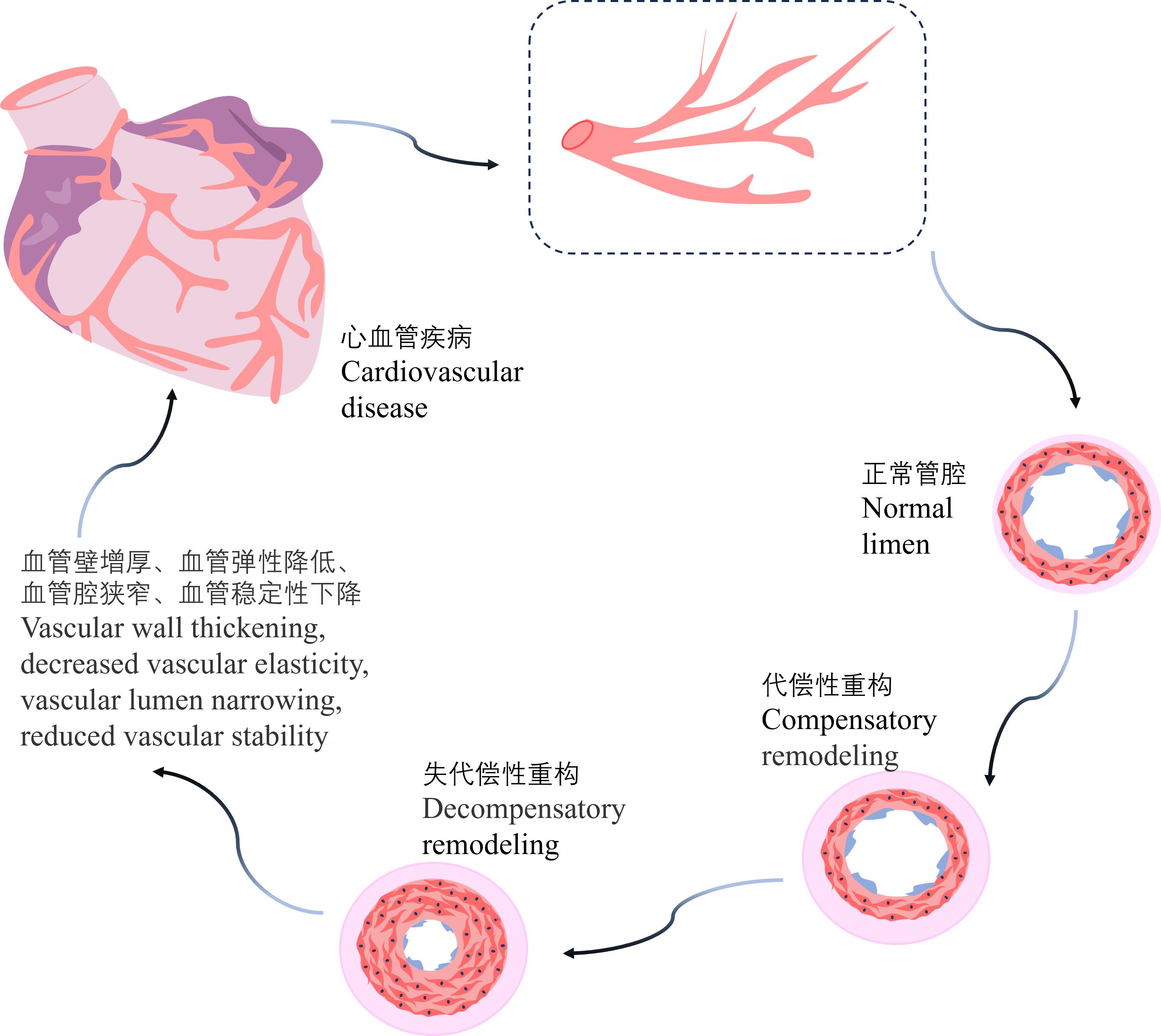
图 1 血管重构示意图
Figure 1 Schematic diagram of vascular remodeling
实验动物 Laboratory animals | 特点 Characteristics | 局限性 Limitations |
|---|---|---|
小鼠 Mice | 体型小;繁殖快;成本低;基因背景明确 | 生理结构与人类不同;寿命短;稳定构建手术模型难度大;血液样本少 |
大鼠 Rats | 便于手术性操作;药物代谢接近人类;器官系统与人类相似;认知和社会行为多样 | 基因编辑难度大 |
兔 Rabbits | 可长期生理监测;免疫系统与人相似;眼睛结构与人接近;皮肤敏感性与人相近 | 繁殖周期长;成本高;个体差异大;易受到环境影响 |
猪 Pigs | 解剖系统与人类相似;寿命长 | 饲养成本高;公众接受度较低;存在伦理问题 |
犬 Dogs | 心血管、消化系统与人类相似;适合长期及慢性病的研究 | 饲养成本高;伦理问题敏感 |
非人灵长类 Non-human primates | 高度拟人性 | 饲养和管理复杂;价格昂贵;伦理问题复杂;个体差异显著 |
表 1 血管重构研究常用实验动物的特点与局限性
Table 1 Characteristics and limitations of commonly used laboratory animals in vascular remodeling research
实验动物 Laboratory animals | 特点 Characteristics | 局限性 Limitations |
|---|---|---|
小鼠 Mice | 体型小;繁殖快;成本低;基因背景明确 | 生理结构与人类不同;寿命短;稳定构建手术模型难度大;血液样本少 |
大鼠 Rats | 便于手术性操作;药物代谢接近人类;器官系统与人类相似;认知和社会行为多样 | 基因编辑难度大 |
兔 Rabbits | 可长期生理监测;免疫系统与人相似;眼睛结构与人接近;皮肤敏感性与人相近 | 繁殖周期长;成本高;个体差异大;易受到环境影响 |
猪 Pigs | 解剖系统与人类相似;寿命长 | 饲养成本高;公众接受度较低;存在伦理问题 |
犬 Dogs | 心血管、消化系统与人类相似;适合长期及慢性病的研究 | 饲养成本高;伦理问题敏感 |
非人灵长类 Non-human primates | 高度拟人性 | 饲养和管理复杂;价格昂贵;伦理问题复杂;个体差异显著 |
成分 Components | 机制 Mechanisms | 目的 Objectives |
|---|---|---|
胆固醇 Cholesterol | 激活炎症反应;加重氧化应激;促进管壁硬化 | 提供胆固醇来源;影响机体血脂水平 |
丙基硫氧嘧啶 Propylthiouracil | 抑制甲状腺功能 | 降低机体胆固醇代谢 |
胆酸钠 Sodium cholate | 提高胆固醇吸收率 | 增加机体胆固醇含量 |
蔗糖 Sucrose | 转化成脂肪 | 提供脂肪来源;改善整体味道;增强大鼠食欲 |
猪油 Lard | — | 提供脂肪来源;刺激大鼠食欲 |
普通饲料 Ordinary feed | — | 提供营养;平衡膳食 |
表 2 血管重构造模用高脂饲料各成分的作用机制及加入目的
Table 2 Mechanisms and objectives of various components in high-fat diet for vascular remodeling modeling
成分 Components | 机制 Mechanisms | 目的 Objectives |
|---|---|---|
胆固醇 Cholesterol | 激活炎症反应;加重氧化应激;促进管壁硬化 | 提供胆固醇来源;影响机体血脂水平 |
丙基硫氧嘧啶 Propylthiouracil | 抑制甲状腺功能 | 降低机体胆固醇代谢 |
胆酸钠 Sodium cholate | 提高胆固醇吸收率 | 增加机体胆固醇含量 |
蔗糖 Sucrose | 转化成脂肪 | 提供脂肪来源;改善整体味道;增强大鼠食欲 |
猪油 Lard | — | 提供脂肪来源;刺激大鼠食欲 |
普通饲料 Ordinary feed | — | 提供营养;平衡膳食 |
| [1] | YE C, ZHENG F, WU N, et al. Extracellular vesicles in vascular remodeling[J]. Acta Pharmacol Sin, 2022, 43(9):2191-2201. DOI:10.1038/s41401-021-00846-7 . |
| [2] | LIU H, SUN M Y, WU N, et al. TGF-β/Smads signaling pathway, Hippo-YAP/TAZ signaling pathway, and VEGF: Their mechanisms and roles in vascular remodeling related diseases[J]. Immun Inflamm Dis, 2023, 11(11): e1060. DOI:10.1002/iid3.1060 . |
| [3] | TOTOŃ-ŻURAŃSKA J, MIKOLAJCZYK T P, SAJU B, et al. Vascular remodelling in cardiovascular diseases: hyperten-sion, oxidation, and inflammation[J]. Clin Sci, 2024, 138(13):817-850. DOI:10.1042/CS20220797 . |
| [4] | 周清辰, 刘坤, 韩数, 等. 动物实验在针灸转化医学中的作用[J]. 中国针灸, 2022, 42(12):1339-1343. DOI:10.13703/j.0255-2930.20220803-k0003 . |
| ZHOU Q C, LIU K, HAN S, et al. Role of animal experiment in acupuncture translational medicine[J]. Zhongguo Zhen Jiu, 2022, 42(12):1339-1343. DOI:10.13703/j.0255-2930.20220803-k0003 . | |
| [5] | SMITH J R, BOLTON E R, DWINELL M R. The rat: a model used in biomedical research[J]. Methods Mol Biol, 2019, 2018:1-41. DOI:10.1007/978-1-4939-9581-3_1 . |
| [6] | PALLOCCA G, LEIST M. On the usefulness of animals as a model system (part Ⅱ): Considering benefits within distinct use domains[J]. ALTEX, 2022, 39(3):531-539. DOI:10.14573/altex.2207111 . |
| [7] | MITRA A, DAS A, GHOSH S, et al. Metformin instigates cellular autophagy to ameliorate high-fat diet-induced pancreatic inflammation and fibrosis/EMT in mice[J]. Biochim Biophys Acta(BBA) Mol Basis Dis, 2024, 1870(7):167313. DOI:10.1016/j.bbadis.2024.167313 . |
| [8] | RAI V. High-fat diet, epigenetics, and atherosclerosis: a narrative review[J]. Nutrients, 2024, 17(1):127. DOI:10.3390/nu17010127 . |
| [9] | 王淑琪, 李慧, 杨晓强, 等. 建立大鼠动脉粥样硬化模型的研究进展[J]. 中国医药导报, 2020, 17(12):45-48, 68. DOI: 10.20047/j.issn1673-7210.2020.12.011 . |
| WANG S Q, LI H, YANG X Q, et al. Progress in the establishment of atherosclerosis models in rats[J]. China Med Her, 2020, 17(12):45-48, 68. DOI: 10.20047/j.issn1673-7210.2020.12.011 . | |
| [10] | ADACHI Y, UEDA K, NOMURA S, et al. Beiging of perivascular adipose tissue regulates its inflammation and vascular remodeling[J]. Nat Commun, 2022, 13(1):5117. DOI:10.1038/s41467-022-32658-6 . |
| [11] | FRAZIER K, KAMBAL A, ZALE E A, et al. High-fat diet disrupts REG3γ and gut microbial rhythms promoting metabolic dysfunction[J]. Cell Host Microbe, 2022, 30(6):809-823.e6. DOI:10.1016/j.chom.2022.03.030 . |
| [12] | DUAN X H, ZHANG L, LIAO Y, et al. Semaglutide alleviates gut microbiota dysbiosis induced by a high-fat diet[J]. Eur J Pharmacol, 2024, 969:176440. DOI:10.1016/j.ejphar.2024. 176440 . |
| [13] | LI X, HUANG G W, ZHANG Y N, et al. Succinate signaling attenuates high-fat diet-induced metabolic disturbance and intestinal barrier dysfunction[J]. Pharmacol Res, 2023, 194:106865. DOI:10.1016/j.phrs.2023.106865 . |
| [14] | WANG Y, BAI M S, PENG Q F, et al. Angiogenesis, a key point in the association of gut microbiota and its metabolites with disease[J]. Eur J Med Res, 2024, 29(1):614. DOI:10.1186/s40001-024-02224-5 . |
| [15] | GAN G W, LIN S H, LUO Y F, et al. Unveiling the oral-gut connection: chronic apical periodontitis accelerates atherosclerosis via gut microbiota dysbiosis and altered metabolites in apoE-/- mice on a high-fat diet[J]. Int J Oral Sci, 2024, 16(1):39. DOI:10.1038/s41368-024-00301-3 . |
| [16] | LAVILLEGRAND J R, AL-RIFAI R, THIETART S, et al. Alternating high-fat diet enhances atherosclerosis by neutrophil reprogramming[J]. Nature, 2024, 634(8033):447-456. DOI:10.1038/s41586-024-07693-6 . |
| [17] | HUMPHREY J D. Mechanisms of vascular remodeling in hypertension[J]. Am J Hypertens, 2021, 34(5):432-441. DOI:10.1093/ajh/hpaa195 . |
| [18] | DRENJANČEVIĆ-PERIĆ I, JELAKOVIĆ B, LOMBARD J H, et al. High-salt diet and hypertension: focus on the renin-angiotensin system[J]. Kidney Blood Press Res, 2011, 34(1):1-11. DOI:10.1159/000320387 . |
| [19] | ZHENG X Y, SEN J B, LI Z X, et al. High-salt diet augments systolic blood pressure and induces arterial dysfunction in outbred, genetically diverse mice[J]. Am J Physiol Heart Circ Physiol, 2023, 324(4): H473-H483. DOI:10.1152/ajpheart. 00415.2022 . |
| [20] | MIHALJ M, ŠTEFANIĆ M, MIHALJEVIĆ Z, et al. Early low-grade inflammation induced by high-salt diet in sprague dawley rats involves Th17/treg axis dysregulation, vascular wall remodeling, and a shift in the fatty acid profile[J]. Cell Physiol Biochem, 2024, 58(1):83-103. DOI:10.33594/000000684 . |
| [21] | LEE M K S, MURPHY A J. A high-salt diet promotes atherosclerosis by altering haematopoiesis[J]. Nat Rev Cardiol, 2023, 20(7):435-436. DOI:10.1038/s41569-023-00879-x . |
| [22] | GRIGOROVA Y N, JUHASZ O, ZERNETKINA V, et al. Aortic fibrosis, induced by high salt intake in the absence of hypertensive response, is reduced by a monoclonal antibody to marinobufagenin[J]. Am J Hypertens, 2016, 29(5):641-646. DOI:10.1093/ajh/hpv155 . |
| [23] | POWER G, PADILLA J. (Re)modeling high-salt diet-induced hypertension in mice[J]. Am J Physiol Heart Circ Physiol, 2023, 324(4): H470-H472. DOI:10.1152/ajpheart.00093.2023 . |
| [24] | YE C, ZHENG F, WU N, et al. Extracellular vesicles in vascular remodeling[J]. Acta Pharmacol Sin, 2022, 43(9):2191-2201. DOI:10.1038/s41401-021-00846-7 . |
| [25] | 曾昭华, Robert M.K.W.Lee, 罗碧辉, 等. 一种新的高盐致高血压动物模型及其血管重构改变[J]. 中国临床药理学与治疗学, 2005, 10(1):24-28. DOI: 10.3969/j.issn.1009-2501.2005.01.006 . |
| ZENG Z H, LEE R M K W, LUO B H, et al. Establishment of new hypertensive rat model induced by high salt diet and research on changes of arterial remodeling in model[J]. Chin J Clin Pharmacol Ther, 2005, 10(1):24-28. DOI: 10.3969/j.issn.1009-2501.2005.01.006 . | |
| [26] | GOHAR E Y, DE MIGUEL C, OBI I E, et al. Acclimation to a high-salt diet is sex dependent[J]. J Am Heart Assoc, 2022, 11(5): e020450. DOI:10.1161/JAHA.120.020450 . |
| [27] | SANZ R L, INSERRA F, GARCÍA MENÉNDEZ S, et al. Metabolic syndrome and cardiac remodeling due to mitochondrial oxidative stress involving gliflozins and sirtuins[J]. Curr Hypertens Rep, 2023, 25(6):91-106. DOI:10.1007/s11906-023-01240-w . |
| [28] | 张伟, 马骁, 程浩, 等. 高糖饮食与炎症性疾病研究进展. 四川大学学报(医学版), 2022, 53(3):538-542. DOI:10.12182/20220560103 . |
| ZHANG W, MA X, CHENG H, et al. Research progress in high-sugar diet and inflammatory diseases[J]. Sichuan Da Xue Xue Bao Yi Xue Ban, 2022, 53(3):538-542. DOI:10.12182/20220560103 . | |
| [29] | TETTAMANZI F, BAGNARDI V, LOUCA P, et al. A high protein diet is more effective in improving insulin resistance and glycemic variability compared to a Mediterranean diet-a cross-over controlled inpatient dietary study[J]. Nutrients, 2021, 13(12):4380. DOI:10.3390/nu13124380 . |
| [30] | ZHANG W, ZHU M Z, LIU X C, et al. Edible bird's nest regulates glucose and lipid metabolic disorders via the gut-liver axis in obese mice[J]. Food Funct, 2024, 15(14):7577-7591. DOI:10.1039/d4fo00563e . |
| [31] | LI S Y, CHEN S, LU X T, et al. Serum trimethylamine-N-oxide is associated with incident type 2 diabetes in middle-aged and older adults: a prospective cohort study[J]. J Transl Med, 2022, 20(1):374. DOI:10.1186/s12967-022-03581-7 . |
| [32] | CHEN C Y, LEU H B, WANG S C, et al. Inhibition of trimethylamine N-oxide attenuates neointimal formation through reduction of inflammasome and oxidative stress in a mouse model of carotid artery ligation[J]. Antioxid Redox Signal, 2023, 38(1-3):215-233. DOI:10.1089/ars.2021.0115 . |
| [33] | HONG Q Q, QUE D D, ZHONG C B, et al. Trimethylamine-N-oxide (TMAO) promotes balloon injury-induced neointimal hyperplasia via upregulating Beclin1 and impairing autophagic flux[J]. Biomed Pharmacother, 2022, 155:113639. DOI:10.1016/j.biopha.2022.113639 . |
| [34] | FAN Z, YANG J, YANG C J, et al. MicroRNA-24 attenuates diabetic vascular remodeling by suppressing the NLRP3/caspase-1/IL-1β signaling pathway[J]. Int J Mol Med, 2022, 49(3):24. DOI: 10.3892/ijmm.2022.5079 . |
| [35] | PENG H Y, LV Y, LI C J, et al. Cathepsin S inhibition in dendritic cells prevents Th17 cell differentiation in perivascular adipose tissues following vascular injury in diabetic rats[J]. J Biochem Mol Toxicol, 2023, 37(9): e23419. DOI:10.1002/jbt.23419 . |
| [36] | BELLOMO R, ZARBOCK A, LANDONI G. Angiotensin Ⅱ[J]. Intensive Care Med, 2024, 50(2):279-282. DOI:10.1007/s00134-023-07290-7 . |
| [37] | CUI X R, WANG Y W, LU H L, et al. ZFP36 regulates vascular smooth muscle contraction and maintains blood pressure[J]. Adv Sci, 2025, 12(3): e2408811. DOI:10.1002/advs.202408811 . |
| [38] | BIWER L A, LU Q, IBARROLA J, et al. Smooth muscle mineralocorticoid receptor promotes hypertension after preeclampsia[J]. Circ Res, 2023, 132(6):674-689. DOI:10.1161/CIRCRESAHA.122.321228 . |
| [39] | AJOOLABADY A, PRATICO D, REN J. Angiotensin Ⅱ: Role in oxidative stress, endothelial dysfunction, and diseases[J]. Mol Cell Endocrinol, 2024, 592:112309. DOI:10.1016/j.mce. 2024.112309 . |
| [40] | ZHOU Q Y, PAN J Q, LIU W, et al. Angiotensin Ⅱ: a novel biomarker in vascular diseases[J]. Clin Chim Acta, 2025, 568:120154. DOI:10.1016/j.cca.2025.120154 . |
| [41] | XU C M, LIU C J, XIONG J H, et al. Cardiovascular aspects of the (pro)renin receptor: Function and significance[J]. FASEB J, 2022, 36(4): e22237. DOI:10.1096/fj.202101649RRR . |
| [42] | LI R L, ZHUO C L, YAN X, et al. Irisin attenuates vascular remodeling in hypertensive mice induced by Ang Ⅱ by suppressing Ca2+-dependent endoplasmic reticulum stress in VSMCs[J]. Int J Biol Sci, 2024, 20(2):680-700. DOI:10.7150/ijbs.84153 . |
| [43] | LIN W T, JIANG Y C, MEI Y L, et al. Endothelial deubiquinatase YOD1 mediates Ang Ⅱ-induced vascular endothelial-mesenchymal transition and remodeling by regulating β-catenin[J]. Acta Pharmacol Sin, 2024, 45(8):1618-1631. DOI:10.1038/s41401-024-01278-9 . |
| [44] | LU Z Y, QI J, YANG B, et al. Diallyl trisulfide suppresses angiotensin Ⅱ-induced vascular remodeling via inhibition of mitochondrial fission[J]. Cardiovasc Drugs Ther, 2020, 34(5):605-618. DOI:10.1007/s10557-020-07000-1 . |
| [45] | PELLICCIA F, ZIMARINO M, NICCOLI G, et al. In-stent restenosis after percutaneous coronary intervention: emerging knowledge on biological pathways[J]. Eur Heart J Open, 2023, 3(5): oead083. DOI:10.1093/ehjopen/oead083 . |
| [46] | CHAKRABORTY A, LI Y M, ZHANG C, et al. Epigenetic induction of smooth muscle cell phenotypic alterations in aortic aneurysms and dissections[J]. Circulation, 2023, 148(12):959-977. DOI:10.1161/CIRCULATIONAHA.123.063332 . |
| [47] | MATSUSHITA K, SATO C, BRUCKERT C, et al. Potential of dapagliflozin to prevent vascular remodeling in the rat carotid artery following balloon injury[J]. Atherosclerosis, 2024, 397:117595. DOI:10.1016/j.atherosclerosis.2024.117595 . |
| [48] | ZHAO H L, WU X J, YANG S M, et al. Formononetin alleviates the inflammatory response induced by carotid balloon injury in rats via the PP2A/MAPK axis[J]. Immunol Invest, 2025, 54(5):729-742. DOI:10.1080/08820139.2025.2470323 . |
| [49] | SOMARATHNA M, ISAYEVA-WALDROP T, AL-BALAS A, et al. A novel model of balloon angioplasty injury in rat arteriovenous fistula[J]. J Vasc Res, 2020, 57(4):223-235. DOI:10.1159/000507080 . |
| [50] | LI Z X, ZHANG Y Q, MA M R, et al. Targeted mitigation of neointimal hyperplasia via magnetic field-directed localization of superparamagnetic iron oxide nanoparticle-labeled endothelial progenitor cells following carotid balloon catheter injury in rats[J]. Biomed Pharmacother, 2024, 177:117022. DOI:10.1016/j.biopha.2024.117022 . |
| [51] | WEI H J, LIU R Y, ZHAO M, et al. Ischemia-Reperfusion accelerates neointimal hyperplasia via IL-1β-mediated pyrop-tosis after balloon injury in the rat carotid artery[J]. Biochem Biophys Rep, 2023, 36:101567. DOI:10.1016/j.bbrep.2023.101567 . |
| [52] | YE J, LYU T J, LI L Y, et al. Ginsenoside Re attenuates myocardial ischemia/reperfusion induced ferroptosis via miR-144-3p/SLC7A11[J]. Phytomedicine, 2023, 113:154681. DOI:10.1016/j.phymed.2023.154681 . |
| [53] | GÜLTEKIN Ç, SAYINER S, ÇETINEL Ş, et al. Does Ambroxol alleviate kidney ischemia-reperfusion injury in rats?[J]. Iran J Basic Med Sci, 2022, 25(8):1037-1041. DOI:10.22038/ijbms. 2022.64330.14148 . |
| [54] | JURCAU A, ARDELEAN A I. Oxidative stress in ischemia/reperfusion injuries following acute ischemic stroke[J]. Biomedicines, 2022, 10(3):574. DOI:10.3390/biomedicines 10030574 . |
| [55] | CHEN W X, ZHANG Y, WANG Z X, et al. Dapagliflozin alleviates myocardial ischemia/reperfusion injury by reducing ferroptosis via MAPK signaling inhibition[J]. Front Pharmacol, 2023, 14:1078205. DOI:10.3389/fphar.2023.1078205 . |
| [56] | TAMARGO I A, BAEK K I, XU C B, et al. HEG1 protects against atherosclerosis by regulating stable flow-induced KLF2/4 expression in endothelial cells[J]. Circulation, 2024, 149(15):1183-1201. DOI:10.1161/CIRCULATIONAHA.123.064735 . |
| [57] | ZHANG L N, PARKINSON J F, HASKELL C, et al. Mechanisms of intimal hyperplasia learned from a murine carotid artery ligation model[J]. Curr Vasc Pharmacol, 2008, 6(1):37-43. DOI:10.2174/157016108783331321 . |
| [58] | WAKAKO A, SADATO A, OEDA M, et al. Development of a model for plaque induction in rat carotid arteries[J]. Asian J Neurosurg, 2023, 18(3):499-507. DOI:10.1055/s-0043-1763522 . |
| [59] | SAITO T, MIKAMI T, HIRANO T, et al. Microbleeds due to reperfusion enhance early seizures after carotid ligation in a rat ischemic model[J]. Neurol Med Chir, 2023, 63(6):228-235. DOI:10.2176/jns-nmc.2022-0372 . |
| [60] | WILLIAMS H, BROWN B A, JOHNSON J L, et al. Use of mouse carotid artery ligation model of intimal thickening to probe vascular smooth muscle cell remodeling and function in atherosclerosis[J]. Methods Mol Biol, 2022, 2419:537-560. DOI:10.1007/978-1-0716-1924-7_33 . |
| [61] | MA Z H, MAO C F, CHEN X, et al. Peptide vaccine against ADAMTS-7 ameliorates atherosclerosis and postinjury neointima hyperplasia[J]. Circulation, 2023, 147(9):728-742. DOI:10.1161/CIRCULATIONAHA.122.061516 . |
| [62] | QI Y, GAZELIUS B, LINDEROTH B, et al. Arterial blood flow and microcirculatory changes in the rat groin flap after ischemia provocation by electrical stimulation of the artery[J]. Microvasc Res, 2001, 62(3):243-251. DOI:10.1006/mvre. 2001.2339 . |
| [63] | YU J Y, WANG W, YANG J N, et al. LncRNA PSR regulates vascular remodeling through encoding a novel protein arteridin[J]. Circ Res, 2022, 131(9):768-787. DOI:10.1161/CIRCRESAHA.122.321080 . |
| [64] | GACH O, FINIANOS L, PALMERS P J, et al. Complex percutaneous coronary intervention assisted by 3-dimensional printing model[J]. JACC Cardiovasc Interv, 2022, 15(13): e159-e161. DOI:10.1016/j.jcin.2022.04.029 . |
| [1] | 曹星新, 李艾亦, 侯婧涵, 李明学, 李艳艳, 靳玮华, 杨凤梅, 段素琴, 和占龙. 黄芪或其成分治疗急性胰腺炎的动物实验Meta分析[J]. 实验动物与比较医学, 2025, 45(5): 561-573. |
| [2] | 王笑铭, 孟晨晨, 范鹿, 李艳阳, 张军平, 吕仕超. 中医证候类动物模型构建方法概述[J]. 实验动物与比较医学, 2025, 45(5): 596-610. |
| [3] | 刘子琪, 李云英, 李钦, 李元涵, 何芳雁, 温伟波. 脾胃虚寒型胃溃疡动物模型研究进展[J]. 实验动物与比较医学, 2025, 45(5): 574-585. |
| [4] | 罗一凡, 张臻玮, 梅璐, 史叶萍, 邢艺彤, 张泽奇, 李楚欣, 韩春霞, 杨平顺, 陈秋生. 特络细胞介导肥胖症大鼠膏摩寡肽中草药复合制剂的减肥作用及其机制[J]. 实验动物与比较医学, 2025, 45(5): 551-560. |
| [5] | 刘洋, 程来洋, 郭中坤. 中医病症结合的围绝经期综合征动物模型研究进展[J]. 实验动物与比较医学, 2025, 45(5): 586-595. |
| [6] | . 椎间盘退行性病变的临床前研究动物模型选择指南(2025年版)[J]. 实验动物与比较医学, 2025, 45(5): 524-541. |
| [7] | 秦超, 李双星, 赵婷婷, 蒋晨晨, 赵晶, 杨艳伟, 林志, 王三龙, 文海若. 药物安全评价用SD大鼠90 d喂养试验的背景数据研究[J]. 实验动物与比较医学, 2025, 45(4): 439-448. |
| [8] | 刘力瑜, 嵇波, 刘小玄, 方洋, 张玲, 郭亭廷, 全烨, 李鹤文, 刘翼天. 大鼠胎儿期肺组织固定方法的探索[J]. 实验动物与比较医学, 2025, 45(4): 432-438. |
| [9] | 赵鑫, 王晨曦, 石文清, 娄月芬. 斑马鱼在炎症性肠病机制及药物研究中的应用进展[J]. 实验动物与比较医学, 2025, 45(4): 422-431. |
| [10] | 刘亚益, 贾云凤, 左一鸣, 张军平, 吕仕超. 心气阴两虚证动物模型的构建方法与评价进展[J]. 实验动物与比较医学, 2025, 45(4): 411-421. |
| [11] | 刘智伟, 杨然, 连浩, 张玉, 金立伦. 秦皮素对碘乙酸钠诱导骨关节炎模型大鼠的软骨保护与抗炎作用[J]. 实验动物与比较医学, 2025, 45(3): 259-268. |
| [12] | 潘颐聪, 蒋汶洪, 胡明, 覃晓. 慢性肾脏病大鼠主动脉钙化模型的术式优化及效果评价[J]. 实验动物与比较医学, 2025, 45(3): 279-289. |
| [13] | 李会萍, 高洪彬, 温金银, 杨锦淳. 疾病动物模型数字化图谱数据库平台的构建与初步应用[J]. 实验动物与比较医学, 2025, 45(3): 300-308. |
| [14] | 姜萌, 郝淑兰, 仝立国, 仲启明, 高振飞, 王永辉, 王晞星, 吉海杰. 长春瑞滨诱导大鼠足背静脉炎模型的动态评价[J]. 实验动物与比较医学, 2025, 45(3): 251-258. |
| [15] | 肖林林, 杨逸萱, 黎珊杉, 罗兰诗雨, 尹思威, 孙俊铭, 施维, 欧阳轶强, 李习艺. 利用脑立体定位技术将人源三突变APP基因导入海马区构建阿尔茨海默病大鼠模型[J]. 实验动物与比较医学, 2025, 45(3): 269-278. |
| 阅读次数 | ||||||
|
全文 |
|
|||||
|
摘要 |
|
|||||
