实验动物与比较医学 ›› 2022, Vol. 42 ›› Issue (5): 409-415.DOI: 10.12300/j.issn.1674-5817.2022.043

• 人类疾病动物模型 • 上一篇    下一篇

不同年龄段食蟹猴骨矿含量与骨密度测定分析

韦祝梅1,2()(), 申果1,2, 李振明3, 曾勇1,2, 季风1,2, 杨继红3()   

  1. 1.广西华仁医学科技集团有限公司, 南宁 530000
    2.广西科学院医学与健康研究院, 南宁 530000
    3.广西玮美生物科技有限公司, 南宁 530021
  • 收稿日期:2022-04-01 修回日期:2022-07-21 出版日期:2022-10-25 发布日期:2022-11-04
  • 通讯作者: 杨继红(1961—)女,博士,教授,从事疾病动物模型研究。E-mail: jihongyn@126.com
  • 作者简介:韦祝梅(1978—),女,兽医师,专业兽医硕士,从事灵长类动物疾病模型研究。E-mail: weizhumei@hrmtgroup.com。ORCID:0000-0002-1846-7501
  • 基金资助:
    国家科技重大专项“重大新药创制”子课题:非人灵长类实验动物(食蟹猴)研发平台(2011ZX09307-303-02)

Measurement and Analysis of Bone Mineral Content and Bone Mineral Density in Healthy Cynomolgus Monkeys at Different Ages

Zhumei WEI1,2()(), Guo SHEN1,2, Zhenming LI3, Yong ZENG1,2, Feng JI1,2, Jihong YANG3()   

  1. 1.Guangxi Huaren Medical Technology Group Co. Ltd. , Nanning 530000, China
    2.Institute of Medicine and Health, Guangxi Academy of Sciences, Nanning 530000, China
    3.Guangxi Weimei Biological Technology Co. , Ltd. , Nanning 530021, China
  • Received:2022-04-01 Revised:2022-07-21 Published:2022-10-25 Online:2022-11-04
  • Contact: YANG Jihong, E-mail: jihongyn@126.com

摘要:

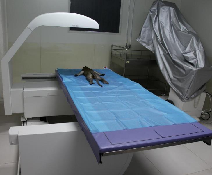
目的 研究食蟹猴在不同生长阶段的骨骼生长发育特点,为使用食蟹猴作为骨骼模型动物的研究提供参考。 方法 选择1~19岁283只食蟹猴,分为雄性和雌性不同年龄段:1岁 ≤ 年龄<3岁组、3岁 ≤ 年龄<5岁组、5岁 ≤ 年龄<7岁组、7岁 ≤ 年龄<9岁组、9岁 ≤ 年龄<11岁组、11岁 ≤ 年龄<13岁组、13岁 ≤ 年龄<15岁组、年龄 ≥ 15岁组。用双能X线骨密度仪(dual energy X-ray bone density instrument,DEXA)测量不同年龄段的雄性和雌性食蟹猴骨密度(bone mineral density,BMD)和骨矿含量(bone mineral content,BMC)。 结果 雄性食蟹猴1~12岁BMC从67 g增加到399 g,BMD从0.32 g/m2增加到0.57 g/m2;12~15岁雄性食蟹猴的BMC和BMD相对稳定,≥15岁BMC维持在(367.51±7.17)g,BMD维持在(0.56±0.06)g/m2。雌性食蟹猴1~10岁BMC从58 g增加到233 g,BMC最高值仅相当于雄性食蟹猴的58%;BMD从0.31 g/m2增加到0.47 g/m2,最高值相当于雄性食蟹猴的80%。雌性食蟹猴从10岁开始BMC显著下降,≥15岁的BMC维持在(166.63±6.21)g,BMD维持在(0.46±0.04)g/m2,分别相当于雄性食蟹猴的45%和80%。 结论 雄性食蟹猴12岁之前BMC和BMD逐年增长,是骨骼发育期;12~15岁的BMC和BMD相对稳定。雌性食蟹猴10岁之前BMC和BMD逐年增长,是骨骼发育期;从10岁开始BMC显著下降,10~15岁BMD相对稳定。雌性食蟹猴骨量峰值和BMD比雄性食蟹猴低。

关键词: 食蟹猴, 骨密度, 骨矿含量, 测定, 分析

Abstract:

Objective To study the skeletal characteristics of cynomolgus monkeys at different ages, and to provide reference values for skeletal research using cynomolgus monkeys as model animals. Methods A total of 283 cynomolgus monkeys aged 1-19 years were selected and divided into 8 groups according to sex and age :1 year≤age<3 years, 3 years≤age<5 years, 5 years≤age<7 years, 7 years≤age<9 years, 9 years≤age<11 years, 11 years≤age<13 years, 13 years≤age<15 years, and age≥15 years groups. Bone mineral density (BMD) and bone mineral content (BMC) were measured using dual-energy X-ray absorptiometry (DEXA). Results BMD increased from 0.32 g/m2 to 0.57 g/m2 and BMC increased from 67 g to 399 g in male cynomolgus monkeys between the age of 1 and 12. While BMC and BMD were relatively stable in the 12~15 years group. BMC and BMD remained at (367.51±7.17) g and (0.56±0.06) g/m2, respectively, in the ≥15 year-old group. In female cynomolgus monkeys, BMC increased from 58 g to 233 g and BMD increased from 0.31 g/m2 to 0.47 g/m2 between the age of 1 and 10. The maximum BMC was only about 58% of that of male cynomolgus monkeys, the maximum BMD was about 80% of that of male cynomolgus monkeys, and BMC decreased significantly after the age of 10 years. BMC remained at (166.63±6.21) g and BMD remained at (0.46±0.04) g/m2 after the age of 15 years, which were 45% and 80%, respectively, of those of male cynomolgus monkeys. Conclusion In male cynomolgus monkeys, as their skeleton develops, BMC and BMD increase continuously until the age of 12 years, and are relatively stable between the age of 12 and 15 years. While in female cynomolgus monkeys, BMC and BMD increase annually until the age of 10 years, but after the age of 10 years, BMC decreases significantly, while BMD remains relatively stable between the age of 10 and 15 years. Peak bone mass of female cynomolgus monkeys is lower than that of male cynomolgus monkeys.

Key words: Cynomolgus monkey, Bone mineral density, Bone mineral content, Measurement, Analysis

中图分类号: