实验动物与比较医学 ›› 2020, Vol. 40 ›› Issue (3): 183-.DOI: 10.3969/j.issn.1674-5817.2020.03.002
柴文君, 孙 磊, 刘晓丽, 潘洪玉, 郭天安, 徐 烨, 闫明霞
收稿日期:2020-04-10
出版日期:2020-06-25
发布日期:2020-12-15
作者简介:柴文君(1987—), 女, 本科, 技术员,兽医师, 主要从事肿瘤动物模型建立的相关工作。E-mail: chaiwj108@163.com
基金资助:CHAI Wenjun, SUN Lei, LIU Xiaoli, PAN Hongyu, GUO Tianan, XU Ye, YAN Mingxia
Received:2020-04-10
Published:2020-06-25
Online:2020-12-15
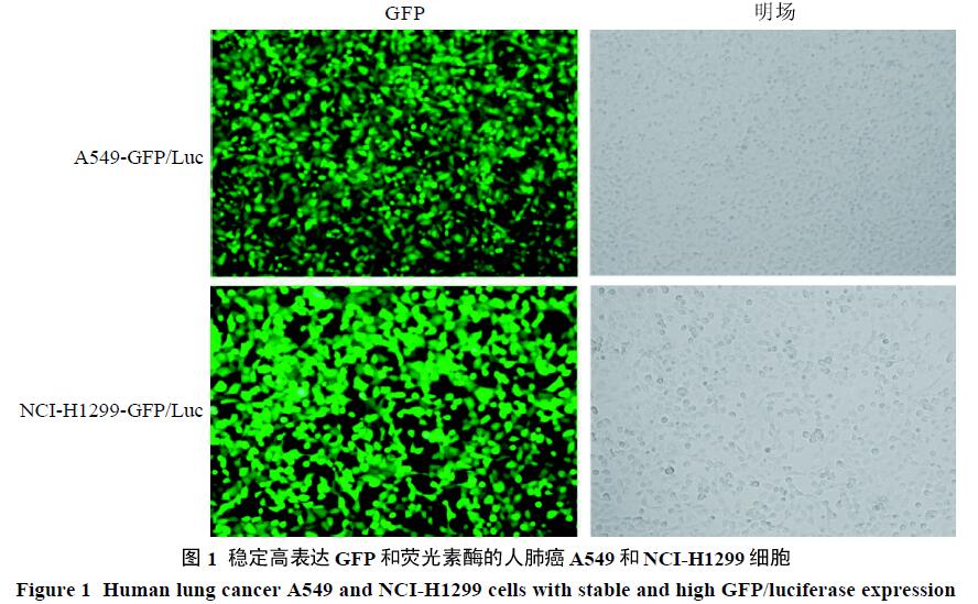
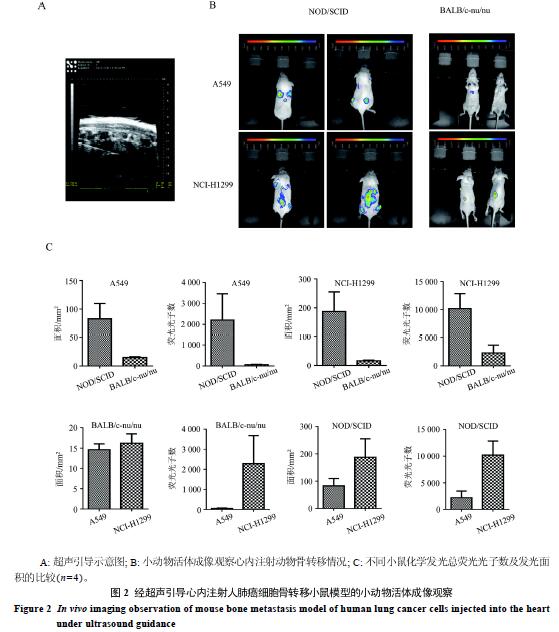
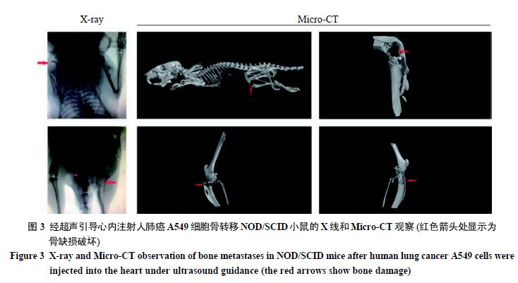
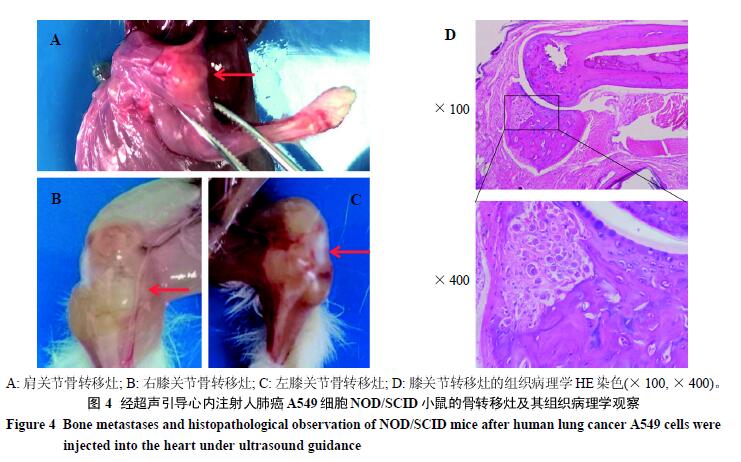
摘要: 目的 通过超声引导建立心内注射人肺癌细胞骨转移小鼠模型。方法 在超声引导下,将人肺癌A549和NCI-H1299细胞分别注入NOD/SCID和BALB/c-nu/nu小鼠左心室,结合小动物活体成像、X线和小动物Micro-CT等多种小动物影像学方法观察模型建立和骨转移发生情况。采用HE染色法进行肿瘤组织的病理学观察。结果 在超声引导下进行心内注射肺癌细胞时动物全部存活。小动物活体成像显示小鼠的中轴骨和四肢骨位置均有发光区域。X线及小动物Micro-CT对小鼠全身骨骼进行三维重构,显示小鼠胫骨平台和肩关节等部位均出现明显的骨质和骨膜缺损及破坏,骨正常组织形态消失,呈溶骨性改变,其影像学表现符合肿瘤性骨破坏的改变。大体解剖可见小鼠的中轴骨和四肢骨关节被肿瘤组织包裹,出现不同程度的骨转移灶。结论 超声引导下心内注射肺癌骨转移模型能够有效提高建模成功率,较真实地模拟临床肺癌患者骨转移的生物学特性,能够为肺癌转移机制研究、抗转移治疗和个体化治疗等研究提供实验工具。
中图分类号:
柴文君,孙 磊,刘晓丽,等. 超声引导下左心室内注射人肺癌细胞建立骨转移小鼠模型[J]. 实验动物与比较医学, 2020, 40(3): 183-. DOI: 10.3969/j.issn.1674-5817.2020.03.002.
CHAI Wenjun,SUN Lei,LIU Xiaoli,et al. Establishment of Bone Metastasis Mouse Models through Injecting Human Lung Cancer Cells into Left Ventricle#br# under Ultrasound Guidance#br#[J]. Laboratory Animal and Comparative Medicine, 2020, 40(3): 183-. DOI: 10.3969/j.issn.1674-5817.2020.03.002.
| [1] | 焦青贞, 吴桂华, 唐雯, 樊帆, 冯凯, 杨春响, 乔建, 邓素芳. 暖通系统暂停送风下实验动物设施氨浓度的动态监测与分析[J]. 实验动物与比较医学, 2025, 45(4): 490-495. |
| [2] | 刘文涛, 罗艳红, 龙永霞, 罗启慧, 陈正礼, 刘丽达. 四川省实验动物设施常见环境问题及检测经验[J]. 实验动物与比较医学, 2025, 45(4): 483-489. |
| [3] | 赵鑫, 王晨曦, 石文清, 娄月芬. 斑马鱼在炎症性肠病机制及药物研究中的应用进展[J]. 实验动物与比较医学, 2025, 45(4): 422-431. |
| [4] | 贡磊磊, 王晓霞, 封学伟, 李心蕾, 赵涵, 张雪艳, 冯欣. 不同浓度环磷酰胺诱导早发性卵巢功能不全小鼠模型及作用机制研究[J]. 实验动物与比较医学, 2025, 45(4): 403-410. |
| [5] | 林振华, 褚祥宇, 魏振西, 董传俊, 赵增琳, 孙晓霞, 李庆雨, 张琪. 椎体成形术用于实验猪体内骨水泥安全性及有效性评价[J]. 实验动物与比较医学, 2025, 45(4): 466-472. |
| [6] | 姜娟, 宋宁, 连文博, 邵丛丛, 顾文文, 石燕. 两种浓度乙醇溶液灌注建立小鼠宫腔粘连模型的组织病理和分子病理表型比较[J]. 实验动物与比较医学, 2025, 45(4): 393-402. |
| [7] | 刘月琴, 薛卫国, 王淑友, 申耀华, 贾术永, 王广军, 宋晓晶. 探头式激光共聚焦成像技术用于小鼠消化道组织形态特征分析[J]. 实验动物与比较医学, 2025, 45(4): 457-465. |
| [8] | 郑卿勇, 杨冬华, 马智超, 周姿余, 陆洋, 王晶宇, 邢丽娜, 康迎英, 杜莉, 赵春香, 狄宝山, 田金徽. 动物实验系统评价与Meta分析报告的规范撰写建议[J]. 实验动物与比较医学, 2025, 45(4): 496-507. |
| [9] | 王庭君, 罗浩, 陈琦. 基于人工智能的实验动物中心信息化升级及应用实践[J]. 实验动物与比较医学, 2025, 45(4): 473-482. |
| [10] | 王娇祥, 张璐, 陈姝含, 角德灵, 赵恒, 魏太云, 郭建雄, 徐凯祥, 魏红江. GTKO/hCD55基因编辑异种器官移植供体猪的构建及功能验证[J]. 实验动物与比较医学, 2025, 45(4): 379-392. |
| [11] | 秦超, 李双星, 赵婷婷, 蒋晨晨, 赵晶, 杨艳伟, 林志, 王三龙, 文海若. 药物安全评价用SD大鼠90 d喂养试验的背景数据研究[J]. 实验动物与比较医学, 2025, 45(4): 439-448. |
| [12] | 刘鹍, 兰青, 易兵, 谢晓婕. 药物非临床生殖毒性试验中动物妊娠的主要难点及应对方法[J]. 实验动物与比较医学, 2025, 45(4): 449-456. |
| [13] | 孙强. 非动物实验替代知多少[J]. 实验动物与比较医学, 2025, 45(4): 508-514. |
| [14] | 陈子宜, 孙红燕, 康品方, 武文娟. 肺动脉高压动物实验模型的研究进展[J]. 实验动物与比较医学, 2025, (): 1-12. |
| [15] | 徐英韬, 王蒙蒙, 林平, 迟海涛, 王怡, 白鹰. 外泌体通过NRF2/SLC7A11/GPX4通路调控铁死亡治疗小鼠缺血性脑卒中[J]. 实验动物与比较医学, 2025, (): 1-11. |
| 阅读次数 | ||||||
|
全文 |
|
|||||
|
摘要 |
|
|||||