实验动物与比较医学

• •    

外泌体通过NRF2/SLC7A11/GPX4通路调控铁死亡治疗小鼠缺血性脑卒中

徐英韬(), 王蒙蒙, 林平, 迟海涛, 王怡, 白鹰()   

  1. 大连大学附属新华医院神经内科, 大连 116023
  • 出版日期:2025-08-04
  • 通讯作者: 白鹰(1965—),女,博士,教授,主任医师,硕士研究生导师,研究方向:缺血性脑卒中与神经保护。E-mail: 1029981825@qq.com。ORCID: 0009-0008-1965-8801
  • 作者简介:徐英韬(1997—),男,硕士研究生,研究方向:缺血性脑卒中与神经保护。E-mail: 951072549@qq.com
    白鹰(1965—),女,博士,教授,主任医师,硕士研究生导师,研究方向:缺血性脑卒中与神经保护。E-mail: 1029981825@qq.com。ORCID: 0009-0008-1965-8801
    第一联系人:

    徐英韬负责初稿写作及修改,动物实验、实验数据的检测及分析;

    王蒙蒙负责动物实验、实验数据的检测及分析;

    林平负责实验数据分析;

    迟海涛负责实验设计;

    王怡负责实验监督与指导;

    白鹰负责项目管理及论文修订。

  • 基金资助:
    大连市科技创新基金“miR-451/Phd3/p53轴抑制缺血性脑卒中铁死亡作用机制及外泌体装载miR-451神经保护治疗研究”(2022JJ13SN063)

Exosomes Improve Ischemic Stroke by Regulation of Ferroptosis Through the NRF2/SLC7A11/GPX4 Pathway in Mice

XU Yingtao(), WANG Mengmeng, LIN Ping, CHI Haitao, WANG Yi, BAI Ying()   

  1. Department of Neurology, Xinhua Hospital Affiliated to Dalian University, Dalian 116023, China
  • Published:2025-08-04
  • Contact: BAI Ying (ORCID: 0009-0008-1965-8801), E-mail: 1029981825@qq.com

摘要:

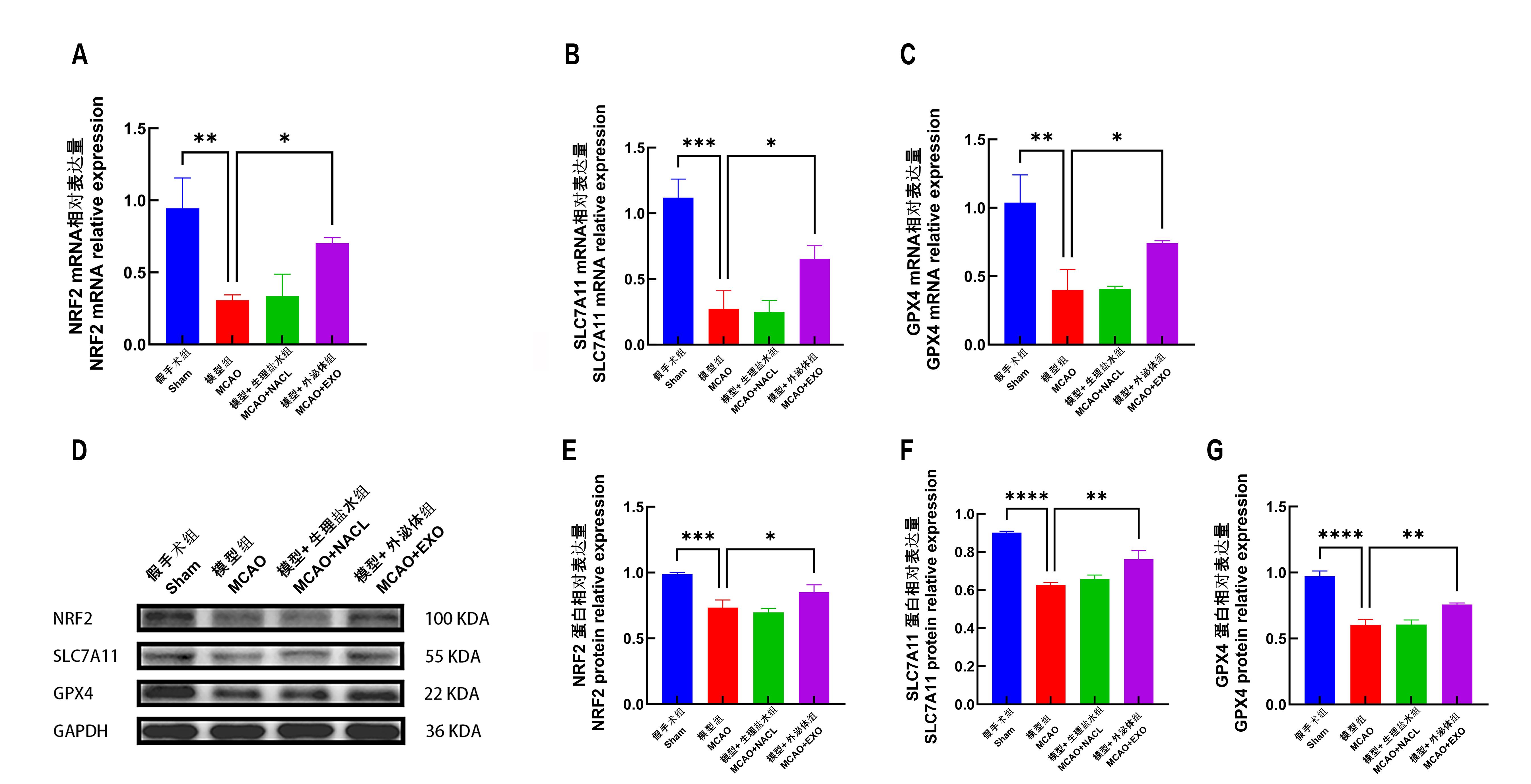
目的 通过电凝法阻断小鼠大脑中动脉(middle cerebral artery,MCA),构建小鼠大脑中动脉闭塞模型(middle cerebral artery occlusion, MCAO),比较模型小鼠和外泌体(exosome,EXO)干预后的模型小鼠神经细胞损伤程度和铁死亡(ferroptosis)相关分子表达量的差异,以探索MCAO发病过程中,外泌体抑制铁死亡,改善缺血性脑卒中的作用机制。 方法 将32只6~8周龄SPF级雄性C57BL/6小鼠随机分为4组,每组8只,假手术组(Sham组)、模型组(MCAO组)、模型+生理盐水组(MCAO+NACL组)、模型+外泌体组(MCAO+EXO组)。利用离心和过滤的方法从人羊膜间充质干细胞(human amniotic mesenchymal stem cells,hAMSCs)的上清培养液中提取外泌体,通过电子显微镜对外泌体的粒径进行分析确认。对MCAO+EXO组的小鼠尾静脉注射1 mL的外泌体,MCAO+NACL组的小鼠尾静脉注射1 mL生理盐水。使用电凝法建立大脑中动脉缺血(MCAO)模型,Sham组暴露大脑中动脉但不实施电凝。采用Longa神经功能缺损评分评价各组小鼠神经功能损害程度,然后进行心脏灌注并取脑。通过TTC 染色法评估各组小鼠脑梗死体积百分比差异;通过HE染色评估各组小鼠脑组织神经细胞的形态特点差异;通过微量法检测各组小鼠脑梗死区及其周围组织亚铁离子(Fe2+)、丙二醛(MDA)、总谷胱甘肽(total glutathione,Total GSH)、氧化型谷胱甘肽(glutathione oxidized,GSSG)和谷胱甘肽(glutathione, GSH)含量的差异;取适量脑梗死区及周围组织,通过实时RT-PCR方法检测各组小鼠铁死亡相关因子:核因子红细胞相关因子2(NRF2)、溶质载体家族7成员11(SLC7A11)、谷胱甘肽过氧化物酶4(GPX4)的mRNA表达;通过Western blot检测各组小鼠脑梗死区及其周围组织中NRF2、SLC7A11和GPX4的蛋白质表达。 结果 相较于MCAO组,MCAO+EXO组的Longa评分显著降低(P<0.01)。TTC染色发现:MCAO组小鼠脑组织出现明显梗死灶,而MCAO+EXO组脑梗死体积百分比显著减少(P<0.001)。HE染色发现:与Sham组相比,MCAO组出现神经细胞胞体空泡变性、细胞核固缩,深染,细胞核结构不清晰,神经细胞排列不整齐。而相较于MCAO组小鼠,MCAO+EXO组小鼠神经细胞结构较完整,细胞核大而规整,位于细胞中央。微量法检测发现:MCAO组脑梗死区及周围组织Fe2+、MDA含量较Sham组显著上升(P<0.001),MCAO+EXO组Fe2+、MDA含量较MCAO组显著下降(P<0.01)。相较于Sham组,MCAO模型组Total GSH、GSSG、GSH含量显著下降(P<0.01),相较于MCAO组,MCAO+EXO组Total GSH、GSH含量显著上升(P<0.001),GSSG含量无显著差异。实时RT-PCR发现:相较于Sham组,MCAO组NRF2、SLC7A11、GPX4的mRNA表达量均显著下降(P<0.01),相较于MCAO组,MCAO+EXO组NRF2、SLC7A11、GPX4的mRNA表达量显著升高(P<0.05)。Western blot发现:相较于Sham组,MCAO组NRF2、SLC7A11、GPX4的蛋白表达量显著下降(P<0.001)。相较于MCAO组,MCAO+EXO组NRF2、SLC7A11、GPX4的蛋白表达量显著上升(P<0.05)。 结论 在小鼠MCAO模型中,尾静脉注射人羊膜间充质干细胞来源外泌体可以改善运动功能、减少梗死面积、保护神经细胞形态、降低神经损伤程度;外泌体可能通过NRF2/SLC7A11/GPX4通路,减少MCAO模型小鼠脑神经细胞铁死亡而发挥对神经细胞的保护作用。

关键词: 动物模型, 缺血性脑卒中, 铁死亡, 外泌体, 小鼠

Abstract:

Objective By using electrocoagulation to block the middle cerebral artery (MCA) in mice, a middle cerebral artery occlusion model (MCAO) was established in mice. The degree of neuronal cell damage and the expression levels of ferroptosis-related molecules in the model mice were compared with those in the model mice after exosome intervention to explore the mechanism by which exosomes regulate ferroptosis to improve ischemic stroke. Methods A total of 32 male SPF-grade C57BL/6 mice at 6-8-week-old were randomly divided into 4 groups, with 8 mice in each group. The sham operation group (Sham group), the model group (MCAO group), the model + normal saline group (MCAO + NACL group), and the model + exosome group (MCAO + EXO group). Exosomes were extracted from the supernatant of human amniotic mesenchymal stem cell culture medium by centrifugation and filtration,and the extracted exosomes were observed under an electron microscope by analyzing particle size (NTA). The MCAO + EXO group mice were intravenously injected with 1 ml of exosomes, and the MCAO + NACL group mice were injected with the same volume of normal saline. The MCAO model was established by electrocoagulation of the middle cerebral artery,while the artery was exposed without electrocoagulation in sham group. The Longa neurological deficit score was used to evaluate the degree of neurological impairment, followed by cardiac perfusion and brain collection. The percentage of cerebral infarction volume was evaluated by TTC staining; the morphological characteristics of nerve cells in the brain were evaluated by H&E staining; the expression levels of ferroptosis-related factors (NRF2, SLC7A11, GPX4) in the cerebral infarction area and its surrounding tissues were examined by RT-qPCR. Results Compared with the MCAO group, the Longa score of the MCAO + EXO group was significantly lower (P < 0.01). TTC staining revealed that there were obvious infarction foci in the brain tissue of the MCAO group, and the percentage of cerebral infarction volume in the MCAO + EXO group was significantly reduced compared with the MCAO group (P < 0.001). HE staining showed that the MCAO group showed vacuolar degeneration of neuronal cell bodies, nuclear shrinkage, dark staining, unclear nuclear structure, and disordered neuronal arrangement,but these alterations were largely restored in the MCAO + EXO group. Critically, the contents of Fe2+ and MDA in the cerebral infarction area and its surrounding tissues of the MCAO group was significantly higher than those of the sham group (P < 0.001),but significantly reduced in the MCAO + EXO group (P < 0.01). In addition, compared with the Sham group, the total of GSH, GSSG and GSH contents were significantly decreased in the MCAO model group (P < 0.01), but the GSH and GSH contents were largely recovered in the MCAO + EXO group (P < 0.001) with unchanged GSSG content. Real-time RT-qPCR revealed that the mRNA expression levels of NRF2, SLC7A11, and GPX4 were significantly decreased in the MCAO group compared with the Sham group, (P < 0.01), but they were significantly increased in the MCAO + EXO group (P < 0.05). Western blot showed that the protein expression levels of NRF2, SLC7A11 and GPX4 were significantly decreased in the MCAO group compared with the Sham group (P < 0.001), and they were also significantly increased in the MCAO + EXO group (P < 0.05). Conclusion In the mouse MCAO model, tail vein injection of exosomes can improve motor function, reduce infarct area, protect neuronal cell morphology, and reduce the degree of nerve cell injury.The infarct area and surrounding tissues of the mouse MCAO model undergoes ferroptosis, and the exosomes can reduce ferroptosis of nerve cells possibly through the NRF2/SLC7A11/GPX4 pathway, protecting the survival of nerve cells in ischemic condition.

Key words: Animal model, Ischemic stroke, Ferroptosis, Exosome, Mouse

中图分类号: