实验动物与比较医学 ›› 2022, Vol. 42 ›› Issue (2): 135-140.DOI: 10.12300/j.issn.1674-5817.2021.093

• 人类疾病动物模型 • 上一篇    下一篇

利格列汀对2型糖尿病大鼠肾组织纤维化过程中骨膜蛋白及转化生长因子-β1的影响

弓慧杰1,2(), 张光明2()   

  1. 1.四川省成都市新都区人民医院内分泌肾病血液内科, 成都 610500
    2.西南医科大学病理生理研究室, 泸州 646000
  • 收稿日期:2021-05-10 修回日期:2021-10-25 出版日期:2022-04-25 发布日期:2022-05-07
  • 通讯作者: 张光明(1970—),男,主任医师,研究方向: 糖尿病微血管并发症。E-mail: 770930017@qq.com
  • 作者简介:弓慧杰(1984—),男,硕士,主治医师,研究方向: 糖尿病肾病微血管并发症。E-mail: 82295788@qq.com

Effects of Inagliptin on the Expression of Periosteal and TGF-β1 in Renal Fibrosis of Type 2 Diabetic Rats

Huijie GONG1,2(), Guangming ZHANG2()   

  1. 1.Department of Endocrinology, Nephropathy and Hematology, Chengdu Xindu District People's Hospital, Chengdu 610500, China
    2.Department of Pathophysiology, Southwest Medical University, Luzhou 646000, China
  • Received:2021-05-10 Revised:2021-10-25 Published:2022-04-25 Online:2022-05-07
  • Contact: ZHANG Guangming, E-mail: 770930017@qq.com

摘要:

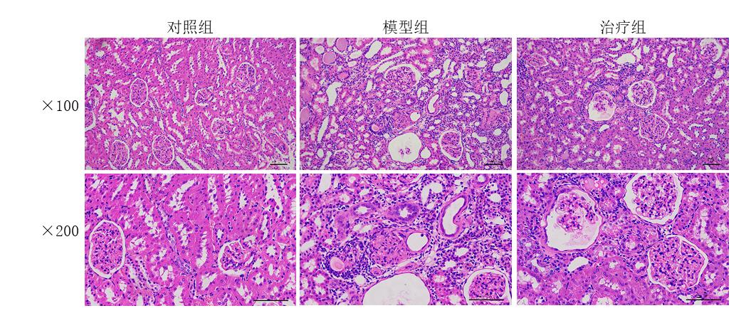
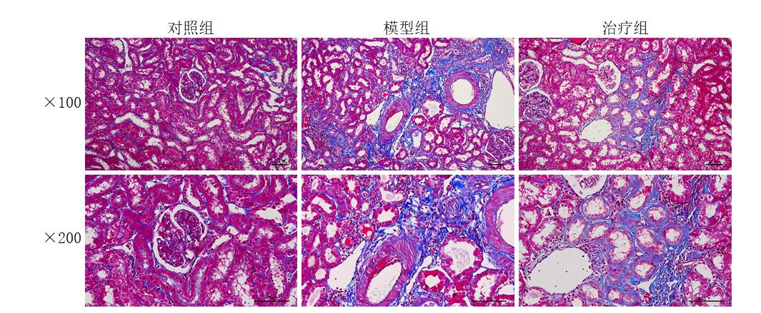
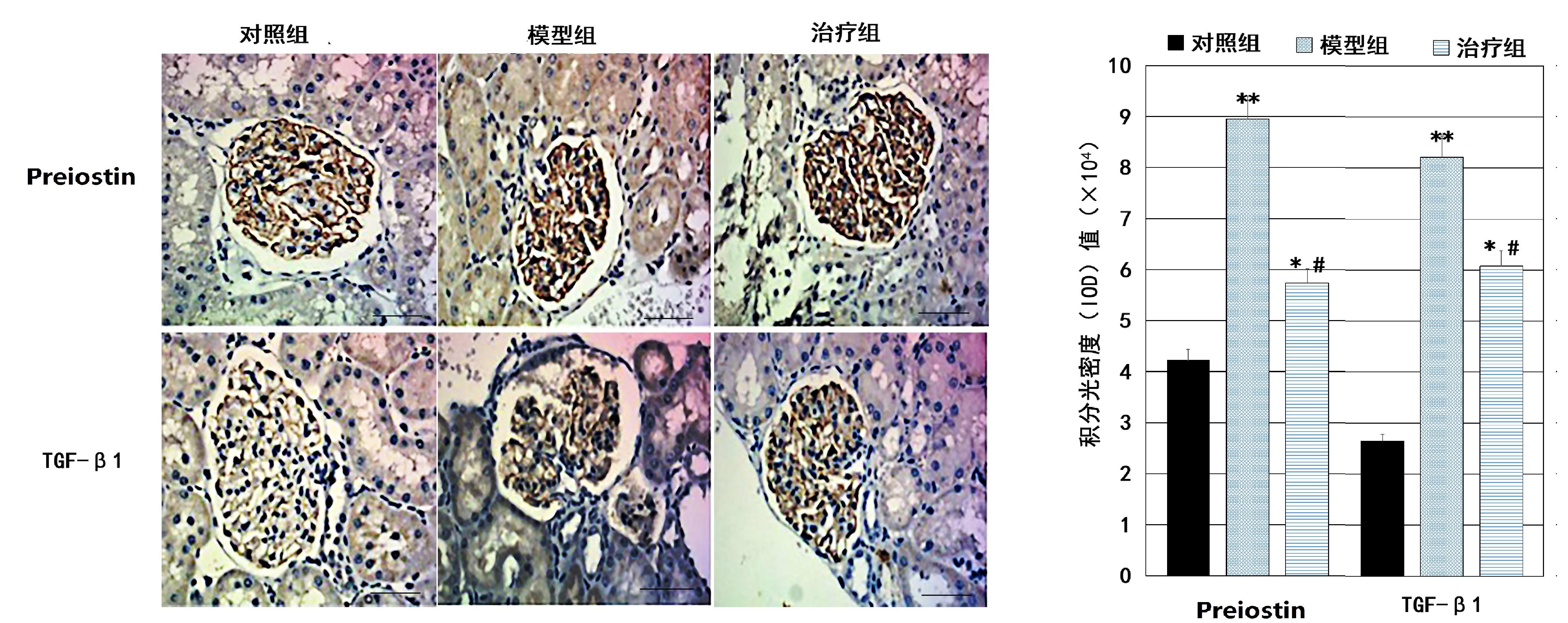
目的 通过观察利格列汀对2型糖尿病大鼠肾组织纤维化过程中骨膜蛋白(periostin)及转化生长因子-β1(transforming growth factor-β1,TGF-β1)的影响,探讨利格列汀治疗糖尿病肾病患者的抗炎、抗纤维化等保护机制。方法 24只SD大鼠采用腹腔注射链脲佐菌素的方式建立2型糖尿病大鼠模型,另取12只SD大鼠注射等剂量的柠檬酸缓冲液作为对照组。糖尿病模型大鼠分为模型组及治疗组。治疗组大鼠予以利格列汀(10 mg?kg-1?d-1)持续灌胃12周,其他2组予以等量生理盐水灌胃。12周后取材,HE染色和Masson染色法观察肾组织病理学变化,分别采用免疫组织化学法和RT-PCR法检测各组大鼠肾组织中骨膜蛋白和TGF-β1蛋白及mRNA的表达水平。结果 与对照组相比,模型组大鼠的肾间质有大量被深染为蓝色的炎性细胞浸润,治疗组则有较少的炎性细胞浸润;模型组的肾间质纤维含量比对照组增多,而治疗组的纤维化不明显。与对照组相比,模型组大鼠的肾脏组织中骨膜蛋白和TGF-β1蛋白表达水平及mRNA含量均明显升高(P<0.05);与模型组相比,治疗组大鼠的肾脏组织中骨膜蛋白和TGF-β1蛋白表达水平及mRNA含量均明显降低(P<0.05)。结论 利格列汀可通过抑制2型糖尿病大鼠肾脏组织中TGF-β1的过表达,下调骨膜蛋白表达,减缓肾组织纤维化进程。

关键词: 利格列汀, 糖尿病肾病, 肾脏纤维化, 骨膜蛋白, 转化生长因子-β1, 大鼠

Abstract:

Objective To analyze the effect of linagliptin on the expression of periostin and transforming growth factor-β1 (TGF-β1) in the process of renal tissue fibrosis in type 2 diabetic rats, and explore the anti-inflammatory and anti-fibrotic protective mechanism of linagliptin on diabetic nephropathy.Methods Totally twenty-four SD rats were injected with streptozotocin in intraperitoneal to replicate the type 2 diabetes rats, and another 12 SD rats were injected with the same dosage of citrate buffer as the control group. The type 2 diabetic rats were randomly divided into two groups: model group and treatment group. The rats in the treatment group were continuously given liggliptin (10 mg?kg-1?d-1) for 12 weeks, and the other two groups were given the same amount of normal saline. The pathological changes of renal were observed by HE staining and Masson staining. Immunohistochemistry and RT-PCR were used to detect the expressions of periostin and TGF-β1 protein and mRNA levels in kidney tissues of rats, respectively.Results HE staining showed that compared with the control group, there were a large number of inflammatory cells in the renal stroma in the model group, while there were less inflammatory cells in the treatment group. Messon staining showed that compared with the control group, there were more collagen fibers in renal interstitial in the model group, while the collagen fibers in the treatment group was not obvious. Immunohistochemistry and RT-PCR showed that compared with control group, the protein and mRNA expressions of periostin and TGF-β1 in rat renal in model group were significantly increased (P< 0.05), while they were decreased in treatment group (P< 0.05).Conclusion Inagliptin maybe reduce the protein expression of periostin by inhibiting the overexpression of TGF-β1 in the kidney of type 2 diabetic rats, and slow down the progression of renal fibrosis.

Key words: Inagliptin, Diabetic nephropathy, Renal fibrosis, Periostin, Transforming growth factor-β1, Rats

中图分类号: