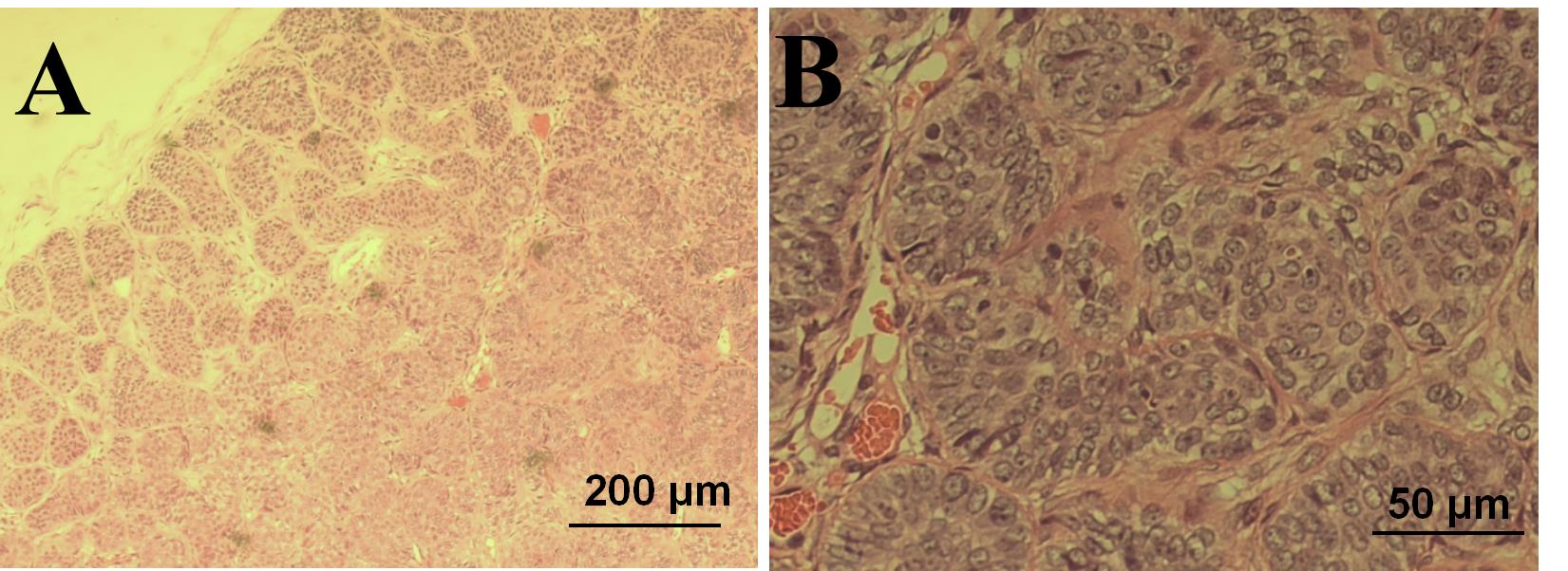
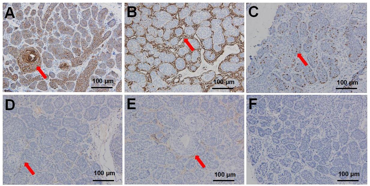
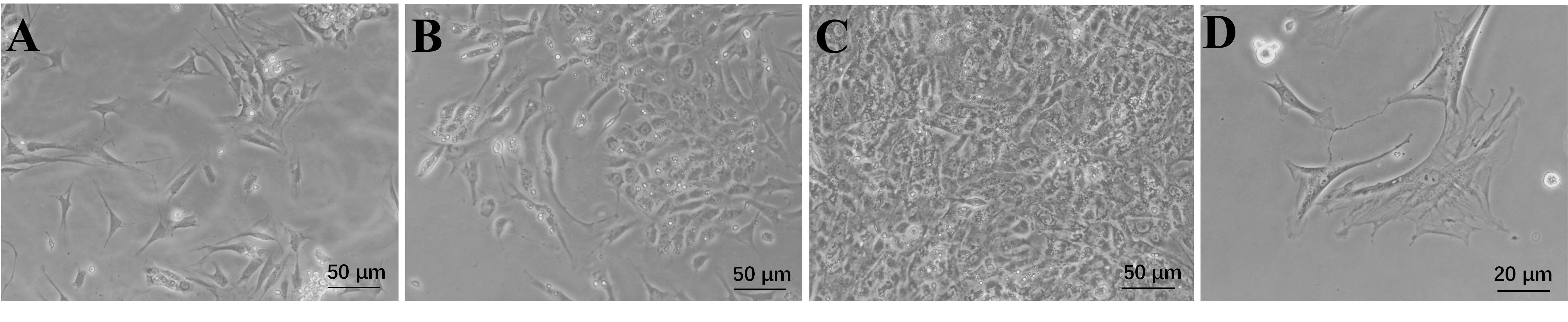
| 1 |
KARIMZADEH I, NAMAZI M R, KARIMZADEH A. Trichoepithelioma: a comprehensive review[J]. Acta Dermatovenerol Croat, 2018, 26(2):162-165.
|
| 2 |
ALSAAD K O, OBAIDAT N A, GHAZARIAN D. Skin adnexal neoplasms: part 1: an approach to tumours of the pilosebaceous unit[J]. J Clin Pathol, 2007, 60(2):129-144. DOI: 10.1136/jcp.2006.040337 .
|
| 3 |
SMOLLER B R. Lever's histopathology of the skin, 10th edition[J]. J Cutan Pathol, 2009, 36(5):605. DOI: 10.1111/j.1600-0560.2008.01213.x .
|
| 4 |
YE M S, ZHANG J Y, YU D D, et al. Comprehensive annotation of the Chinese tree shrew genome by large-scale RNA sequencing and long-read isoform sequencing[J]. Zool Res, 2021, 42(6):692-709. DOI: 10.24272/j.issn.2095-8137.2021.272 .
|
| 5 |
曾雯, 雷玲, 赵铖. 树鼩用于构建自身免疫性疾病动物模型展望[J]. 中国免疫学杂志, 2022, 38(15):1918-1921. DOI: 10.3969/j.issn.1000-484X.2022.15.024 .
|
|
ZENG W, LEI L, ZHAO C. Prospects of tree shrews used to establish animal models of autoimmune diseases[J]. Chin J Immunol, 2022, 38(15):1918-1921. DOI: 10.3969/j.issn.1000-484X.2022.15.024 .
|
| 6 |
LI R F, ZANIN M, XIA X S, et al. The tree shrew as a model for infectious diseases research[J]. J Thorac Dis, 2018, 10(S9): S2272-S2279. DOI: 10.21037/jtd.2017.12.121 .
|
| 7 |
贾杰, 代解杰. 树鼩在生物医学研究中的优势与挑战[J]. 实验动物与比较医学, 2019, 39(1):3-8. DOI: 10.3969/j.issn.1674-5817.2019.01.002 .
|
|
JIA J, DAI J J. Advantages and challenges of tree shrews in biomedical research[J]. Lab An Comp Med, 2019, 39(1): 3-8. DOI: 10.3969/j.issn.1674-5817.2019.01.002 .
|
| 8 |
徐新峰. 犬乳腺肿瘤细胞的分离、培养及鉴定[D]. 石河子: 石河子大学, 2014.
|
|
XU X F. Isolation, culture and identification of canine breast tumor cells[D]. Shihezi: Shihezi University, 2014.
|
| 9 |
MIGDEN M R, CHANG A L S, DIRIX L, et al. Emerging trends in the treatment of advanced basal cell carcinoma[J]. Cancer Treat Rev, 2018, 64:1-10. DOI: 10.1016/j.ctrv.2017.12.009 .
|
| 10 |
PATTERSON J W. Weedon's skin pathology[M]. 4th ed. Amsterdam: Elsevier, 2015.
|
| 11 |
LEAR J T, CORNER C, DZIEWULSKI P, et al. Challenges and new horizons in the management of advanced basal cell carcinoma: a UK perspective[J]. Br J Cancer, 2014, 111(8):1476-1481. DOI: 10.1038/bjc.2014.270 .
|
| 12 |
孙彩虹, 张彩萍, 丁克云, 等. 基底细胞癌72例临床特征分析[J]. 皮肤性病诊疗学杂志, 2015, 22(2): 99-101. DOI: 10.3969/j.issn.1674-8468.2015.02.004 .
|
|
SUN C H, ZHANG C P, DING K Y, et al. Analysis of the clinical feature of 72 cases with basal cell carcinoma[J]. J Diagn Ther Derm Venereol, 2015, 22(2): 99-101. DOI: 10.3969/j.issn.1674-8468.2015.02.004 .
|
| 13 |
吴海建. BCL-2和CK15在毛发上皮瘤和基底细胞癌中的表达及其意义[D]. 石家庄: 河北医科大学, 2015.
|
|
WU H J. Expression and significance of BCL-2 and CK15 in trichoepithelioma and basa cell carcinoma[D]. Shijiazhuang: Hebei Medical University, 2015.
|
| 14 |
CARRASQUILLO O Y, CRUZVAL-O'REILLY E, SÁNCHEZ J E, et al. Differentiation of basal cell carcinoma and trichoepithelioma: an immunohistochemical study[J]. Am J Dermatopathol, 2020, 43(3):191-197. DOI: 10.1097/dad.0000000000001783 .
|
| 15 |
SMOLLER B R, VAN DE RIJN M, LEBRUN D, et al. Bcl-2 expression reliably distinguishes trichoepitheliomas from basal cell carcinomas[J]. Br J Dermatol, 1994, 131(1):28-31. DOI: 10.1111/j.1365-2133.1994.tb08453.x .
|
| 16 |
SWANSON P E, FITZPATRICK M M, RITTER J H, et al. Immunohistologic differential diagnosis of basal cell carcinoma, squamous cell carcinoma, and trichoepithelioma in small cutaneous biopsy specimens[J]. J Cutan Pathol, 1998, 25(3):153-159. DOI: 10.1111/j.1600-0560.1998.tb01708.x .
|
| 17 |
ARITS A H M M, PARREN L J M T, VAN MARION A M W, et al. Basal cell carcinoma and trichoepithelioma: a possible matter of confusion[J]. Int J Dermatol, 2008, 47():13-17. DOI: 10.1111/j.1365-4632.2008.03951.x .
|
| 18 |
刘金丽, 张弛, 薛浩伟, 等. 127例皮肤基底细胞癌患者的临床观察[J]. 安徽医科大学学报, 2021, 56(4):659-662. DOI: 10.19405/j.cnki.issn1000-1492.2021.04.031 .
|
|
LIU J L, ZHANG C, XUE H W, et al. Analysis of clinical feature of basal cell carcinoma in 127 patients[J]. Acta Univ Med Anhui, 2021, 56(4):659-662. DOI: 10.19405/j.cnki.issn1000-1492.2021.04.031 .
|
| 19 |
SARI ASLANI F, AKBARZADEH-JAHROMI M, JOWKAR F. Value of CD10 expression in differentiating cutaneous basal from squamous cell carcinomas and basal cell carcinoma from trichoepithelioma[J]. Iran J Med Sci, 2013, 38(2):100-106.
|
| 20 |
ASTARCI H M, GURBUZ G A, SENGUL D, et al. Significance of androgen receptor and CD10 expression in cutaneous basal cell carcinoma and trichoepithelioma[J]. Oncol Lett, 2015, 10(6):3466-3470. DOI: 10.3892/ol.2015.3804 .
|


 ), 梁亮1, 曹颖颖1, 李竹欣1, 王青1, 陶俊宇1,2, 运晨霞1,3, 冷静1,2,3(
 )(
)(