


实验动物与比较医学 ›› 2023, Vol. 43 ›› Issue (5): 574-578.DOI: 10.12300/j.issn.1674-5817.2023.095
王成稷1, 王珏1, 王海杰1, 陆炜晟1, 史岩2, 顾正页1, 万鸣秋1, 沈如凌1( )(
)( )
)
收稿日期:2023-06-30
修回日期:2023-10-02
出版日期:2023-10-25
发布日期:2023-10-25
通讯作者:
沈如凌(1981—),女,博士,副研究员,从事模式动物模型研发及表型研究、肿瘤免疫和神经免疫方向。E-mail:shenruling@slarc.org.cn。ORCID:0000-0002-7529-810X作者简介:王成稷(1989—),男,学士,实验师,从事动物实验及解剖学研究。E-mail:wangchengji@slarc.org.cn;共同第一作者
王珏(1984—),女,学士,实验师,从事动物实验及分子生物学研究方向。E-mail:wangjue@slarc.org.cn 共同第一作者
基金资助:
Chengji WANG1, Jue WANG1, Haijie WANG1, Weisheng LU1, Yan SHI2, Zhengye GU1, Mingqiu WAN1, Ruling SHEN1()()
Received:2023-06-30
Revised:2023-10-02
Published:2023-10-25
Online:2023-10-25
Contact:
SHEN Ruling (ORCID: 0000-0002-7529-810X), E-mail: shenruling@slarc.org.cn摘要:
目的 优化乳胶灌注技术,并将该技术应用于小鼠头面部静脉血管模型制作。 方法 将9只8周龄、体质量(25.0±1.3)g的雄性C57BL/6小鼠随机分成60%乳胶生理盐水组、60%乳胶肝素组和30%乳胶肝素组,相应试剂灌注完成后将标本浸泡于4 ℃甲醛固定液中,24 h后解剖观察并测量颈外血管直径。 结果 于小鼠颈外静脉灌注乳胶灌注液200 μL后,各组小鼠的眶上静脉、眶下静脉、颞静脉、面后静脉、咬肌静脉、颈外静脉等均得到灌注,通过对灌注后面部血管末端分支、舌下静脉及舌尖小静脉的灌注程度比较后发现,30%乳胶肝素组的灌注效果最佳,其次为60%乳胶肝素组,而60%乳胶生理盐水组的灌注效果最差。 结论 优化后的乳胶灌注技术可有效灌注小鼠头面部的静脉血管,该技术可为小鼠面部静脉血管的走向及形态研究提供很好的参考。
中图分类号:
王成稷,王珏,王海杰,等. 乳胶血管灌注技术制作小鼠头面部静脉血管模型方法初探[J]. 实验动物与比较医学, 2023, 43(5): 574-578. DOI: 10.12300/j.issn.1674-5817.2023.095.
Chengji WANG,Jue WANG,Haijie WANG,et al. Application of Optimized Latex Perfusion Technique in the Establishment of Craniofacial Venous Model in Mice[J]. Laboratory Animal and Comparative Medicine, 2023, 43(5): 574-578. DOI: 10.12300/j.issn.1674-5817.2023.095.
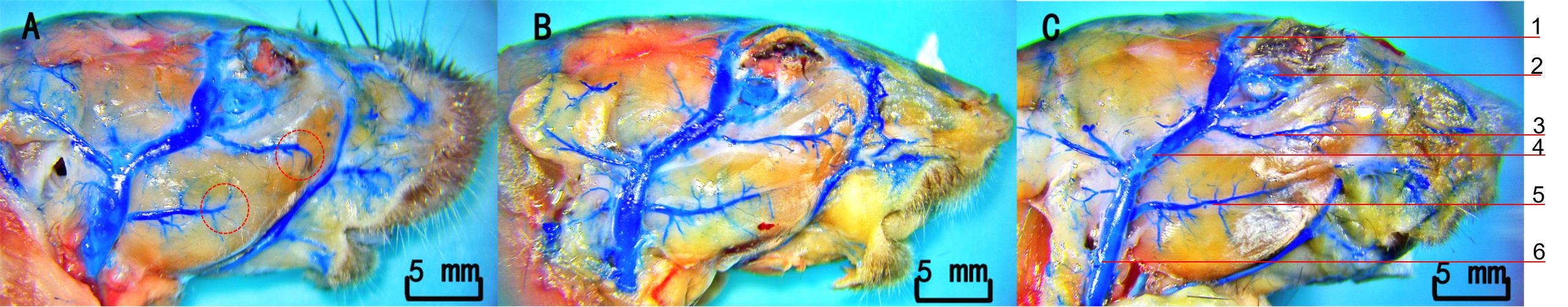
图1 小鼠头面部静脉的乳胶灌注结果注:A,60%乳胶生理盐水组;B,60%乳胶肝素组;C,30%乳胶肝素组。1,上睑静脉;2,眼眶静脉窦;3,面横静脉;4,颞浅静脉;5,咬肌静脉;6,颈外静脉。
Figure 1 Results of latex perfusion in the head and facial veins of miceNote:A, 60% latex saline group; B, 60% latex heparin group; C, 30% latex heparin group. 1, Superior palpebral vein; 2, Orbital venous sinus; 3, Facial transverse vein; 4, Superficial temporal vein; 5, Masseteric vein; 6, External jugular vein.
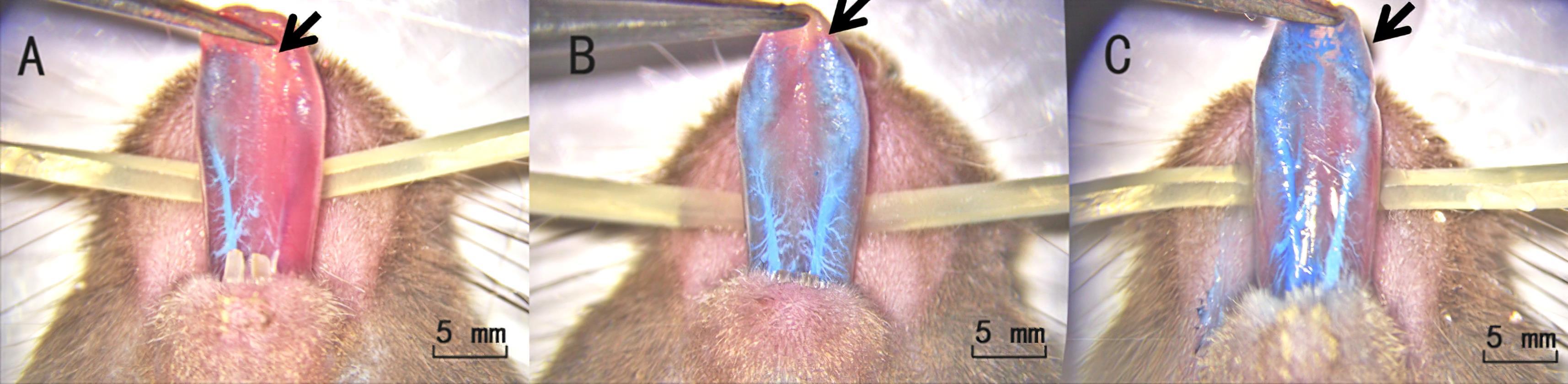
图2 各组小鼠舌静脉灌注效果对比注:A,60%乳胶生理盐水组;B,60%乳胶肝素组;C,30%乳胶肝素组。
Figure 2 Comparison of lingual vein perfusion effects among different groups of miceNote: A, 60% latex saline group; B, 60% latex heparin group; C, 30% latex heparin group.
| 1 | 余琛琳, 赵乐, 汤球, 等. 一项小鼠采血新技术: 颌下静脉丛采血法[J]. 中国比较医学杂志, 2008, 18(12):53-54, 95. DOI: 10.3969/j.issn.1671-7856.2008.12.014 . |
| YU C L, ZHAO L, TANG Q, et al. A new method of blood collection from the submandibular vein plexus in mice[J]. Chin J Comp Med, 2008, 18(12):53-54, 95. DOI: 10.3969/j.issn.1671-7856.2008.12.014 . | |
| 2 | 车兆义, 邹悦, 宋清斌. 大鼠实验中几种常用的采血方法探讨[J]. 局解手术学杂志, 2008, 17(2): 84-85. DOI: 10.3969/j.issn.1672-5042.2008.02.005 . |
| CHE Z Y, ZOU Y, SONG Q B. Several experiments in rat blood collection methods commonly used[J]. J Reg Anat Oper Surg, 2008, 17(2): 84-85. DOI: 10.3969/j.issn.1672-5042.2008.02.005 . | |
| 3 | 杨健莉, 刘佳, 郑志红. 常用实验大小鼠采血方法及其对实验动物福利的影响[J]. 中国比较医学杂志, 2019, 29(1): 90-94. DOI: 10.3969/j.issn.1671-7856.2019.01.016 . |
| YANG J L, LIU J, ZHENG Z H. Comparison and analysis of blood sampling methods from rats and mice[J]. Chin J Comp Med, 2019, 29(1): 90-94. DOI: 10.3969/j.issn.1671-7856.2019.01.016 . | |
| 4 | 邹天乐, 龚瑜, 林爱娣, 等. 硫酸钡血管造影术的优化[J]. 中国临床解剖学杂志, 2010, 28(1):104-106. DOI: 10.13418/j.issn.1001-165x.2010.01.017 . |
| ZOU T L, GONG Y, LIN A D, et al. Modified Barium Sulfate-latex injection technique for angiography[J]. Chin J Clin Anat, 2010, 28(1):104-106. DOI: 10.13418/j.issn.1001-165x.2010.01.017 . | |
| 5 | 钱凤涛, 王成稷, 王恺, 等. 优化硫酸钡造影剂对小鼠血管造影效果观察[J]. 实验动物与比较医学, 2019, 39(4): 319-322. DOI: 10.3969/j.issn.1674-5817.2019.04.012 . |
| QIAN F T, WANG C J, WANG K, et al. Study on angiography in mice with Barium sulfate[J]. Lab Anim Comp Med, 2019, 39(4): 319-322. DOI: 10.3969/j.issn.1674-5817.2019.04.012 . | |
| 6 | 杨曦, 徐永清, 何晓清, 等. 数字化技术制备大鼠跨区穿支皮瓣微小血管模型的实验研究[J]. 中国修复重建外科杂志, 2017, 31(12):1485-1489. |
| YANG X, XU Y Q, HE X Q, et al. Establishment of micro-vessels model of cross-boundary perforator flap in rat via digital technology[J]. Chin J Reparative Reconstr Surg, 2017, 31(12):1485-1489. | |
| 7 | 张习高, 陈超, 王建武, 等. 采用颈内静脉逆行插管行乳胶灌注翼丛标本制作法[J]. 解剖学杂志, 2003, 26(4): 359. DOI: 10.3969/j.issn.1001-1633.2003.04.035 . |
| ZHANG X G, CHEN C, WANG J W, et al. Preparation of pterygium plexus by retrograde intubation of internal jugular vein[J]. Chin J Anat, 2003, 26(4): 359. DOI: 10.3969/j.issn.1001-1633.2003.04.035 . | |
| 8 | 甘承, 田佳, 刘立强, 等. 人体头面部解剖标本的防腐固定技术及血管内乳胶灌注技术研究[J]. 中国美容医学, 2015, 24(12):44-49. DOI: 10.15909/j.cnki.cn61-1347/r.000515 . |
| GAN C, TIAN J, LIU L Q, et al. Research of anticorrosion and latex perfusion technology in human craniofacial specimens[J]. Chin J Aesthetic Med, 2015, 24(12):44-49. DOI: 10.15909/j.cnki.cn61-1347/r.000515 . | |
| 9 | 罗东辉, 罗坤. 乳胶灌注和硅胶灌注在头颅血管教学标本制作中的对比研究[J]. 新疆医学, 2014, 44(9): 171-173. |
| LUO D H, LUO K. Comparative study of latex perfusion and silica gel perfusion in making teaching specimens of cranial blood vessels[J]. Xinjiang Med J, 2014, 44(9): 171-173. | |
| 10 | 周文逊, 王刚. 乳胶灌注在心的血管标本制作中的应用[J]. 中国医药导报, 2010, 7(36): 134. DOI: 10.3969/j.issn.1673-7210.2010.36.071 . |
| ZHOU W X, WANG G. Application of latex perfusion in blood vessel specimen making of heart[J]. China Med Her, 2010, 7(36): 134. DOI: 10.3969/j.issn.1673-7210.2010.36.071 . | |
| 11 | 成亮, 陈铿, 柴益民, 等. 手指末节指掌侧浅静脉的显微解剖及临床应用[J]. 中华显微外科杂志, 2011, 34(2):131-133, F0003. |
| CHENG L, CHEN K, CHAI Y M, et al. Microanatomy study and clinical application of superficial palmar digital veins in fingertip replantation[J]. Chin J Microsurg, 2011, 34(2):131-133, F0003. | |
| 12 | ONISHI A, ANGE K ST, DORDICK J S, et al. Heparin and anticoagulation[J]. Front Biosci (Landmark Ed), 2016, 21(7):1372-1392. DOI: 10.2741/4462 . |
| 13 | 漆光平, 杜亚政, 潘爱华, 等. 乳胶血管灌注的技术探讨[J]. 湖南医科大学学报, 2000, 25(4):411-412. |
| QI G P, DU Y Z, PAN A H, et al. Filling blood vessels with latex and its application[J]. Bull Hunan Med Univ, 2000, 25(4):411-412. |
| [1] | 焦青贞, 吴桂华, 唐雯, 樊帆, 冯凯, 杨春响, 乔建, 邓素芳. 暖通系统暂停送风下实验动物设施氨浓度的动态监测与分析[J]. 实验动物与比较医学, 2025, 45(4): 490-495. |
| [2] | 刘文涛, 罗艳红, 龙永霞, 罗启慧, 陈正礼, 刘丽达. 四川省实验动物设施常见环境问题及检测经验[J]. 实验动物与比较医学, 2025, 45(4): 483-489. |
| [3] | 赵鑫, 王晨曦, 石文清, 娄月芬. 斑马鱼在炎症性肠病机制及药物研究中的应用进展[J]. 实验动物与比较医学, 2025, 45(4): 422-431. |
| [4] | 贡磊磊, 王晓霞, 封学伟, 李心蕾, 赵涵, 张雪艳, 冯欣. 不同浓度环磷酰胺诱导早发性卵巢功能不全小鼠模型及作用机制研究[J]. 实验动物与比较医学, 2025, 45(4): 403-410. |
| [5] | 林振华, 褚祥宇, 魏振西, 董传俊, 赵增琳, 孙晓霞, 李庆雨, 张琪. 椎体成形术用于实验猪体内骨水泥安全性及有效性评价[J]. 实验动物与比较医学, 2025, 45(4): 466-472. |
| [6] | 姜娟, 宋宁, 连文博, 邵丛丛, 顾文文, 石燕. 两种浓度乙醇溶液灌注建立小鼠宫腔粘连模型的组织病理和分子病理表型比较[J]. 实验动物与比较医学, 2025, 45(4): 393-402. |
| [7] | 刘月琴, 薛卫国, 王淑友, 申耀华, 贾术永, 王广军, 宋晓晶. 探头式激光共聚焦成像技术用于小鼠消化道组织形态特征分析[J]. 实验动物与比较医学, 2025, 45(4): 457-465. |
| [8] | 郑卿勇, 杨冬华, 马智超, 周姿余, 陆洋, 王晶宇, 邢丽娜, 康迎英, 杜莉, 赵春香, 狄宝山, 田金徽. 动物实验系统评价与Meta分析报告的规范撰写建议[J]. 实验动物与比较医学, 2025, 45(4): 496-507. |
| [9] | 王庭君, 罗浩, 陈琦. 基于人工智能的实验动物中心信息化升级及应用实践[J]. 实验动物与比较医学, 2025, 45(4): 473-482. |
| [10] | 王娇祥, 张璐, 陈姝含, 角德灵, 赵恒, 魏太云, 郭建雄, 徐凯祥, 魏红江. GTKO/hCD55基因编辑异种器官移植供体猪的构建及功能验证[J]. 实验动物与比较医学, 2025, 45(4): 379-392. |
| [11] | 秦超, 李双星, 赵婷婷, 蒋晨晨, 赵晶, 杨艳伟, 林志, 王三龙, 文海若. 药物安全评价用SD大鼠90 d喂养试验的背景数据研究[J]. 实验动物与比较医学, 2025, 45(4): 439-448. |
| [12] | 刘鹍, 兰青, 易兵, 谢晓婕. 药物非临床生殖毒性试验中动物妊娠的主要难点及应对方法[J]. 实验动物与比较医学, 2025, 45(4): 449-456. |
| [13] | 孙强. 非动物实验替代知多少[J]. 实验动物与比较医学, 2025, 45(4): 508-514. |
| [14] | 陈子宜, 孙红燕, 康品方, 武文娟. 肺动脉高压动物实验模型的研究进展[J]. 实验动物与比较医学, 2025, (): 1-12. |
| [15] | 徐英韬, 王蒙蒙, 林平, 迟海涛, 王怡, 白鹰. 外泌体通过NRF2/SLC7A11/GPX4通路调控铁死亡治疗小鼠缺血性脑卒中[J]. 实验动物与比较医学, 2025, (): 1-11. |
| 阅读次数 | ||||||
|
全文 |
|
|||||
|
摘要 |
|
|||||