实验动物与比较医学 ›› 2020, Vol. 40 ›› Issue (6): 477-482.DOI: 10.3969/j.issn.1674-5817.2020.06.003

• 论著 • 上一篇    下一篇

兔斯氏艾美耳球虫感染模型及巢式PCR诊断方法的建立

温福利   

  1. 联勤保障部队第九〇〇医院基础医学实验室,福州 350025
  • 收稿日期:2020-06-24 出版日期:2020-12-25 发布日期:2020-12-30
  • 作者简介:温福利(1987-), 男, 主管技师, 主要从事实验动物寄生虫研究。E-mail: wen_fuli@163.com
  • 基金资助:
    军队实验动物专项课题(SYDW[2018]15); 联勤保障部队第九〇〇医院杰出青年培育专项

Establishment of Eimeria stiedai Infected Rabbit Model and Nested PCR Assay

WEN Fuli   

  1. Laboratory of Basic Medicine, 900 Hospital of the Joint Logistics Team, PLA, Fuzhou 350025, China
  • Received:2020-06-24 Online:2020-12-25 Published:2020-12-30

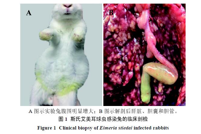
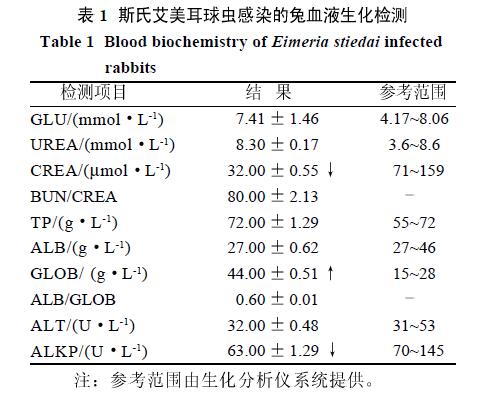
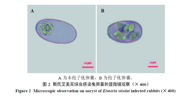
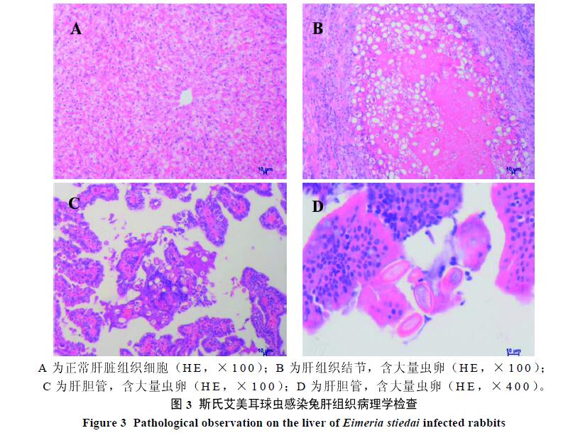
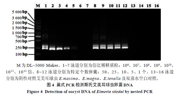
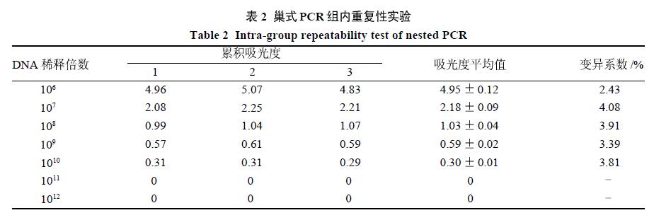
摘要: 目的 建立兔斯氏艾美耳球虫(Eimeria stiedai,E.stiedai)感染模型和巢式PCR诊断方法。方法 利用剖检、显微镜观察、血液生化和病理组织学检查等方法对建立的兔E.stiedai模型进行鉴定。通过收集卵囊,提取E.stiedai的DNA,设计特异性引物,建立E.stiedai巢式PCR检测方法。结果 临床剖检:兔腹围明显增大,解剖见肝脏肿大,肝脏表面和实质布满白色及淡黄色结节,胆囊和胆管肿大,胆汁呈淡黄色。显微镜观察:虫卵大小为(31.72~38.43)?m×(18.10~22.69)?m。血液生化检查:球蛋白(GLOB)指标偏高,肌酐(CREA)和碱性磷酸酶(ALKP)指标偏低,其余检测指标均在参考值范围内。病理组织学检查:肝组织和胆管中见大量粉红色E.stiedai虫卵。巢式PCR检查:最低检测限为1个卵囊DNA样本和1.14×10³拷贝数质粒,阴性对照和空白对照均未出现条带,重复性实验变异性系数<5%。结论 成功构建兔E.stiedai感染模型,建立的巢式PCR诊断方法可扩增出E. stiedai的特异片段,敏感性强,重复性好。

关键词: 斯氏艾美耳球虫, 病理组织学, 巢式PCR,

Abstract: Objective To establish an infection model and nested PCR assay method for Eimeria stiedai (E.stiedai) in rabbit. Methods The established infection model of E.stiedai in rabbits was identified by clinical examination, microscopic observation, blood biochemistry and histopathology. By collecting oocysts, DNA of E.stiedai was extracted, and the specific primers were designed to establish nested PCR for detection of E.stiedai. Results Clinical necropsy: the abdominal circumference of the rabbit was obviously enlarged. The liver enlargement and a large number of white and light yellow nodules were found by biopsy, while the gallbladder and bile duct were swollen, and the bile was pale yellow. Microscopic examination: the size of the eggs was (31.72-38.43) ?m×(18.10-22.69) ?m. Blood biochemical examination: the content of globulin (GLOB) was increased, while the levels of creatinine (CREA) and alkaline phosphatase (ALKP) were decreased, and the rest of the detection indicators were in the reference range. Histopathological examination: a large number of pink E.stiedai eggs were seen in the liver tissue and the bile duct. Nested PCR: the lower limit of detection was 1 oocyst DNA sample and 1.14×10³ copies of plasmids, and the negative control and blank control showed no bands. The variability coefficient was less than 5%. Conclusion The E.stiedai infection model in rabbits can be successfully constructed, and the nested PCR assay can amplify the specific fragments of E.stiedai with high sensitivity and good repeatability.

Key words: Eimeria stiedai, Histopathology, Nested PCR, Rabbits

中图分类号: