
Laboratory Animal and Comparative Medicine ›› 2025, Vol. 45 ›› Issue (4): 466-472.DOI: 10.12300/j.issn.1674-5817.2025.006
• Animal Experimental Techniques and Methods • Previous Articles Next Articles
LIN Zhenhua, CHU Xiangyu( )(
)( ), WEI Zhenxi, DONG Chuanjun, ZHAO Zenglin, SUN Xiaoxia, LI Qingyu, ZHANG Qi
Received:2025-01-14
Revised:2025-04-19
Online:2025-08-25
Published:2025-09-01
Contact:
CHU Xiangyu
CLC Number:
LIN Zhenhua,CHU Xiangyu,WEI Zhenxi,et al. Evaluation of the Safety and Efficacy of Bone Cement in Experimental Pigs Using Vertebroplasty[J]. Laboratory Animal and Comparative Medicine, 2025, 45(4): 466-472. DOI: 10.12300/j.issn.1674-5817.2025.006.
Add to citation manager EndNote|Ris|BibTeX
URL: https://www.slarc.org.cn/dwyx/EN/10.12300/j.issn.1674-5817.2025.006
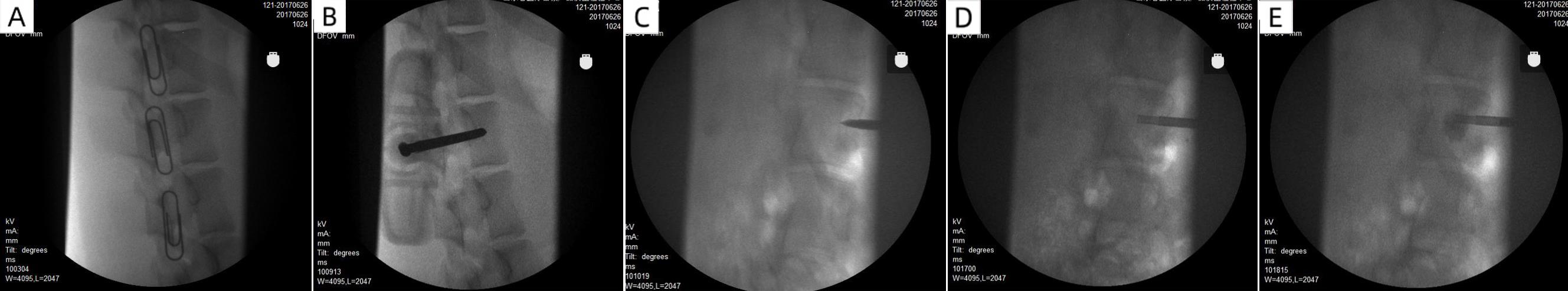
Figure 1 X-ray imaging pictures of pig percutaneous vertebroplastyNote: A, Locate the puncture site; B, Puncture needle reaches the target point (lateral position); C, Puncture needle reaches the target point (anteroposterior position); D, Bone cement injector enters the vertebral body; E, Bone cement is injected into the porcine vertebral body.
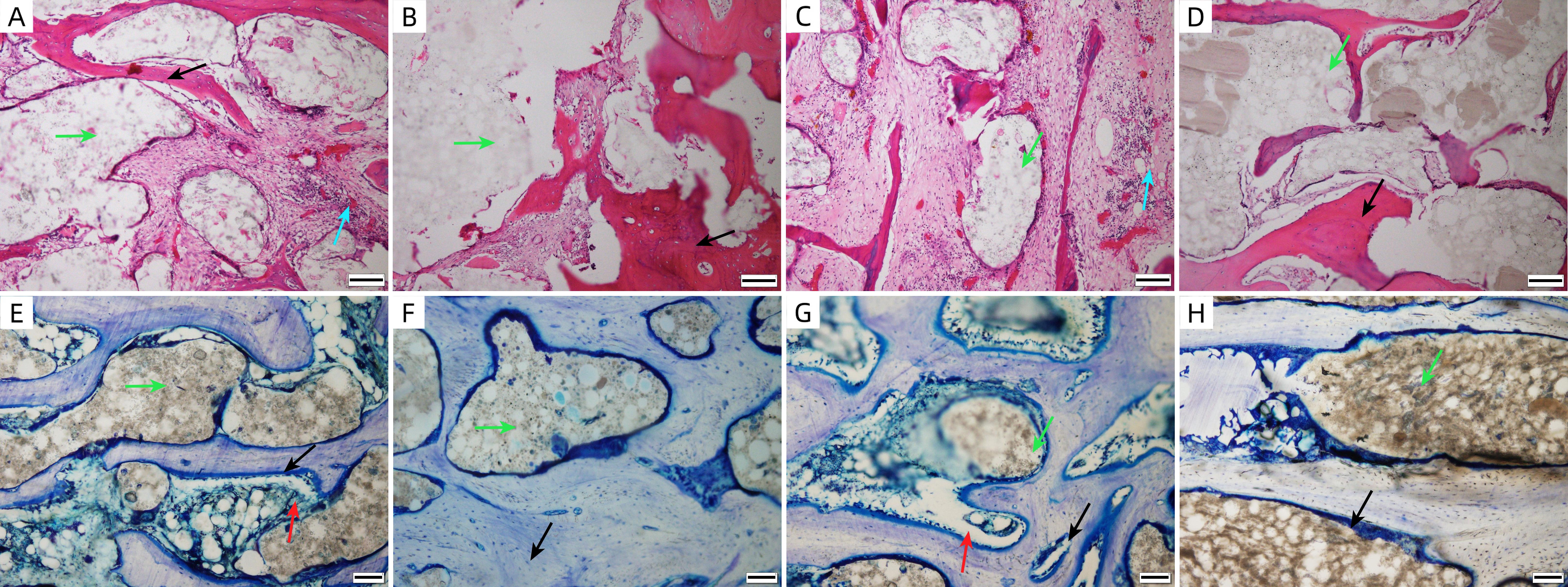
Figure 2 HE and toluidine blue staining results of bone cement implantation site tissues (×100)Note: A, Group A at 4 weeks post-surgery (HE staining); B, Group A at 26 weeks post-surgery (HE staining); C, Group B at 4 weeks post-surgery (HE staining); D, Group B at 26 weeks post-surgery (HE staining); E, Group A at 4 weeks post-surgery (toluidine blue staining); F, Group A at 26 weeks post-surgery (toluidine blue staining); G, Group B at 4 weeks post-surgery (toluidine blue staining); H, Group B at 26 weeks post-surgery (toluidine blue staining). The red arrows indicate the osteoblast, the green arrows indicate the bone cement, the black arrows indicate the bone trabecula, the blue arrows indicate the area of inflammatory cell infiltration and the scale bars in all figures are 200 μm.
| [1] | 陈继良, 许庆山, 王旭, 等. 经皮椎体成形术治疗骨质疏松性椎体压缩性骨折伴椎体内裂隙样变的疗效观察[J]. 中国微创外科杂志, 2018, 18(2):138-142. DOI: 10.3969/j.issn.1009-6604.2018.02.013 . |
| CHEN J L, XU Q S, WANG X, et al. Clinical effect of percutaneous vertebroplasty for osteoporotic vertebral compression fractures combined with intravertebral vacuum cleft[J]. Chin J Minim Invasive Surg, 2018, 18(2):138-142. DOI: 10.3969/j.issn.1009-6604.2018.02.013 . | |
| [2] | 于振和, 李东君. 经皮穿刺椎体后凸成形术治疗骨质疏松性椎体压缩性骨折的疗效观察[J]. 中国微创外科杂志, 2015, 15(6):555-557. DOI: 10.3969/j.issn.1009-6604.2015.06.022 . |
| YU Z H, LI D J. Efficacy of percutaneous kyphoplasty for osteoporosis vertebral compression fractures[J]. Chin J Minim Invasive Surg, 2015, 15(6):555-557. DOI: 10.3969/j.issn.1009-6604.2015.06.022 . | |
| [3] | 黎双庆, 杨波, 杨逸禧, 等. 经皮穿刺椎体成形术治疗骨质疏松性严重椎体压缩性骨折[J]. 中国微创外科杂志, 2015, 15(9):818-821. DOI: 10.3969/j.issn.1009-6604.2015.09.014 . |
| LI S Q, YANG B, YANG Y X, et al. Percutaneous vertebroplasty for severe osteoporotic vertebral compression fractures[J]. Chin J Minim Invasive Surg, 2015, 15(9):818-821. DOI: 10.3969/j.issn.1009-6604.2015.09.014 . | |
| [4] | 孟仪, 于一民, 许卫兵, 等. 经皮椎体成形术治疗骨质疏松性椎体压缩骨折的疗效和安全性评价[J]. 中国医科大学学报, 2018, 47(10):948-951. DOI: 10.12007/j.issn.0258-4646.2018.10.020 . |
| MENG Y, YU Y M, XU W B, et al. Safety and long-term outcome of percutaneous vertebroplasty for osteoporotic vertebral compression fractures[J]. J China Med Univ, 2018, 47(10):948-951. DOI: 10.12007/j.issn.0258-4646.2018.10.020 . | |
| [5] | 姜嘉伟, 张金龙, 徐冠华, 等. 经皮椎体-间盘成形术治疗胸腰段严重压缩的骨质疏松性椎体骨折的疗效分析[J]. 中华创伤骨科杂志, 2023(1):25-30.DOI: 10.3760/cma.j.cn115530-20221116-00584 . |
| JIANG J W, ZHANG J L, XU G H, et al. Percutaneous vertebral-disc plasty for very severe osteoporotic vertebral compre-ssion fractures[J]. Chin J Orthop Trauma, 2023(1):25-30.DOI: 10.3760/cma.j.cn115530-20221116-00584 . | |
| [6] | SUN Y, ZHANG Y, MA H N, et al. Therapeutic efficacy and safety of percutaneous curved vertebroplasty in osteoporo-tic vertebral compression fractures: a systematic review and meta-analysis[J]. Orthop Surg, 2023, 15(10):2492-2504. DOI: 10.1111/os.13800 . |
| [7] | SUN H, LI C. Comparison of unilateral and bilateral percutaneous vertebroplasty for osteoporotic vertebral com-pression fractures: a systematic review and meta-analysis[J]. J Orthop Surg Res, 2016, 11(1):156. DOI: 10.1186/s13018-016-0479-6 . |
| [8] | 胡浩,曹开学,黄攀,等. 超高龄骨质疏松性胸腰椎压缩骨折经皮椎体成形术术后邻椎再骨折的危险因素分析[J]. 中国骨伤, 2022, 35(8):710-714. DOI:10.12200/j.issn.1003-0034.2022.08.002 |
| HU H, CAO K X, HUANG P, et al. Risk factors of adjacent vertebral refracture after percutaneous vertebroplasty for osteoporotic vertebral compression fractures in super-old patients[J]. Zhongguo Gu Shang, 2022, 35(8):710-714. DOI: 10.12200/j.issn.1003-0034.2022.08.002 . | |
| [9] | 王孝林, 权正学, 王南, 等. 网袋加压椎体成形术治疗骨质疏松性椎体压缩性骨折[J]. 中国微创外科杂志, 2017, 17(9):822-826. DOI: 10.3969/j.issn.1009-6604.2017.09.017 . |
| WANG X L, QUAN Z X, WANG N, et al. Vesselplasty for osteoporotic vertebral compression fracture[J]. Chin J Minim Invasive Surg, 2017, 17(9):822-826. DOI: 10.3969/j.issn.1009-6604.2017.09.017 . | |
| [10] | 李天任, 邵安良, 魏利娜, 等. 医疗器械临床前动物试验研究的考虑要点[J]. 中国药事, 2019, 33(10):1121-1128. DOI: 10.16153/j.1002-7777.2019.10.007 . |
| LI T R, SHAO A L, WEI L N, et al. Essential considerations on preclinical animal study for medical devices[J]. Chin Pharm Aff, 2019, 33(10):1121-1128. DOI: 10.16153/j.1002-7777.2019.10.007 . | |
| [11] | 国家药品监督管理局.医疗器械临床前动物研究第1部分:通用要求:YY/T 1754.1-2020[s].北京:中国标准出版社, 2020. |
| National Medical Products Administration. Preclinical animal studies for medicaldevices part 1: general requirements: [S]. Beijing: Standards press of China, 2020. | |
| [12] | WANG S, WANG H, NIU L. Clinical efficacy of PVP and PKP in the treatment of OVCFs after bilateral resection of ovarian cancer[J]. Oncol Lett, 2018, 16(1):151-156. DOI: 10.3892/ol.2018.8658 . |
| [13] | ENGLAND R W, GONG A N, LI T B, et al. Clinical outcomes and safety of the SpineJack vertebral augmentation system for the treatment of vertebral compression fractures in a United States patient population[J]. J Clin Neurosci, 2021, 89:237-242. DOI: 10.1016/j.jocn.2021.04.031 . |
| [14] | 李庆达, 杨俊松, 高林, 等. 经皮椎体成形术与非手术治疗急性症状性骨质疏松性胸腰椎骨折分型Ⅰ型骨折的疗效比较[J]. 中华创伤杂志, 2021(6):541-548. DOI: 10.3760/cma.j.cn501098-20210120-00058 . |
| LI Q D, YANG J S, GAO L, et al. Comparison of curative efficacy of percutaneous vertebroplasty and non-surgical treatment of type Ⅰ acute symptomatic osteoporotic thora-columbar fracture[J]. Chin J Trauma, 2021(6):541-548. DOI: 10.3760/cma.j.cn501098-20210120-00058 . | |
| [15] | 彭亚, 肖辉灯, 祝永刚, 等. 单侧经皮椎体后凸成形术联合过伸复位法治疗新鲜骨质疏松性椎体压缩性骨折的效果观察[J]. 中国综合临床, 2021, 37(5):438-443. DOI: 10.3760/cma.j.cn101721-20210513-00010 . |
| PENG Y, XIAO H D, ZHU Y G, et al. Effect of unilateral percutaneous kyphoplasty combined with hyperextension reduction in the treatment of fresh osteoporotic vertebral compression fractures[J]. Clin Med China, 2021, 37(5):438-443. DOI: 10.3760/cma.j.cn101721-20210513-00010 . | |
| [16] | 唐本强, 王彦辉, 许崧杰, 等. 骨质疏松性椎体压缩骨折经皮椎体强化术后椎体内骨水泥分布类型的研究进展[J]. 中华骨科杂志, 2022(5):320-330. DOI: 10.3760/cma.j.cn121113-20210129-00089 . |
| TANG B Q, WANG Y H, XU S J, et al. The distribution of bone cement in the vertebral body after percutaneous vertebral augmentation for osteoporotic vertebral compression fractures[J]. Chin J Orthop, 2022(5):320-330. DOI: 10.3760/cma.j.cn121113-20210129-00089 . | |
| [17] | 刘训伟, 孔小燕, 钟建, 等. 骨填充网袋修复椎体压缩骨折的生物力学变化[J]. 中国组织工程研究, 2014, 18(16):2487-2492. DOI: 10.3969/j.issn.2095-4344.2014.16.005 . |
| LIU X W, KONG X Y, ZHONG J, et al. Bone filling mesh container repairs vertebral compression fractures: biomecha-nical changes[J]. Chin J Tissue Eng Res, 2014, 18(16):2487-2492. DOI: 10.3969/j.issn.2095-4344.2014.16.005 . | |
| [18] | 郭松, 付强, 杭栋华, 等. Mazor脊柱机器人辅助改良经皮椎体成形术治疗腰椎骨质疏松性骨折的疗效分析[J]. 中国脊柱脊髓杂志, 2021, 31(9):818-824. DOI: 10.3969/j.issn.1004-406X.2021.09.06 . |
| GUO S, FU Q, HANG D H, et al. Effectiveness of Mazor spine robot-assisted percutaneous vertebroplasty with modified approach in treating lumbar osteoporotic vertebral com-pression fractures[J]. Chin J Spine Spinal Cord, 2021, 31(9):818-824. DOI: 10.3969/j.issn.1004-406X.2021.09.06 . | |
| [19] | GUO Z, WANG W, GAO W S, et al. Comparison the clinical outcomes and complications of high-viscosity versus low-viscosity in osteoporotic vertebral compression fractures[J]. Medicine (Baltimore), 2017, 96(48): e8936. DOI: 10.1097/md.0000000000008936 . |
| [20] | 邹华章, 廖威明, 段昕, 等. 新型可注射磷酸钙骨水泥在椎体后凸成形术中的生物力学评价[J]. 中华生物医学工程杂志, 2011(2):151-155.DOI:10.3760/cma.j.issn.1674-1927.2011.02.012 |
| ZOU H Z, LIAO W M, DUAN X, et al. Biomechanical evaluation of a novel injectable calcium phosphate cement for use in kyphoplasty[J]. Chin J Biomed Eng, 2011(2):151-155.DOI:10.3760/cma.j.issn.1674-1927.2011.02.012 . | |
| [21] | WEISSKOPF M, OHNSORGE J A, NIETHARD F U. Intraver-tebral pressure during vertebroplasty and balloon kypho-plasty: an in vitro study[J]. Spine (Phila Pa 1976), 2008, 33(2):178-182. DOI: 10.1097/brs.0b013e3181606139 . |
| [22] | BERLEMANN U, FERGUSON S J, NOLTE L P, et al. Adjacent vertebral failure after vertebroplasty. A biomechanical investigation[J]. J Bone Joint Surg Br, 2002, 84(5):748-752. DOI: 10.1302/0301-620x.84b5.11841 . |
| [23] | 许晨辉, 何阿祥, 谢栋, 等. 介孔钙硅酸盐改性硫酸钙骨水泥的体内降解及成骨性能[J]. 中国矫形外科杂志, 2016, 24(6):544-551. DOI: 10.3977/j.issn.1005-8478.2016.06.14 . |
| XU C H, HE A X, XIE D, et al. In vivo degradability and osteogenesis of m-CS/CSC bone cement[J]. Orthop J China, 2016, 24(6):544-551. DOI: 10.3977/j.issn.1005-8478.2016.06.14 . | |
| [24] | 曹天庆, 程朋真, 杨柳, 等. 系统性生物力学评价大鼠股骨缺损修复进程的实验研究[J]. 中华创伤骨科杂志, 2018(3):247-253. DOI: 10.3760/cma.j.issn.1671-7600.2018.03.012 . |
| CAO T Q, CHENG P Z, YANG L, et al. Systematic evaluation of repairing femoral defects by biomechanical measurements in rats[J]. Chin J Orthop Trauma, 2018(3):247-253. DOI: 10.3760/cma.j.issn.1671-7600.2018.03.012 . | |
| [25] | 朱爱国, 张烽, 葛勇, 等. 硫酸钙骨水泥增强椎弓根螺钉置入骨质疏松椎体的瞬时稳定性[J]. 中国组织工程研究, 2014, 18(26):4195-4199. DOI: 10.3969/j.issn.2095-4344.2014.26.018 . |
| ZHU A G, ZHANG F, GE Y, et al. Calcium sulfate cement augments transient stability of pedicle screw in osteoporotic vertebral body[J]. Chin J Tissue Eng Res, 2014, 18(26):4195-4199. DOI: 10.3969/j.issn.2095-4344.2014.26.018 . |
| [1] | WANG Jiaoxiang, ZHANG Lu, CHEN Shuhan, JIAO Deling, ZHAO Heng, WEI Taiyun, GUO Jianxiong, XU Kaixiang, WEI Hongjiang. Construction and Functional Validation of GTKO/hCD55 Gene-Edited Xenotransplant Donor Pigs [J]. Laboratory Animal and Comparative Medicine, 2025, 45(4): 379-392. |
| [2] | LIU Yuanyuan, XIN Wenshui, CHAO Zhe, CAO Zongxi, CAI Yifei, LI Qiang, LI Lingwei, LIU Guangliang. Identification and Analysis of MHCⅡ Genes in Wuzhishan Pigs [J]. Laboratory Animal and Comparative Medicine, 2025, 45(3): 340-348. |
| [3] | LIU Yishu, CAI Liping. Advances and Challenges of Using Experimental Pigs in Da Vinci Surgical Robot Training [J]. Laboratory Animal and Comparative Medicine, 2024, 44(6): 667-674. |
| [4] | Jiaoxiang WANG, Yan WANG, Ke HU, Kaixiang XU, Taiyun WEI, Deling JIAO, Heng ZHAO, Hongye ZHAO, HongJiang WEI. Establishment of PCR Identification Method for Pig Blood Type [J]. Laboratory Animal and Comparative Medicine, 2023, 43(6): 585-594. |
| [5] | Laien XUE. Anesthetic Effects of Zoletil and Xylazine on Bama Mini-pigs [J]. Laboratory Animal and Comparative Medicine, 2022, 42(1): 26-30. |
| [6] | XIAO Pan, NIU Tingxian, LUO Xiaohong, WANG Hongyi, GUO Xiaoyu, LU Lu, FENG Xiaoming. Changes of C-reactive Protein and Inflammatory Factors in Juema Pig Model with High Altitude Multiple Organ Dysfunction Syndrome [J]. Laboratory Animal and Comparative Medicine, 2021, 41(3): 238-243. |
| [7] | ZHANG Hui, WU Yanjun, YAO Jin, LU Taofeng, WANG Guoqi, ZHAO Hai, YAO Gang, WU Shuguang. Therapeutic Effect of Wuling Powder on Guizhou Mini-pigs of Hyperlipidemia Model [J]. Laboratory Animal and Comparative Medicine, 2021, 41(2): 138-142. |
| [8] | HUANG Peidi, LI Haiwen, ZHENG Huantian, LI Jian. Observation of LumianningⅡ or Ketamine Combined with Propofol in Experimental Pigs Undergoing Natural Orifice Transluminal Endoscopic Surgery#br# [J]. Laboratory Animal and Comparative Medicine, 2020, 40(3): 242-. |
| [9] | YANG Li-chang, ZHOU Wen-bing, DING Jun, ZHOU Xin-chu, XIE Dong, YANG Wei-min. Investigation on Electrocardiogram of Bama Minipigs by Telemetry [J]. Laboratory Animal and Comparative Medicine, 2017, 37(5): 410-413. |
| [10] | CHEN Ming-fei, YAO Gang, ZHAO Hai, QIAN Ning, WU Shu-guang. Primiparous Reproductive Performance of Outbred Guizhou Miniature Pigs and its Correlation and Path Analysis [J]. Laboratory Animal and Comparative Medicine, 2017, 37(4): 324-327. |
| [11] | YANG Li-chang, ZHOU Wen-bing, DING Jun, ZHOU Xin-chu, XIE Dong, YANG Wei-min. Reproductive Performance of Three Breeds of Minipigs [J]. Laboratory Animal and Comparative Medicine, 2017, 37(1): 50-54. |
| [12] | ZHANG Yue-han, WEN Jing, ZHU Hai-tao, ZHAO Guang-zong, LI Xiao-yu. Intragastric Estrogen Administration Induced Guinea Pig Model of Uterine Leiomyoma [J]. Laboratory Animal and Comparative Medicine, 2016, 36(3): 195-199. |
| [13] | PAN Jin-chun, MIN Fan-gui, WANG Xi-long, CHEN Rui-ai, WANG Feng-guo, LUO Shu-ming. Establishment of Peritoneal Adhesion Model in Wuzhishan Minipigs [J]. Laboratory Animal and Comparative Medicine, 2014, 34(4): 299-302. |
| [14] | TANG Hua, BI Wen-hao, AI Wei, LI De-sheng, PAN Guang-hui, SHI Jun-wen, LIU Wei, HUANG Wei, XIAO Dong, GU Wei-wang. Isolation and Culture of Bone Marrow Mesenchymal Stem Cells (MSCs) of Tibetan miniature pig, and Lentivirus-mediated EGFP Gene Transfer into MSCs [J]. Laboratory Animal and Comparative Medicine, 2012, 32(2): 105-110. |
| [15] | WANG Xi-long, MIN Fan-gui, PAN Jin-chun, YUAN Wen, CHEN Rui-ai, HUANG Bao-song, WANG Feng-guo. Selection and Breeding of Experimental Wuzhishan Minipigs [J]. Laboratory Animal and Comparative Medicine, 2011, 31(5): 345-348. |
| Viewed | ||||||
|
Full text |
|
|||||
|
Abstract |
|
|||||