

实验动物与比较医学 ›› 2026, Vol. 46 ›› Issue (5): 650-659.DOI: 10.12300/j.issn.1674-5817.2026.010
许浩莀, 乐卿清, 何西文, 蔡兵, 杨隆河( )(
)( )
)
收稿日期:2026-01-08
修回日期:2026-06-16
出版日期:2026-10-25
发布日期:2026-10-16
通讯作者:
杨隆河(1986—),男,博士,副研究员,研究方向:海洋药物药理及功能食品研究。E-mail:longheyang@tio.org.cn。ORCID: 0009-0006-6101-2066作者简介:基金资助:
XU Haochen, LE Qingqing, HE Xiwen, CAI Bing, YANG Longhe()()
Received:2026-01-08
Revised:2026-06-16
Published:2026-10-25
Online:2026-10-16
Correspondence to:
摘要:
目的 为探明蓝光诱导小鼠视网膜损伤的时间依赖性,确立适宜的动物模型,并评价含叶黄素酯、DHA藻油、黑果枸杞等药食同源原料的复合配方对视网膜的保护作用。 方法 将56只SPF级雄性C57BL/6J小鼠中的32只随机分为对照组、模型组(1 h、3 h、6 h照射)(n=8),以确定蓝光最佳照射时间。首先采用467 nm发光二极管[(5 000±200) lx]蓝光分别照射小鼠1 h、3 h、6 h,然后分别用苏木精-伊红染色切片检测小鼠视网膜组织病理学改变及厚度,用试剂盒方法检测血清丙二醛(malondialdehyde,MDA)和谷胱甘肽(glutathione,GSH)水平,使用实时荧光定量反转录PCR检测视网膜炎症因子mRNA表达量。确定3 h为适宜模型后,其余24只小鼠用于护眼复合配方对蓝光视网膜损伤的干预实验,以复合配方(1 000 mg·kg⁻¹·d⁻¹)持续灌胃小鼠14 d,再行3 h蓝光照射,以同样的方法评估以上指标。 结果 随照射时间的延长,小鼠视网膜各层的厚度逐渐变薄(P<0.05至P<0.01),细胞排列紊乱;血清MDA含量逐步升高(P<0.01),而GSH含量逐步降低(P<0.01);视网膜组织白细胞介素-1β(interleukin-1β,IL-1β)、白细胞介素-6(interleukin-6,IL-6)和肿瘤坏死因子-α(tumor necrosis factor-α,TNF-α)的mRNA表达水平随着照射时间延长逐渐上调,其中1 h照射组指标变化较小(P>0.05),6 h照射组变化最显著(P<0.000 1),因此选择各项指标差异显著且适中的3 h(P<0.001)蓝光照射作为后续造模条件。护眼复合配方干预后,与模型组相比,小鼠血清MDA含量显著降低(P<0.01),而血清GSH含量显著升高(P<0.05);视网膜中炎症因子IL-1β(P<0.000 1)、IL-6(P<0.001)和TNF-α(P<0.01)表达量显著下调;视网膜总厚度显著增加(P<0.01),且内丛状层(P<0.05)、内核层(P<0.05)、外丛状层(P<0.05)与外核层(P<0.01)厚度均显著增加;组织病理学显示,其视网膜及细胞排列的规律性与致密程度均明显优于模型组。 结论 3 h特定照度的蓝光照射可建立稳定的小鼠视网膜损伤模型;护眼复合配方通过抗氧化-抗炎途径缓解损伤,为相关功能食品的开发提供实验依据。
中图分类号:
许浩莀,乐卿清,何西文,等. 护眼复合配方对蓝光损伤模型小鼠视网膜的功效评估[J]. 实验动物与比较医学, 2026, 46(5): 650-659. DOI: 10.12300/j.issn.1674-5817.2026.010.
XU Haochen,LE Qingqing,HE Xiwen,et al. Evaluation of the Efficacy of an Eye-Protective Compound Formula on the Retina in a Mouse Model of Blue Light-Induced Damage[J]. Laboratory Animal and Comparative Medicine, 2026, 46(5): 650-659. DOI: 10.12300/j.issn.1674-5817.2026.010.
实时荧光定量反转录PCR所用引物序列
The sequences of primers used in real-time fluorescent quantitative reverse transcription PCR
| 基因名称 Gene name | 引物序列 Primer sequences (5' - 3') |
|---|---|
| IL-1β | F-GAAATGCCACCTTTTGACAGTG R-TGGATGCTCTCATCAGGACAG |
| IL-6 | F-AATTAAGCCTCCGACTTGTGAAG R-CTTCCATCCAGTTGCCTTCTTG |
| TNF-α | F-GTAGCAAACCACCAAGTGGAGG R-CAGCCTTGTCCCTTGAAGAGAA |
| GAPDH | F-GGTGAAGGTCGGTGTGAACG R-CTCGCTCCTGGAAGATGGTG |
表1
实时荧光定量反转录PCR所用引物序列
Table 1
The sequences of primers used in real-time fluorescent quantitative reverse transcription PCR
| 基因名称 Gene name | 引物序列 Primer sequences (5' - 3') |
|---|---|
| IL-1β | F-GAAATGCCACCTTTTGACAGTG R-TGGATGCTCTCATCAGGACAG |
| IL-6 | F-AATTAAGCCTCCGACTTGTGAAG R-CTTCCATCCAGTTGCCTTCTTG |
| TNF-α | F-GTAGCAAACCACCAAGTGGAGG R-CAGCCTTGTCCCTTGAAGAGAA |
| GAPDH | F-GGTGAAGGTCGGTGTGAACG R-CTCGCTCCTGGAAGATGGTG |
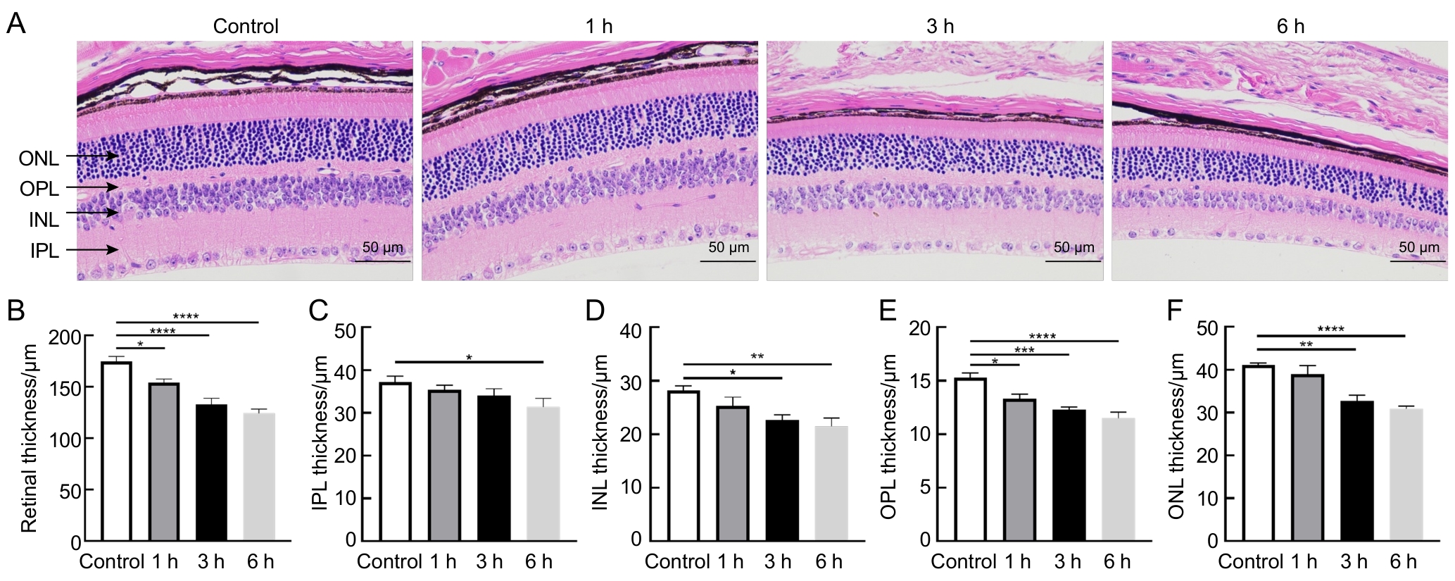
图1
Figure 1
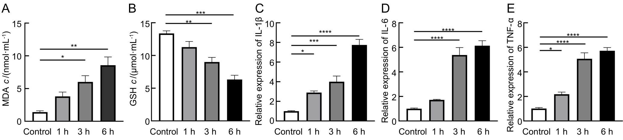
图2
Figure 2
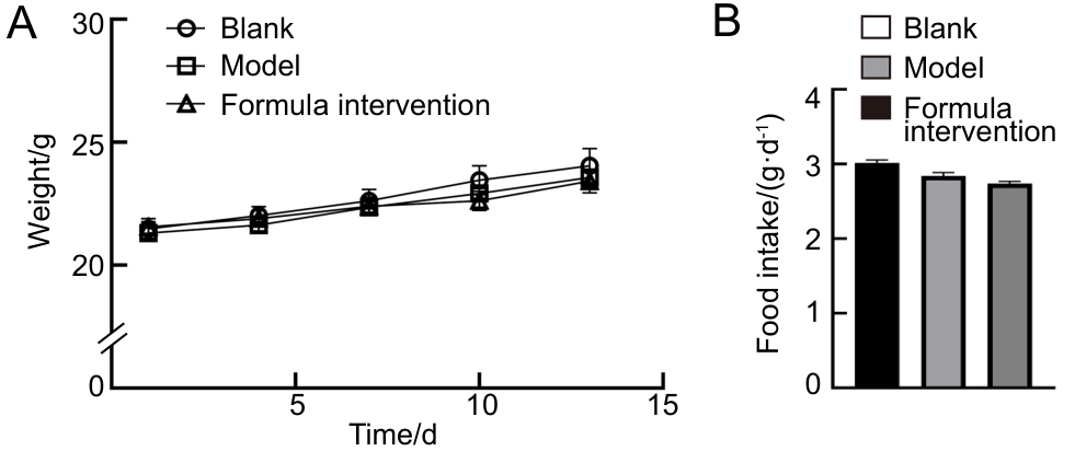
图3
Figure 3
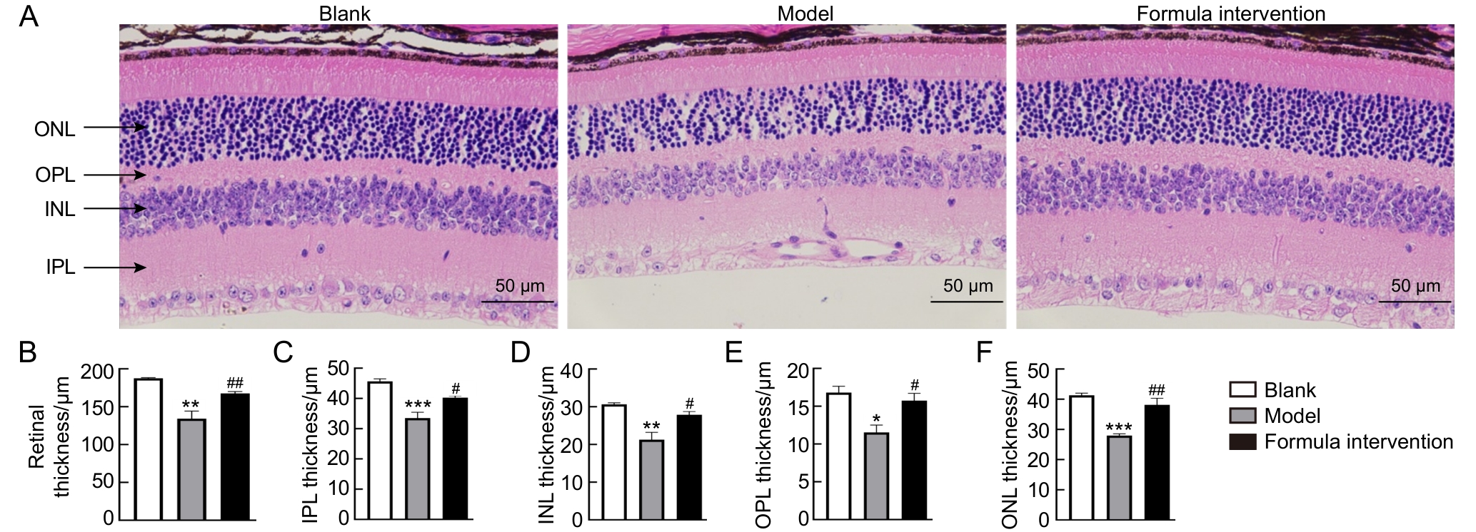
图4
Figure 4

图5
Figure 5
| [1] |
ZHOU Z X, LI S Z, YANG Q S, et al. BlingLife®-marigold extract alleviates blue light-induced retinal mitochondria oxidative stress and senescence by activating NRF2/HO-1 signaling[J]. J Microbiol Biotechnol, 2025, 35: e2411037. DOI: 10.4014/jmb. 2411.11037.
|
| [2] |
CHAKRAVARTHY H, GEORGYEV V, WAGEN C, et al. Blue light-induced phototoxicity in retinal cells: implications in age-related macular degeneration[J]. Front Aging Neurosci, 2024, 16: 1509434. DOI: 10.3389/fnagi.2024.1509434.
|
| [3] |
YEH W J, CHIEN P T, WEN Y T, et al. A comprehensive review of experimental models for investigating blue light-induced ocular damage: Insights into parameters, limitations, and new opportunities[J]. Exp Eye Res, 2024, 249: 110142. DOI: 10. 1016/j.exer.2024.110142.
|
| [4] |
ZHANG S Y, REN J P, GHAI R T. Global burden of low vision and blindness due to age-related macular degeneration from 1990 to 2021 and projections for 2050[J]. BMC Public Health, 2024, 24(1): 3510. DOI: 10.1186/s12889-024-21047-x.
|
| [5] |
THOMAS C J, MIRZA R G, GILL M K. Age-related macular degeneration[J]. Med Clin North Am, 2021, 105(3): 473-491. DOI: 10.1016/j.mcna.2021.01.003.
|
| [6] |
GARCÍA-ROMERA M C, PONCE-GARCÍA V, TORRES-PAREJO Ú, et al. Effect of exposure to blue light from electronic devices and the Mediterranean diet on macular pigment[J]. J Clin Med, 2024, 13(24): 7688. DOI: 10.3390/jcm13247688.
|
| [7] |
KIM S H, KIM Y K, SHIN Y I, et al. Nighttime outdoor artificial light and risk of age-related macular degeneration[J]. JAMA Netw Open, 2024, 7(1): e2351650. DOI: 10.1001/jamanet workopen.2023.51650.
|
| [8] |
SINGH S, KELLER P R, BUSIJA L, et al. Blue-light filtering spectacle lenses for visual performance, sleep, and macular health in adults[J]. Cochrane Database Syst Rev, 2023(8): CD013244. DOI: 10.1002/14651858.cd013244.pub2.
|
| [9] |
MOISEEV R V, MORRISON P W J, STEELE F, et al. Penetration enhancers in ocular drug delivery[J]. Pharmaceutics, 2019, 11(7): 321. DOI: 10.3390/pharmaceutics11070321.
|
| [10] |
YEH W J, YAN C, WU C H. Photoprotective effects of phytochemicals on blue light-induced retinal damage: current evidence and future perspectives[J]. Nutrients, 2025, 17(2): 331. DOI: 10.3390/nu17020331.
|
| [11] |
WON J H, SITNIKOV D, HONG J N. Protective effects of carotenoids against blue light induced-cellular damage in human retinal pigment epithelium[J]. Food Sci Biotechnol, 2025, 34(8): 1713-1723. DOI: 10.1007/s10068-024-01757-z.
|
| [12] |
ACAR N. The implication of omega-3 polyunsaturated fatty acids in retinal physiology[J]. Oléagineux Corps Gras Lipides, 2007, 14(3-4): 194-197. DOI: 10.1051/ocl.2007.0121.
|
| [13] |
LIU Y X, ZHANG D, WU Y P, et al. Docosahexaenoic acid aggravates photooxidative damage in retinal pigment epithelial cells via lipid peroxidation[J]. J Photochem Photobiol B, 2014, 140: 85-93. DOI: 10.1016/j.jphotobiol.2014. 07.016.
|
| [14] |
LI P, LI Z G, SUN Q X, et al. Protective effect and mechanism of Lycium ruthenicum Murray anthocyanins against retinal damage induced by blue light exposure[J]. J Food Sci, 2024, 89(8): 5113-5129. DOI: 10.1111/1750-3841.17184.
|
| [15] |
庞雨菲. 黑果枸杞多酚对丙烯酰胺诱导大鼠纹状体氧化应激损伤的保护作用及机制[D]. 银川: 宁夏医科大学, 2023. DOI: 10.27258/d.cnki.gnxyc.2023.000305.
|
|
PANG Y F. Protective effect and mechanism of polyphenols from Lycium ruthenicum Murray on acrylamide-induced oxidative stress injury in rat striatum [D]. Yinchuan: Ningxia Medical University, 2023. DOI: 10.27258/d.cnki.gnxyc.2023.000305.
|
|
| [16] |
OUYANG X L, YANG J, HONG Z X, et al. Mechanisms of blue light-induced eye hazard and protective measures: a review[J]. Biomed Pharmacother, 2020, 130: 110577. DOI: 10.1016/j.biopha.2020.110577.
|
| [17] |
娄解南, 谭书明, 谢国芳, 等. 刺梨多酚对丙烯酰胺致肝脏氧化损伤的保护作用[J]. 食品安全质量检测学报, 2023, 14(17): 122-128. DOI: 10.19812/j.cnki.jfsq11-5956/ts.2023.17.012.
|
|
LOU J N, TAN S M, XIE G F, et al. Protective effect of Rosa roxburghii Tratt.polyphenols against acrylamide-induced hepatic oxidative damage[J]. J Food Saf Qual, 2023, 14(17): 122-128. DOI: 10.19812/j.cnki.jfsq11-5956/ts.2023.17.012.
|
|
| [18] |
张丹, 葛颖颖, 杨琼琼, 等. 刺梨果抗氧化活性成分与体外抗氧化活性研究[C]//中国食品科学技术学会第十五届年会论文摘要集. 2018: 283-284.
|
|
ZHANG D, GE Y Y, YANG Q Q, et al. Study on antioxidant active components and in vitro antioxidant activity of Rosa roxburghii fruit[C]//Abstracts of the 15th Annual Meeting of Chinese Institute of Food Science and Technology. 2018: 283-284.
|
|
| [19] |
SKIBSTED L H. Anthocyanidins regenerating xanthophylls: a quantum mechanical approach to eye health[J]. Curr Opin Food Sci, 2018, 20: 24-29. DOI: 10.1016/j.cofs.2018.02.014.
|
| [20] |
TÄHTIVAARA L, THAPA R, PARTANEN P, et al. Comparison of blue light-induced damage (BLD) in albino and pigmented mice[J]. Invest Ophthalmol Vis Sci, 2025, 66(8): 2528.
|
| [21] |
KING A, GOTTLIEB E, BROOKS D G, et al. Mitochondria-derived reactive oxygen species mediate blue light-induced death of retinal pigment epithelial cells[J]. Photochem Photobiol, 2004, 79(5): 470-475. DOI: 10.1111/j.1751-1097.2004.tb00036.x.
|
| [22] |
KAYNEZHAD P, FOSBURY R, HOGG C, et al. Near infrared spectroscopy reveals instability in retinal mitochondrial metabolism and haemodynamics with blue light exposure at environmental levels[J]. J Biophotonics, 2022, 15(4): e202100283. DOI: 10.1002/jbio.202100283.
|
| [23] |
ROEHLECKE C, SCHUMANN U, ADER M, et al. Stress reaction in outer segments of photoreceptors after blue light irradiation[J]. PLoS One, 2013, 8(9): e71570. DOI: 10.1371/journal.pone.0071570.
|
| [24] |
马修彬, 陈松. 蓝光对视网膜色素上皮细胞的损伤机制[J]. 国际眼科纵览, 2007, 31(6): 415-419. DOI: 10.3760/cma.j.issn.1673-5803.2007.06.015.
|
|
MA X B, CHEN S. Mechanism of blue-light-induced damage to the retinal pigment epithelium cells[J]. International Rev Ophthalmol, 2007, 31(6): 415-419. DOI: 10.3760/cma.j.issn.1673-5803.2007.06.015.
|
|
| [25] |
NAKAMURA M, KUSE Y, TSURUMA K, et al. The involvement of the oxidative stress in murine blue LED light-induced retinal damage model[J]. Biol Pharm Bull, 2017, 40(8): 1219-1225. DOI: 10.1248/bpb.b16-01008.
|
| [26] |
KAIDZU S, OKUNO T, TANITO M, et al. Structural and functional change in albino rat retina induced by various visible light wavelengths[J]. Int J Mol Sci, 2022, 23(1): 309. DOI: 10.3390/ijms23010309.
|
| [27] |
JI Z Z, ZHANG D C, WANG Y M, et al. The role of the SIRT1/NF-κB/NLRP3 pathway in the pyroptosis of lens epithelial cells under shortwave blue light radiation[J]. Exp Eye Res, 2024, 246: 110019. DOI: 10.1016/j.exer.2024.110019.
|
| [28] |
YANG Y C, YANG R, DENG F Q, et al. Immunoactivation by cutaneous blue light irradiation inhibits remote tumor growth and metastasis[J]. ACS Pharmacol Transl Sci, 2024, 7(4): 1055-1068. DOI: 10.1021/acsptsci.3c00355.
|
| [29] |
GRIMM C, WENZEL A, WILLIAMS T, et al. Rhodopsin-mediated blue-light damage to the rat retina: effect of photoreversal of bleaching[J]. Invest Ophthalmol Vis Sci, 2001,42(2): 497-505.
|
| [30] |
KAMOSHITA M, TODA E, OSADA H, et al. Lutein acts via multiple antioxidant pathways in the photo-stressed retina[J]. Sci Rep, 2016, 6: 30226. DOI: 10.1038/srep30226.
|
| [31] |
KIM J, CHO K, CHOUNG S Y. Protective effect of Prunella vulgaris var. L extract against blue light induced damages in ARPE-19 cells and mouse retina[J]. Free Radic Biol Med, 2020, 152: 622-631. DOI: 10.1016/j.freeradbiomed.2019.12.003.
|
| [32] |
ORGANISCIAK D T, VAUGHAN D K. Retinal light damage: Mechanisms and protection[J]. Prog Retin Eye Res, 2010, 29(2): 113-134. DOI: 10.1016/j.preteyeres.2009.11.004.
|
| [33] |
JAADANE I, BOULENGUEZ P, CHAHORY S, et al. Retinal damage induced by commercial light emitting diodes (LEDs)[J]. Free Radic Biol Med, 2015, 84: 373-384. DOI: 10.1016/j.freeradbiomed.2015.03.034.
|
| [34] |
SHANG Y M, WANG G S, SLINEY D, et al. White light–emitting diodes (LEDs) at domestic lighting levels and retinal injury in a rat model[J]. Environ Health Perspect, 2014, 122(3): 269-276. DOI: 10.1289/ehp.1307294.
|
| [35] |
OGAWA K, KUSE Y, TSURUMA K, et al. Protective effects of bilberry and lingonberry extracts against blue light-emitting diode light-induced retinal photoreceptor cell damage in vitro[J]. BMC Complement Altern Med, 2014, 14: 120. DOI: 10.1186/1472-6882-14-120.
|
| [36] |
WU J M, SEREGARD S, ALGVERE P V. Photochemical damage of the retina[J]. Surv Ophthalmol, 2006, 51(5): 461-481. DOI: 10.1016/j.survophthal.2006.06.009.
|
| [37] |
DABOUZ R, CHENG C W H, ABRAM P, et al. An allosteric interleukin-1 receptor modulator mitigates inflammation and photoreceptor toxicity in a model of retinal degeneration[J]. J Neuroinflammation, 2020, 17(1): 359. DOI: 10.1186/s12974-020-02032-8.
|
| [38] |
朱恒杏, 邵孟茹, 洪天畅, 等. 黑枸杞花青素、EGCG对中波紫外线诱导HaCaT细胞氧化损伤的协同保护作用[J]. 现代食品科技, 2024, 40(8): 106-115. DOI: 10.13982/j.mfst.1673-9078.2024.8.0918.
|
|
ZHU H X, SHAO M R, HONG T C, et al. Synergistic protective effects of the anthocyanins from black wolfberry and EGCG against the UVB-induced oxidative damage in HaCaT cells[J]. Mod Food Sci Technol, 2024, 40(8): 106-115. DOI: 10.13982/j.mfst.1673-9078.2024.8.0918.
|
|
| [39] |
邓楷, 欧阳健, 胡娜, 等. 黑果枸杞花青素结构差异对其稳定性及细胞抗氧化活性的影响[J]. 天然产物研究与开发, 2022, 34(2): 213-219. DOI: 10.16333/j.1001-6880.2022.2.005.
|
|
DENG K, OUYANG J, HU N, et al. Effects of structural difference on stability and cellular antioxidative activity of anthocyanins from Lycium ruthenicum Murr[J]. Nat Prod Res Dev, 2022, 34(2): 213-219. DOI: 10.16333/j.1001-6880.2022.2.005.
|
|
| [40] |
李婧, 苏蓉, 刘振华, 等. 黑果枸杞果实中抗炎活性成分的筛选及作用机制研究[J]. 中国野生植物资源, 2023, 42(5): 9-15, 38. DOI: 10.3969/j.issn.1006-9690.2023.05.002.
|
|
LI J, SU R, LIU Z H, et al. Research on the screening and mechanism of active ingredients for anti-inflammatory in Lycium ruthenicum Murr. fruit[J]. Chin Wild Plant Resour, 2023, 42(5): 9-15, 38. DOI: 10.3969/j.issn.1006-9690.2023.05.002.
|
|
| [41] |
薛才华, 武强, 王梦杰, 等. 黑枸杞花青素对巨噬细胞RAW264.7炎性与自噬交互作用的影响[J]. 中国畜牧杂志, 2021, 57(12): 244-249. DOI: 10.19556/j.0258-7033.20210220-06.
|
|
XUE C H, WU Q, WANG M J, et al. Effect of L. ruthenicum on the interaction between inflammation and autophagy of macrophage RAW264.7[J]. Chin J Anim Sci, 2021, 57(12): 244-249. DOI: 10.19556/j.0258-7033.20210220-06.
|
|
| [42] |
BERNSTEIN P S. The role of lutein and zeaxanthin in protection against age-related macular degeneration[J]. Acta Hortic, 2015(1106): 153-160. DOI: 10.17660/ActaHortic.2015. 1106.24.
|
| [43] |
ZHANG H J, LIU X B, CHEN X M, et al. Lutein delays photoreceptor degeneration in a mouse model of retinitis pigmentosa[J]. Neural Regen Res, 2022, 17(7): 1596-1603. DOI: 10.4103/1673-5374.330622.
|
| [44] |
BRAND A, BAUER N G, HALLOTT A, et al. Membrane lipid modification by polyunsaturated fatty acids sensitizes oligodendroglial OLN-93 cells against oxidative stress and promotes up-regulation of heme oxygenase-1 (HSP32)[J]. J Neurochem, 2010, 113(2): 465-476. DOI: 10.1111/j.1471-4159.2010.06611.x.
|
| [45] |
EGGERS B, STEPIEN J, REKER A K, et al. The protective effect of docosahexaenoic acid on mitochondria in SH-SY5Y model of rotenone-induced toxicity[J]. Metabolites, 2025, 15(1): 29. DOI: 10.3390/metabo15010029.
|
| [46] |
BORGONOVI S M, IAMETTI S, DI NUNZIO M. Docosahexaenoic acid as master regulator of cellular antioxidant defenses: a systematic review[J]. Antioxidants, 2023, 12(6): 1283. DOI: 10.3390/antiox12061283.
|
| [1] | 江淼淼, 方金萍, 黄宗炫, 厉嘉敏, 廖欢, 张文强, 余佳颖, 杨燕燕, 连大卫. 葛根芩连汤缓解糖尿病小鼠心肌损伤的作用机制研究[J]. 实验动物与比较医学, 2026, 46(5): 629-639. |
| [2] | 李燕平, 黄姗, 姚政源, 石怡敏, 隋华秀, 黄侯丹, 俞燕萍, 米燕, 林大晖. 艾黄素对臭氧暴露诱发小鼠肺部炎症的保护作用及机制研究[J]. 实验动物与比较医学, 2026, 46(5): 640-649. |
| [3] | 张娇玲, 林阿萍, 许诗荧, 潘德全, 蔡毅君. 小鼠胚胎移植技术的优化及应用[J]. 实验动物与比较医学, 2026, 46(5): 750-758. |
| [4] | 钟秋明, 钱丽萍, 蔡媛媛, 刘德强, 林玮, 俞春英, 谢金东, 王训立, 吴胜, 陈碧茵, 黄镇. 环磷酰胺/紫杉醇诱导化疗脱发模型小鼠的性别间差异对比分析[J]. 实验动物与比较医学, 2026, 46(5): 677-688. |
| [5] | 王娟, 徐佳慧, 田韵远, 张萌萌, 李敏, 王四旺, 李瑶. 两种雌性小鼠溃疡性结肠炎模型的比较与行为学观察[J]. 实验动物与比较医学, 2026, 46(3): 332-343. |
| [6] | 李嘉菲, 张朕豪, 王硕, 田歌, 温爽, 闫玉雪, 崔然, 叶振, 崔永春. 自主神经调节剂对房颤模型大鼠心房电重塑及组织病理变化的影响[J]. 实验动物与比较医学, 2026, 46(3): 321-331. |
| [7] | 李龙雪, 万崇凡, 张琦, 雷茹婷, 王潇玥, 程乐妍, 赖琦, 刘荣华, 刘漩, 徐铁龙. 基于miRNA测序分析清肺排毒汤防治小鼠急性肺损伤的分子机制研究[J]. 实验动物与比较医学, 2026, 46(3): 311-320. |
| [8] | 艾秀峰, 张利棕, 方明笋, 吕东颖, 陈楚, 蔡兆伟, 王德军. 16S rRNA测序分析饮用含氯水对大鼠和小鼠肠道菌群的影响差异[J]. 实验动物与比较医学, 2026, 46(3): 437-445. |
| [9] | 唐小杭, 谷颖敏, 吕阳阳, 黄明姝, 田雪松. 一种自研固定液用于制备大鼠眼球切片的组织学染色效果评价[J]. 实验动物与比较医学, 2026, 46(2): 261-270. |
| [10] | 吴宪文, 刘丽丽, 陈烨, 徐国恒. 繁育体系中育成期小鼠换笼周期及刨花垫料使用量的优化研究[J]. 实验动物与比较医学, 2026, 46(2): 251-260. |
| [11] | 徐英韬, 王蒙蒙, 林平, 迟海涛, 王怡, 白鹰. 外泌体通过NRF2/SLC7A11/GPX4通路调控铁死亡治疗小鼠缺血性脑卒中[J]. 实验动物与比较医学, 2026, 46(1): 20-31. |
| [12] | 刘畅, 项雪松, 贺辉煌, 陈晓青, 邱文洪. 2,4-二硝基氟苯诱导特应性皮炎氧化应激模型的建立及评价[J]. 实验动物与比较医学, 2026, 46(1): 46-54. |
| [13] | 赵鑫, 王晨曦, 石文清, 娄月芬. 斑马鱼在炎症性肠病机制及药物研究中的应用进展[J]. 实验动物与比较医学, 2025, 45(4): 422-431. |
| [14] | 姜娟, 宋宁, 连文博, 邵丛丛, 顾文文, 石燕. 两种浓度乙醇溶液灌注建立小鼠宫腔粘连模型的组织病理和分子病理表型比较[J]. 实验动物与比较医学, 2025, 45(4): 393-402. |
| [15] | 刘智伟, 杨然, 连浩, 张玉, 金立伦. 秦皮素对碘乙酸钠诱导骨关节炎模型大鼠的软骨保护与抗炎作用[J]. 实验动物与比较医学, 2025, 45(3): 259-268. |
| 阅读次数 | ||||||
|
全文 |
|
|||||
|
摘要 |
|
|||||
