


• •
许浩莀( ), 乐卿清, 何西文, 蔡兵, 杨隆河(
 )()
)()
出版日期:2026-09-07
作者简介:许浩莀(1999—),男,硕士,研究方向:海洋功能食品研究。E-mail: 15739171206@163.com
 |
|
杨隆河(1986—)男,博士,副研究员,研究方向:海洋药物药理及功能食品研究。E-mail: longheyang@tio.org.cn。ORCID:0009-0006-6101-2066
基金资助:
XU Haochen(), LE Qingqing, HE Xiwen, CAI Bing, YANG Longhe()()
Published:2026-09-07
Correspondence to:
摘要:
目的 探明蓝光诱导小鼠视网膜损伤的时间依赖性,确立适宜动物模型,并评价含叶黄素酯、DHA藻油、黑果枸杞等药食同源原料的复合配方对视网膜的保护作用。 方法 SPF级雄性C57BL/6J小鼠随机分为对照组、模型组(1 h、3 h、6 h照射)及3 h配方干预组(n=8)。首先采用467 nm LED(5 000±200 lx)蓝光分别照射1、3、6 h,然后分别用苏木精-伊红染色切片检测视网膜组织病理及厚度、试剂盒方法检测血清丙二醛(Malondialdehyde,MDA)和谷胱甘肽(Glutathione,GSH),使用RT-qPCR检测视网膜炎症因子mRNA表达。确定3 h为适宜模型后,以复合配方(1 000 mg·kg⁻¹·d⁻¹)灌胃14 d,再行3 h蓝光照射,同法评估上述指标。 结果 随照射时间延长,视网膜各层厚度逐渐变薄(P<0.01至P<0.000 1),细胞排列紊乱;血清MDA含量逐步升高(P<0.01),GSH含量逐步降低(P<0.01);视网膜组织白介素-1β(Interleukin-1 beta,IL-1β)、白细胞介素-6(Interleukin-6,IL-6)和肿瘤坏死因子-α(Tumor Necrosis Factor-alpha,TNF-α)基因表达随着照射时间延长显著上调,其中1 h照射组指标变化较小,6 h照射组变化最显著(P<0.000 1),故选择各项指标差异显著且适中的3 h(P<0.001)蓝光照射作为后续造模条件。配方干预后,与模型组相比,小鼠血清MDA含量显著降低(P<0.01),血清GSH含量显著升高(P<0.05);视网膜中炎症因子IL-1β(P<0.000 1)、IL-6(P<0.001)和TNF-α(P<0.01)表达量显著下调;视网膜总厚度显著增加(P<0.01),且内丛状层(P<0.05)、内核层(P<0.05)、外丛状层(P<0.05)与外核层(P<0.01)均较模型组显著增加,且组织病理学显示视网膜及细胞排列的规律性与致密程度明显优于模型组。 结论 3 h特定照度的蓝光照射可建立稳定的小鼠视网膜损伤模型;复合配方通过抗氧化-抗炎途径缓解损伤,为相关功能食品开发提供实验依据。
中图分类号:
许浩莀,乐卿清,何西文,等.护眼复合配方对蓝光损伤小鼠视网膜的功效评估[J]. 实验动物与比较医学.. DOI: 10.12300/j.issn.1674-5817.2026.010.
XU Haochen,LE Qingqing,HE Xiwen,et al. Evaluation of the Efficacy of an Eye-Protective Compound Formulation on the Retina of Mice with Blue Light-Induced Damage[J]. Laboratory Animal and Comparative Medicine. DOI: 10.12300/j.issn.1674-5817.2026.010.
RT-qPCR所用引物序列
The sequences of primers used in RT-qPCR
| 基因名称 Gene | 引物序列 Sequences (5' - 3') |
|---|---|
| IL-1β | F-GAAATGCCACCTTTTGACAGTG R-TGGATGCTCTCATCAGGACAG |
| IL-6 | F-AATTAAGCCTCCGACTTGTGAAG R-CTTCCATCCAGTTGCCTTCTTG |
| TNF-α | F-GTAGCAAACCACCAAGTGGAGG R-CAGCCTTGTCCCTTGAAGAGAA |
| GAPDH | F-GGTGAAGGTCGGTGTGAACG R-CTCGCTCCTGGAAGATGGTG |
表1
RT-qPCR所用引物序列
Table 1
The sequences of primers used in RT-qPCR
| 基因名称 Gene | 引物序列 Sequences (5' - 3') |
|---|---|
| IL-1β | F-GAAATGCCACCTTTTGACAGTG R-TGGATGCTCTCATCAGGACAG |
| IL-6 | F-AATTAAGCCTCCGACTTGTGAAG R-CTTCCATCCAGTTGCCTTCTTG |
| TNF-α | F-GTAGCAAACCACCAAGTGGAGG R-CAGCCTTGTCCCTTGAAGAGAA |
| GAPDH | F-GGTGAAGGTCGGTGTGAACG R-CTCGCTCCTGGAAGATGGTG |
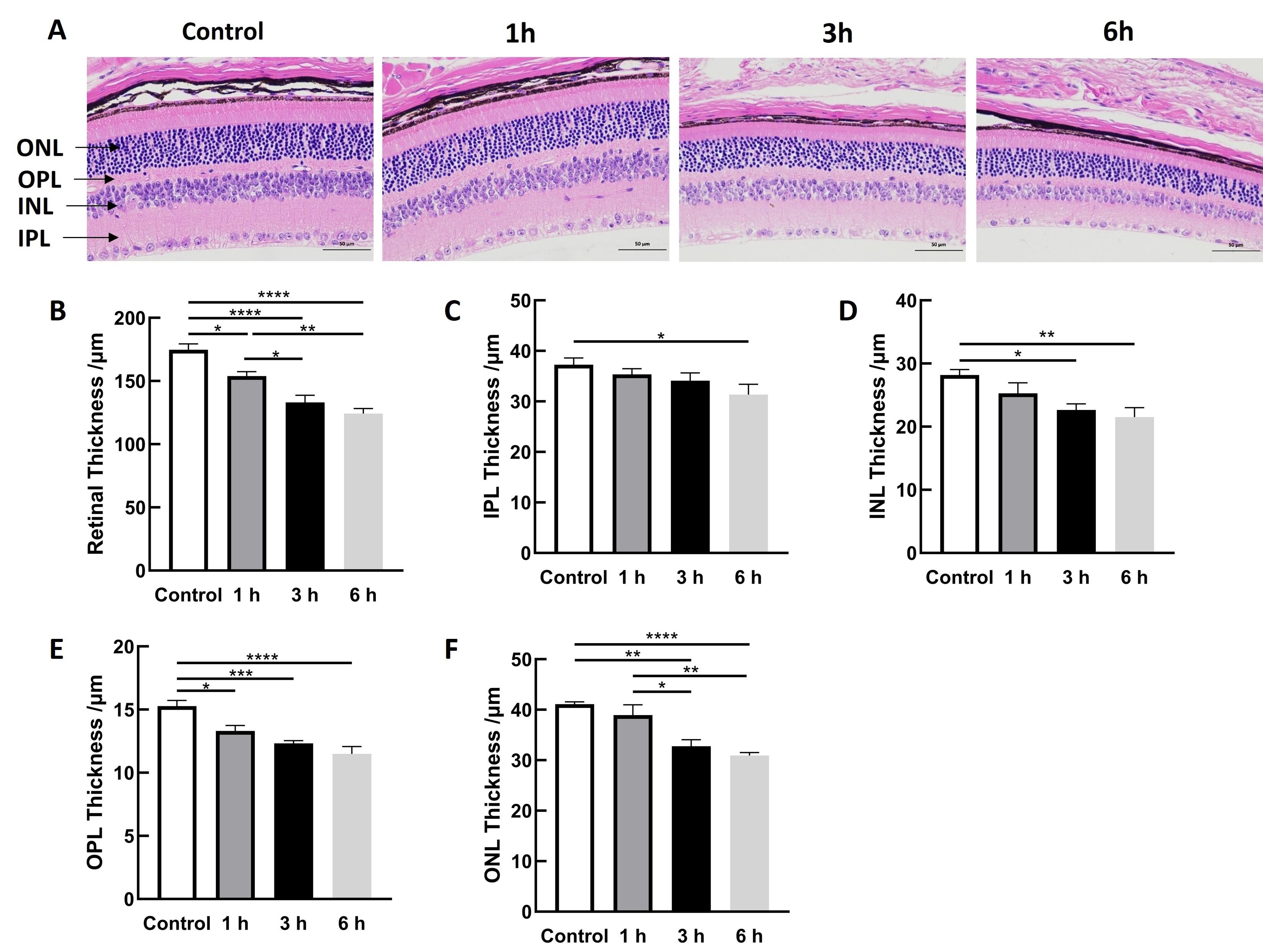
图1
Figure 1
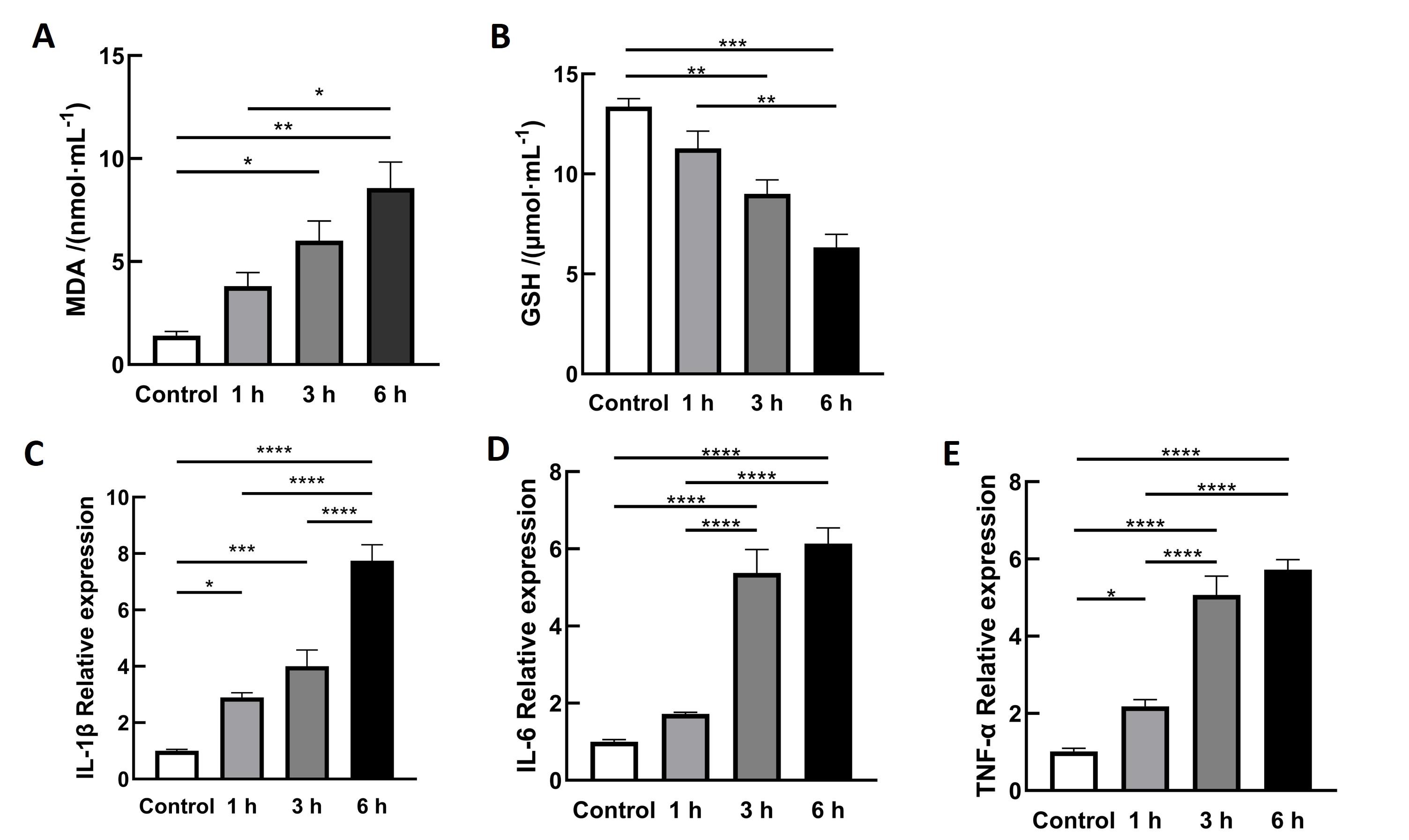
图2
Figure 2
图3
Figure 3
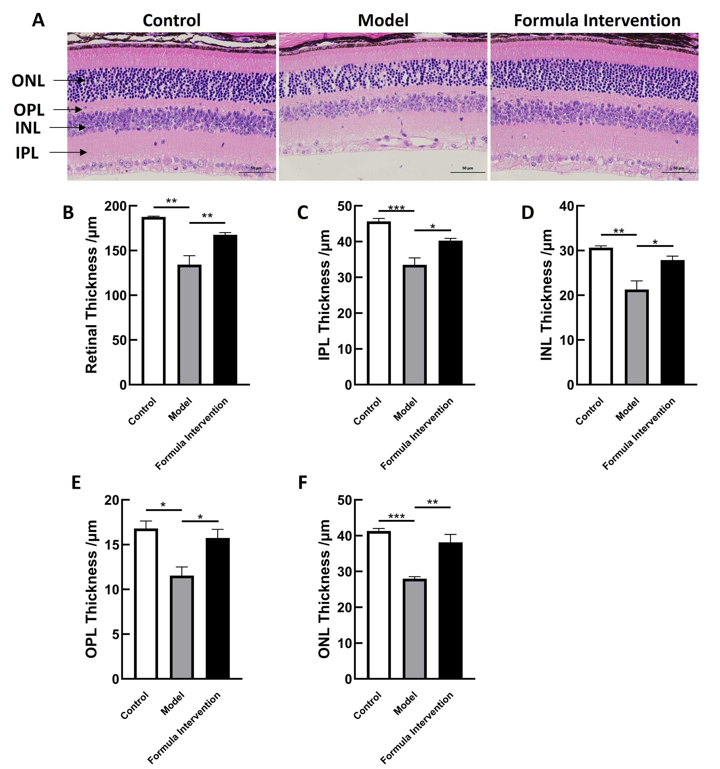
图4
Figure 4
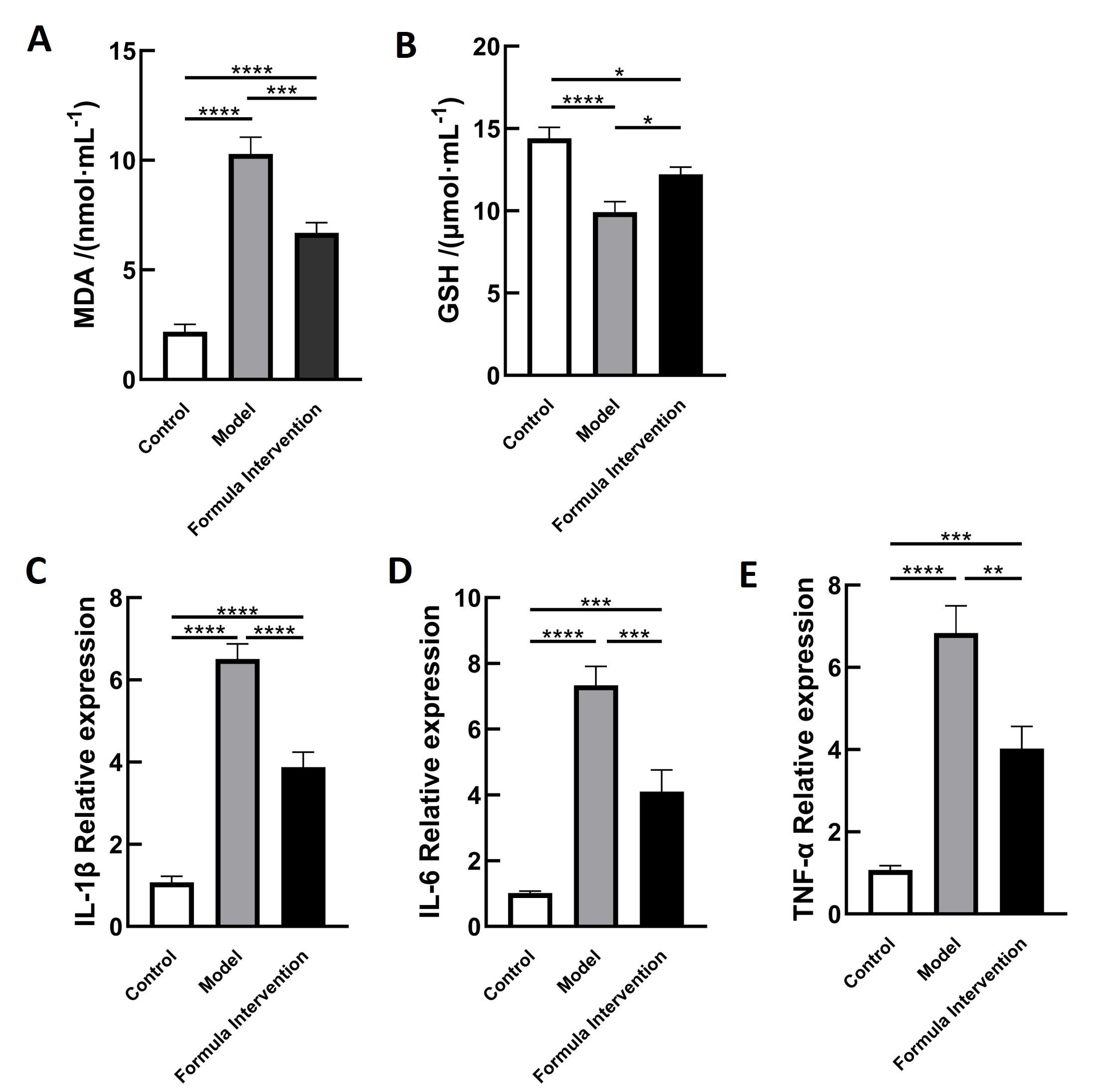
图5
Figure 5
| [1] |
|
| [2] |
|
| [3] |
|
| [4] |
|
| [5] |
|
| [6] |
|
| [7] |
|
| [8] |
|
| [9] |
|
| [10] |
|
| [11] |
|
| [12] |
|
| [13] |
|
| [14] |
庞雨菲. 黑果枸杞多酚对丙烯酰胺诱导大鼠纹状体氧化应激损伤的保护作用及机制[D]. 银川: 宁夏医科大学, 2023.
|
|
|
|
| [15] |
|
| [16] |
娄解南, 谭书明, 谢国芳, 等. 刺梨多酚对丙烯酰胺致肝脏氧化损伤的保护作用[J]. 食品安全质量检测学报, 2023, 14(17): 122-128.
|
|
|
|
| [17] |
张丹, 葛颖颖, 杨琼琼, 等. 刺梨果抗氧化活性成分与体外抗氧化活性研究[C]//中国食品科学技术学会第十五届年会论文摘要集. 2018: 283-284.
|
|
|
|
| [18] |
|
| [19] |
|
| [20] |
|
| [21] |
|
| [22] |
|
| [23] |
马修彬, 陈松. 蓝光对视网膜色素上皮细胞的损伤机制[J]. 国际眼科纵览, 2007, 31(6): 415-419. DOI: 10.3760/cma.j.issn.1673-5803.2007.06.015 .
|
|
|
|
| [24] |
|
| [25] |
|
| [26] |
|
| [27] |
|
| [28] |
|
| [29] |
|
| [30] |
|
| [31] |
|
| [32] |
|
| [33] |
|
| [34] |
|
| [35] |
|
| [36] |
|
| [37] |
朱恒杏, 邵孟茹, 洪天畅, 等. 黑枸杞花青素、EGCG对中波紫外线诱导HaCaT细胞氧化损伤的协同保护作用[J]. 现代食品科技, 2024, 40(8): 106-115. DOI: 10.13982/j.mfst.1673-9078.2024.8.0918 .
|
|
|
|
| [38] |
邓楷, 欧阳健, 胡娜, 等. 黑果枸杞花青素结构差异对其稳定性及细胞抗氧化活性的影响[J]. 天然产物研究与开发, 2022, 34(2): 213-219. DOI: 10.16333/j.1001-6880.2022.2.005 .
|
|
|
|
| [39] |
李婧, 苏蓉, 刘振华, 等. 黑果枸杞果实中抗炎活性成分的筛选及作用机制研究[J]. 中国野生植物资源, 2023, 42(5): 9-15, 38. DOI: 10.3969/j.issn.1006-9690.2023.05.002 .
|
|
|
|
| [40] |
薛才华, 武强, 王梦杰, 等. 黑枸杞花青素对巨噬细胞RAW264.7炎性与自噬交互作用的影响[J]. 中国畜牧杂志, 2021, 57(12): 244-249.
|
|
|
|
| [41] |
|
| [42] |
|
| [43] |
|
| [44] |
|
| [45] |
|
| [1] | 强苏静, 李冬婷, 徐瑾华, 蔡莉, 韦思羽, 邹嘉镭, 丁玉强. 实验大鼠和小鼠中肺炎克雷伯菌复合体的鉴定与分析[J]. 实验动物与比较医学, 2026, 46(4): 564-572. |
| [2] | 赵玉磊, 韩冰冰, 王兴鑫, 杨广建, 王雅杰, 唐晓勇. 病证结合肺癌小鼠模型研究进展[J]. 实验动物与比较医学, 2026, 46(4): 531-540. |
| [3] | 龚志群, 饶希午, 张天弋, 吴荣琛, 叶明楠, 蒋春曦, 王苏美, 朱燕娟, 柴小姝. 免疫检查点抑制剂相关性肺炎小鼠模型的构建与评价[J]. 实验动物与比较医学, 2026, 46(4): 498-506. |
| [4] | 杜红枝, 范瑾, 魏晓露, 王连嵋, 刘艳, 陈韡亚, 陈腾飞, 高云航, 宋玲, 张广平, 侯红平. 多技术联合评估缺氧性肺动脉高压小鼠模型[J]. 实验动物与比较医学, 2026, 46(4): 476-486. |
| [5] | 王娟, 徐佳慧, 田韵远, 张萌萌, 李敏, 王四旺, 李瑶. 两种雌性小鼠溃疡性结肠炎模型的比较与行为学观察[J]. 实验动物与比较医学, 2026, 46(3): 332-343. |
| [6] | 李嘉菲, 张朕豪, 王硕, 田歌, 温爽, 闫玉雪, 崔然, 叶振, 崔永春. 自主神经调节剂对房颤模型大鼠心房电重塑及组织病理变化的影响[J]. 实验动物与比较医学, 2026, 46(3): 321-331. |
| [7] | 李龙雪, 万崇凡, 张琦, 雷茹婷, 王潇玥, 程乐妍, 赖琦, 刘荣华, 刘漩, 徐铁龙. 基于miRNA测序分析清肺排毒汤防治小鼠急性肺损伤的分子机制研究[J]. 实验动物与比较医学, 2026, 46(3): 311-320. |
| [8] | 艾秀峰, 张利棕, 方明笋, 吕东颖, 陈楚, 蔡兆伟, 王德军. 16S rRNA测序分析饮用含氯水对大鼠和小鼠肠道菌群的影响差异[J]. 实验动物与比较医学, 2026, 46(3): 437-445. |
| [9] | 周忆旻, 张昕宇, 杨佳诺, 刘梦佳, 司灿灿, 叶海绿, 孙文超, 兰添. MHV、MPV和Reo-3三重TaqMan定量PCR检测方法的建立[J]. 实验动物与比较医学, 2026, 46(3): 408-415. |
| [10] | 刘赛, 付彬, 李思迪, 陈志达, 张悦, 郭中坤, 王永安, 王可洲. Adra2a通过MAPK信号通路介导LPS诱导的Lbp-/-小鼠肝细胞炎性反应[J]. 实验动物与比较医学, 2026, 46(2): 212-221. |
| [11] | 赵赫, 张涛, 李丽, 肖宇宙, 安学芳, 张帆. 猪抑制素多克隆抗体对C57BL/6J小鼠超数排卵的增效作用[J]. 实验动物与比较医学, 2026, 46(2): 271-278. |
| [12] | 唐小杭, 谷颖敏, 吕阳阳, 黄明姝, 田雪松. 一种自研固定液用于制备大鼠眼球切片的组织学染色效果评价[J]. 实验动物与比较医学, 2026, 46(2): 261-270. |
| [13] | 崔畅婉, 路一平, 于淼, 王爽, 吴思, 孙峥嵘. 溶质载体家族7成员5抑制剂JPH203对单侧输尿管梗阻诱导小鼠肾纤维化的抑制作用[J]. 实验动物与比较医学, 2026, 46(2): 205-211. |
| [14] | 吴宪文, 刘丽丽, 陈烨, 徐国恒. 繁育体系中育成期小鼠换笼周期及刨花垫料使用量的优化研究[J]. 实验动物与比较医学, 2026, 46(2): 251-260. |
| [15] | 荣文双, 牛源菲, 刘美婷, 杨梦园, 崔爽, 马丽娜, 富尧, 王连嵋, 曹俊岭. 抗原类型对诱导型干燥综合征小鼠建模的影响[J]. 实验动物与比较医学, 2026, 46(2): 178-190. |
| 阅读次数 | ||||||
|
全文 |
|
|||||
|
摘要 |
|
|||||
