



实验动物与比较医学 ›› 2026, Vol. 46 ›› Issue (4): 487-497.DOI: 10.12300/j.issn.1674-5817.2025.180
雷建军, 索晨浩, 张贺( )
)
收稿日期:2025-11-05
修回日期:2026-01-23
出版日期:2026-08-25
发布日期:2026-08-22
作者简介:雷建军(1992—),学士,技师,研究方向:脑科学动物模型的制作与研究。E-mail:398116613@qq.com基金资助:
LEI Jianjun, SUO Chenhao, ZHANG He()
Received:2025-11-05
Revised:2026-01-23
Published:2026-08-25
Online:2026-08-22
Correspondence to:
ZHANG He (ORCID: 0000-0002-3830-9228), E-mail: alwayszhh@163.com摘要:
目的 探讨白藜芦醇对脑缺血再灌注大鼠模型血脑屏障损伤的保护作用,并明确该作用是否由C-C基序趋化因子配体3(C-C motif chemokine ligand 3,CCL3)/C-C趋化因子受体5(C-C chemokine receptor type 5,CCR5)信号通路介导。 方法 将100只6~8周龄SPF级雄性SD大鼠随机分为假手术(Sham)组、模型(MCAO)组、白藜芦醇治疗(Res)组、白藜芦醇+CCR5激动剂(Res+Agonist)组和白藜芦醇+CCR5抑制剂(Res+Antagonist)组。Sham组只暴露颈动脉不做缺血处理;MCAO组、Res组、Res+Agonist组和Res+Antagonist组均采用大脑中动脉闭塞法构建大鼠脑缺血模型,缺血45 min后进行再灌注。完成再灌注后,Res组、Res+Agonist组和Res+Antagonist组即刻腹腔注射白藜芦醇(60 mg/kg),Sham组和MCAO组注射等体积生理盐水;30 min后,Res+Agonist组尾静脉注射CCR5激动剂(20 μg/kg),Res+Antagonist组尾静脉注射CCR5抑制剂(1 mg/kg),Res组、Sham组及MCAO组尾静脉注射等体积生理盐水。此后,每24 h给药(包括白藜芦醇、CCR5激动剂、CCR5抑制剂和生理盐水)1次,连续3 d。造模完成后,通过2,3,5-氯化三苯基四氮唑(2,3,5-triphenyltetrazolium chloride,TTC)染色法评估脑梗死体积占比;采用伊文思蓝染色法检测血脑屏障通透性;采用ELISA检测血清中促炎性细胞因子白细胞介素-1β(interleukin-1β,IL-1β)、肿瘤坏死因子-α(tumor necrosis factor alpha,TNF-α)与CCL3水平;通过HE染色和尼氏染色观察脑组织病理变化;采用免疫荧光染色法检测脑组织中小胶质细胞标志物离子钙结合适配器分子1(ionized calcium-binding adapter molecule 1,Iba-1)与CCR5的荧光强度;采用蛋白质印迹法检测脑组织中CCL3/CCR5信号通路相关蛋白(CCL3、CCR5)、血清促炎性细胞因子[磷酸化核因子-κB(phosphorylated nuclear factor-kappa B,p-NF-κB)]和血脑屏障相关蛋白[基质金属蛋白酶9(matrix metalloproteinase 9,MMP9)及闭合蛋白(occludin)]的表达水平。 结果 与Sham组相比,MCAO组大鼠大脑的各项损伤指标[脑梗死体积占比、脑组织伊文思蓝渗出率、血清促炎性细胞因子(IL-1β、TNF-α、CCL3)含量、脑组织Iba-1和CCR5的阳性表达情况、脑组织CCL3/CCR5信号通路相关蛋白(CCL3、CCR5)表达水平和血脑屏障相关蛋白(p-NF-κB和MMP9)表达水平]均显著升高(P<0.001),occludin表达水平显著降低(P<0.001),HE染色和尼氏染色结果显示其脑组织发生明显损伤。与MCAO组相比,Res组大鼠的上述各项损伤指标均显著下降(P<0.01),occludin表达水平显著升高(P<0.01),HE染色和尼氏染色结果显示其脑损伤得到一定的改善。与Res组相比,Res+Agonist组大鼠的上述损伤指标均显著升高(P<0.05),occludin表达水平显著降低(P<0.05),脑组织病理损伤显著加重;而Res+Antagonist组大鼠的上述损伤指标均显著下降(P<0.05),occludin表达水平显著升高(P<0.05),脑组织病理损伤进一步缓解。 结论 白藜芦醇通过靶向调控CCL3/CCR5信号通路减轻缺血再灌注损伤中的外周炎症反应,从而发挥血脑屏障保护作用。
中图分类号:
雷建军,索晨浩,张贺. 白藜芦醇通过调控CCL3/CCR5信号通路减轻脑缺血再灌注大鼠模型的血脑屏障损伤[J]. 实验动物与比较医学, 2026, 46(4): 487-497. DOI: 10.12300/j.issn.1674-5817.2025.180.
LEI Jianjun,SUO Chenhao,ZHANG He. Resveratrol Alleviates Blood‑Brain Barrier Damage in Cerebral Ischemia‑Reperfusion Rat Models through Modulation of the CCL3/CCR5 Signaling Pathway[J]. Laboratory Animal and Comparative Medicine, 2026, 46(4): 487-497. DOI: 10.12300/j.issn.1674-5817.2025.180.
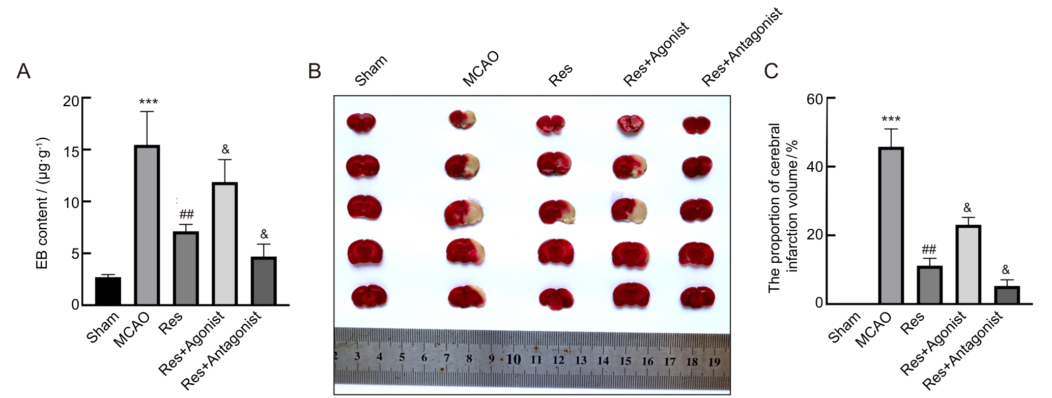
图1 白藜芦醇对大脑中动脉闭塞大鼠脑损伤的影响
Figure 1 Effects of resveratrol on brain injury in middle cerebral artery occlusion rats
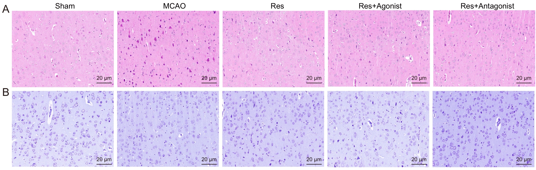
图2 白藜芦醇对大脑中动脉闭塞大鼠脑组织神经元损伤的影响
Figure 2 Effects of resveratrol on neuronal damage in the brain tissue of middle cerebral artery occlusion rats
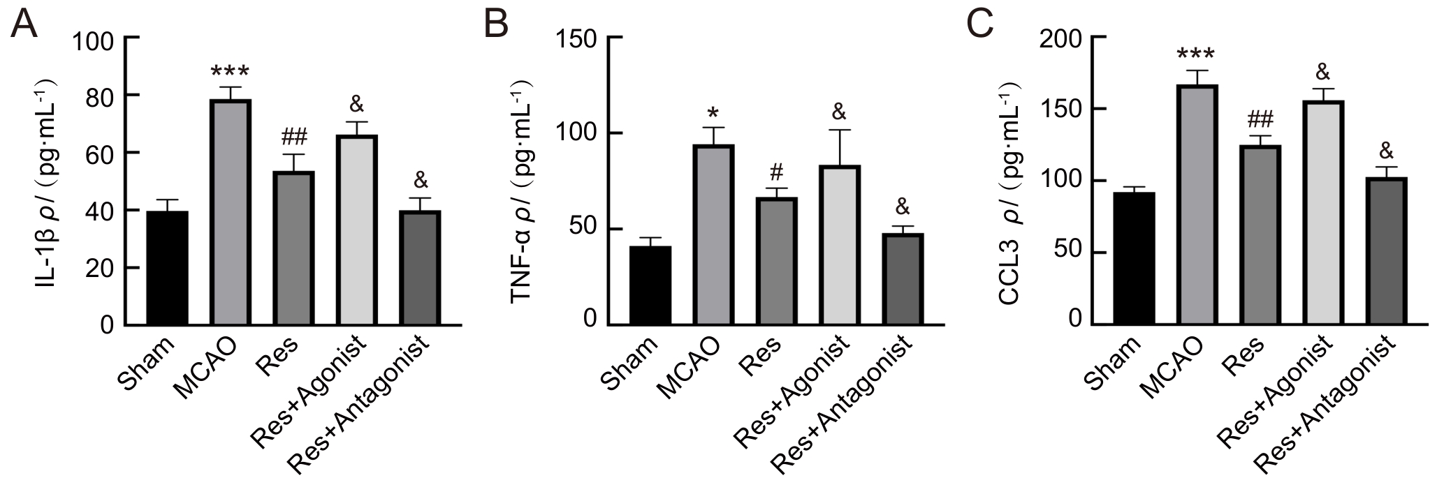
图3 白藜芦醇对大脑中动脉闭塞大鼠外周血促炎性细胞因子的影响
Figure 3 Effects of resveratrol on pro-inflammatory cytokines in peripheral blood of rats with middle cerebral artery occlusion
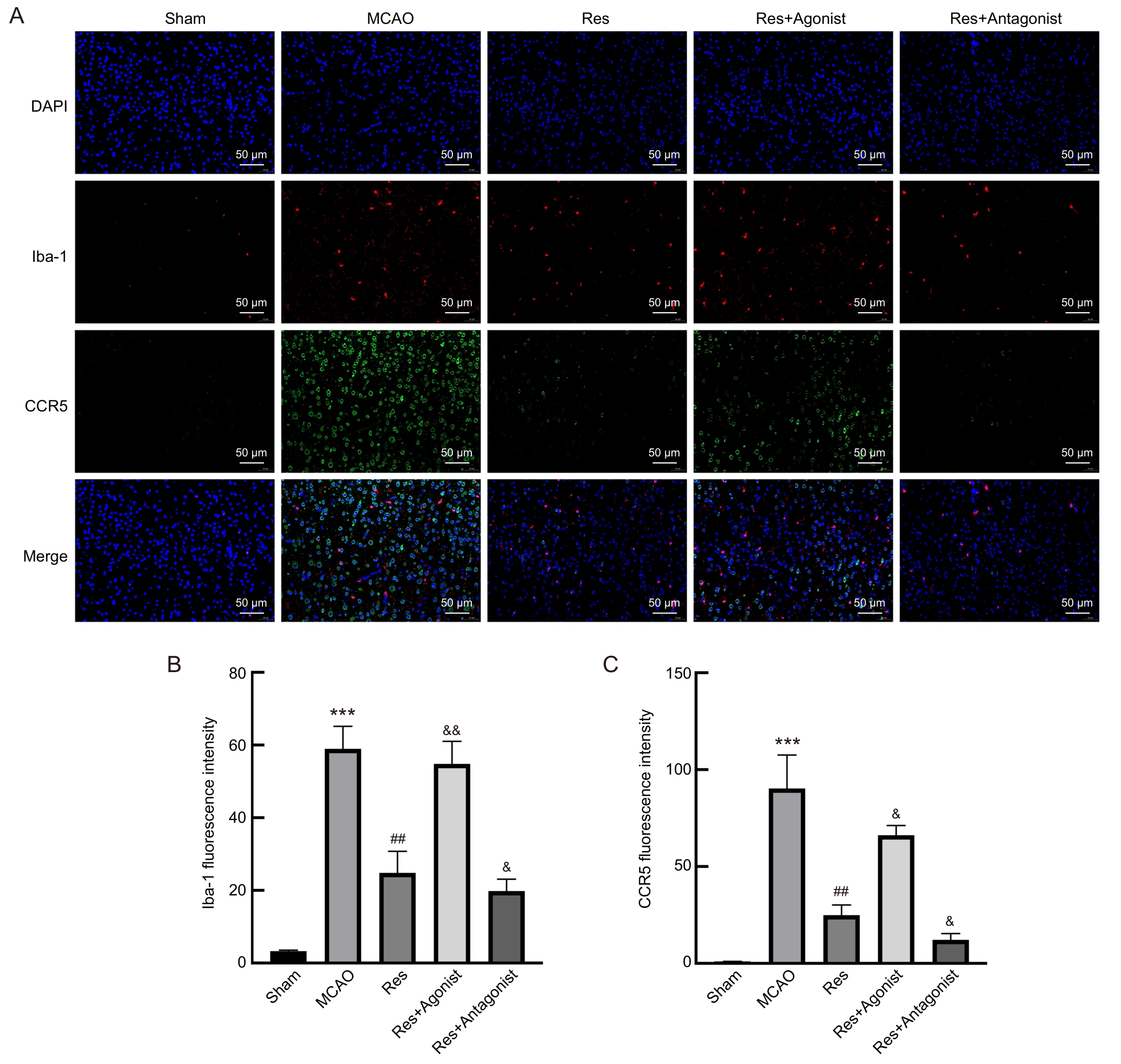
图4 白藜芦醇对大脑中动脉闭塞大鼠小胶质细胞活化的影响
Figure 4 Effects of resveratrol on microglial activation in rats with middle cerebral artery occlusion
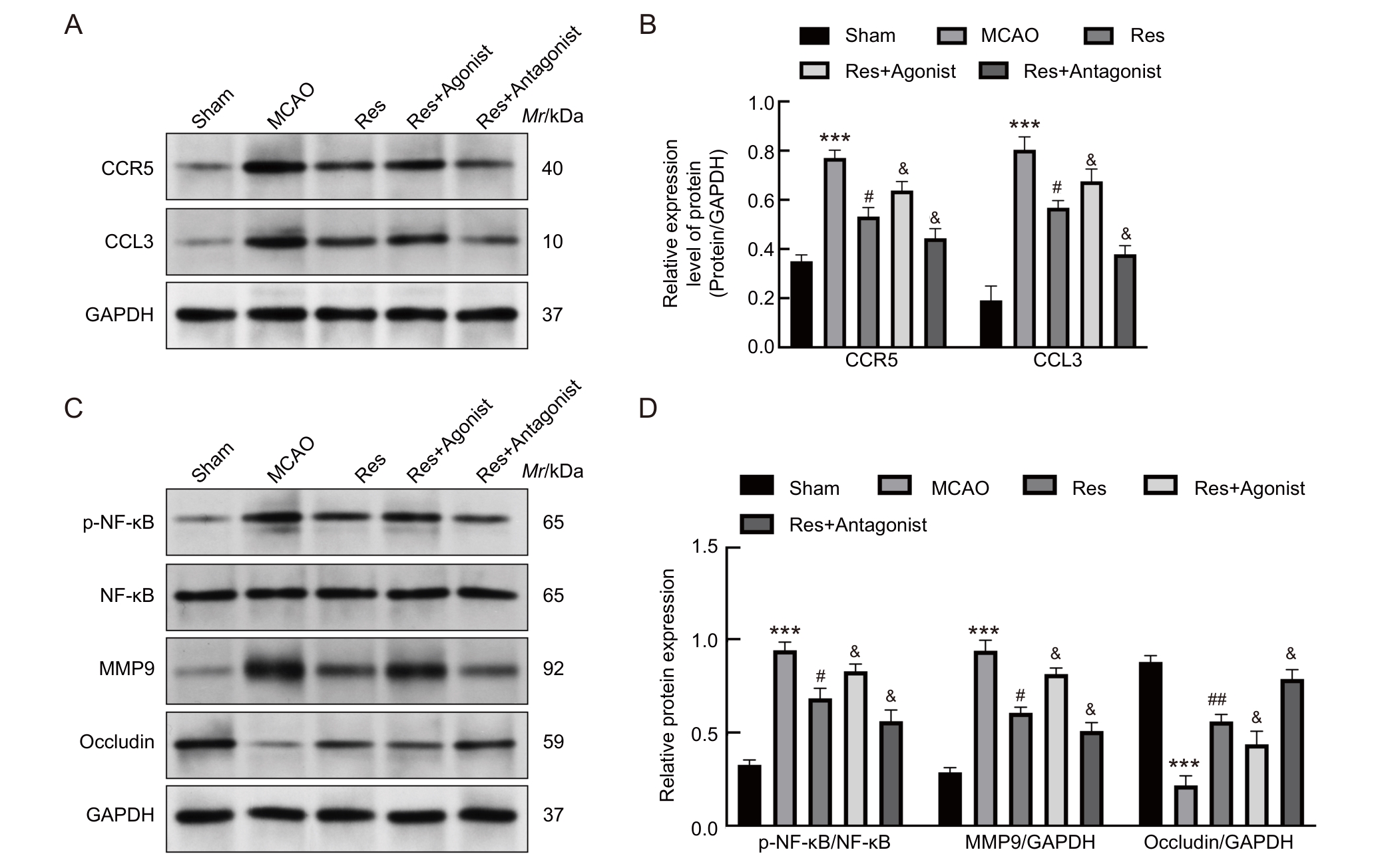
图5 白藜芦醇治疗对大脑中动脉闭塞大鼠脑组织中CCL3/CCR5信号通路和血脑屏障相关蛋白的影响
Figure 5 Effects of resveratrol treatment on CCL3/CCR5 signaling pathway and blood-brain barrier-related proteins in brain tissues of rats with middle cerebral artery occlusion
| [1] | 刘佳, 叶岩荣, 沈赟, 等. 银杏内酯B通过调控脑内T细胞特性及与胶质细胞间相互作用促进缺血性脑卒中小鼠的神经功能恢复[J]. 实验动物与比较医学, 2024, 44(2): 139-148. DOI: 10.12300/j.issn.1674-5817.2023.121 . |
| LIU J, YE Y R, SHEN Y, et al. Ginkgolide B promotes neural function recovery of ischemic stroke mice by regulating characteristics of brain T cells and their interactions with glial cells[J]. Lab Anim Comp Med, 2024, 44(2): 139-148. DOI: 10.12300/j.issn.1674-5817.2023.121 . | |
| [2] | RAFFAELE S, CLAUSEN B H, MANNELLA F C, et al. Characterisation of GPR17-expressing oligodendrocyte precursors in human ischaemic lesions and correlation with reactive glial responses[J]. J Pathol, 2025, 265(2): 226-243. DOI: 10.1002/path.6381 . |
| [3] | ALIDADI M, OMIDI N, ABDI M, et al. Melatonin ameliorates astrogliosis and microgliosis in a cuprizone demyelinating mouse model[J]. Biochem Biophys Rep, 2025, 41: 101929. DOI: 10.1016/j.bbrep.2025.101929 . |
| [4] | LIU X, TAO X W, XIONG Z, et al. Mechanism of HDAC2 regulating Nrf2 acetylation level in neuronal ferroptosis of neonatal rats with hypoxic-ischemic brain injury[J]. Brain Inj, 2025, 39(8): 635-645. DOI: 10.1080/02699052.2025.2468309 . |
| [5] | SHI K B, LI H D, CHANG T, et al. Bone marrow hematopoiesis drives multiple sclerosis progression[J]. Cell, 2022, 185(13): 2234-2247.e17. DOI: 10.1016/j.cell.2022.05.020 . |
| [6] | KNOX E G, ABURTO M R, CLARKE G, et al. The blood-brain barrier in aging and neurodegeneration[J]. Mol Psychiatry, 2022, 27(6): 2659-2673. DOI: 10.1038/s41380-022-01511-z . |
| [7] | AKINLAJA Y O, NISHIYAMA A. Glial modulation of synapse development and plasticity: oligodendrocyte precursor cells as a new player in the synaptic quintet[J]. Front Cell Dev Biol, 2024, 12: 1418100. DOI: 10.3389/fcell.2024.1418100 . |
| [8] | HUNTEMER-SILVEIRA A, PATIL N, BRICKNER M A, et al. Strategies for oligodendrocyte and myelin repair in traumatic CNS injury[J]. Front Cell Neurosci, 2021, 14: 619707. DOI: 10.3389/fncel.2020.619707 . |
| [9] | WANG F, YE Z H, YIN X J, et al. Electroacupuncture at 5/100 Hz alleviates neuropathic pain in rats by inhibiting the CCL3/CCR5 axis in the spinal cord[J]. Acupunct Med, 2025, 43(2): 95-103. DOI: 10.1177/09645284251327197 . |
| [10] | JAFARI M, DAMANI R. Blood pressure variability and outcome after acute intracerebral hemorrhage[J]. J Neurol Sci, 2020, 413: 116766. DOI: 10.1016/j.jns.2020.116766 . |
| [11] | BAI M L, SUN R Z, CAO B, et al. Monocyte-related cytokines/chemokines in cerebral ischemic stroke[J]. CNS Neurosci Ther, 2023, 29(12): 3693-3712. DOI: 10.1111/cns.14368 . |
| [12] | ESCOBAR-PESO A, MARTÍNEZ-ALONSO E, MASJUAN J, et al. Development of pharmacological strategies with therapeutic potential in ischemic stroke[J]. Antioxidants, 2023, 12(12): 2102. DOI: 10.3390/antiox12122102 . |
| [13] | 牟凌梅, 李雪梅, 文君. 白藜芦醇调节TREM-1表达减轻大鼠脑缺血再灌注损伤后细胞焦亡[J]. 四川医学, 2025, 46(6): 628-634. DOI: 10.16252/j.cnki.issn1004-0501-2025.06.006 . |
| MOU L M, LI X M, WEN J. Resveratrol regulates TREM-1 expression to alleviate cell pyroptosis after cerebral ischemia-reperfusion injury in rats[J]. Sichuan Med J, 2025, 46(6): 628-634. DOI: 10.16252/j.cnki.issn1004-0501-2025.06.006 . | |
| [14] | 张小良, 高赛红, 杨迎春, 等. 白藜芦醇激活自噬抑制焦亡减轻脑缺血再灌注损伤[J]. 中国临床药理学与治疗学, 2024, 29(11): 1240-1248. DOI: 10.12092/j.issn.1009-2501.2024.11.005 . |
| ZHANG X L, GAO S H, YANG Y C, et al. Resveratrol activates autophagy and inhibits pyroptosis to alleviate cerebral ischemia-reperfusion injury[J]. Chin J Clin Pharmacol Ther, 2024, 29(11): 1240-1248. DOI: 10.12092/j.issn.1009-2501.2024.11.005 . | |
| [15] | AN Y, SU G, CHEN W, et al. Research progress on the mechanisms of microglial extracellular vesicles affecting the prognosis of ischemic stroke[J]. Neurochem Int, 2025, 185: 105949. DOI: 10.1016/j.neuint.2025.105949 . |
| [16] | LÓPEZ-MORALES M A, CASTELLÓ-RUIZ M, BURGUETE M C, et al. Effect and mechanisms of resveratrol in animal models of ischemic stroke: a systematic review and Bayesian meta-analysis[J]. J Cereb Blood Flow Metab, 2023, 43(12): 2013-2028. DOI: 10.1177/0271678x231206236 . |
| [17] | WANG C X, WANG J Y, ZHU Z C, et al. Spotlight on pro-inflammatory chemokines: regulators of cellular communication in cognitive impairment[J]. Front Immunol, 2024, 15: 1421076. DOI: 10.3389/fimmu.2024.1421076 . |
| [18] | GUO Q X, DU A N, WANG J Y, et al. Integrated bioinformatic analyses reveal immune molecular markers and regulatory networks for cerebral ischemia-reperfusion[J]. Front Biosci (Landmark Ed), 2023, 28(8): 179. DOI: 10.31083/j.fbl2808179 . |
| [19] | DASDELEN M F, CAGLAYAN A B, ER S, et al. Social isolation initiated post-weaning augments ischemic brain injury by promoting pro-inflammatory responses[J]. Exp Neurol, 2024, 375: 114729. DOI: 10.1016/j.expneurol.2024.114729 . |
| [20] | FESTA B P, SIDDIQI F H, JIMENEZ-SANCHEZ M, et al. Microglial-to-neuronal CCR5 signaling regulates autophagy in neurodegeneration[J]. Neuron, 2023, 111(13): 2021-2037.e12. DOI: 10.1016/j.neuron.2023.04.006 . |
| [21] | WANG L F, LI W J, ZHANG X Y, et al. Resveratrol prevents age-related heart impairment through inhibiting the Notch/NF-κB pathway[J]. Food Sci Nutr, 2024, 12(2): 1035-1045. DOI: 10.1002/fsn3.3817 . |
| [22] | BASHEER N, MUHAMMADI M K, FREITES C L, et al. TLR4-mediated chronic neuroinflammation has no effect on tangle pathology in a tauopathy mouse model[J]. Front Aging Neurosci, 2024, 16: 1468602. DOI: 10.3389/fnagi.2024.1468602 . |
| [23] | LI W B, ZHANG Y D, YANG F, et al. Molecular mechanism by which TRPC6 regulates calcium signaling and neuroinflammation in the onset and development of ischemic stroke: a review[J]. Altern Ther Health Med, 2024, 30(8): 170-175. |
| [24] | DENG L, ZHANG J Y, CHEN S, et al. miR-671-5p upregulation attenuates blood–brain barrier disruption in the ischemia stroke model via the NF-кB/MMP-9 signaling pathway[J]. Mol Neurobiol, 2023, 60(7): 3824-3838. DOI: 10.1007/s12035-023-03318-7 . |
| [25] | DENG H J, XUE P F, ZHOU X G, et al. CCL4/CCR5 regulates chondrocyte biology and OA progression[J]. Cytokine, 2024, 183: 156746. DOI: 10.1016/j.cyto.2024.156746 . |
| [1] | 强苏静, 李冬婷, 徐瑾华, 蔡莉, 韦思羽, 邹嘉镭, 丁玉强. 实验大鼠和小鼠中肺炎克雷伯菌复合体的鉴定与分析[J]. 实验动物与比较医学, 2026, 46(4): 564-572. |
| [2] | 汤建平, 赵丽亚, 赵莹. 常用近交系大鼠微卫星遗传位点筛选及分析[J]. 实验动物与比较医学, 2026, 46(3): 388-396. |
| [3] | 李嘉菲, 张朕豪, 王硕, 田歌, 温爽, 闫玉雪, 崔然, 叶振, 崔永春. 自主神经调节剂对房颤模型大鼠心房电重塑及组织病理变化的影响[J]. 实验动物与比较医学, 2026, 46(3): 321-331. |
| [4] | 艾秀峰, 张利棕, 方明笋, 吕东颖, 陈楚, 蔡兆伟, 王德军. 16S rRNA测序分析饮用含氯水对大鼠和小鼠肠道菌群的影响差异[J]. 实验动物与比较医学, 2026, 46(3): 437-445. |
| [5] | 宋静, 杨宗统, 李晓晶, 李自发, 苏凤云, 徐东川, 隋在云. 泻白散对过敏性哮喘大鼠肺、肠组织形态结构及PI3K和Akt表达水平的影响[J]. 实验动物与比较医学, 2026, 46(2): 191-204. |
| [6] | 唐小杭, 谷颖敏, 吕阳阳, 黄明姝, 田雪松. 一种自研固定液用于制备大鼠眼球切片的组织学染色效果评价[J]. 实验动物与比较医学, 2026, 46(2): 261-270. |
| [7] | 徐英韬, 王蒙蒙, 林平, 迟海涛, 王怡, 白鹰. 外泌体通过NRF2/SLC7A11/GPX4通路调控铁死亡治疗小鼠缺血性脑卒中[J]. 实验动物与比较医学, 2026, 46(1): 20-31. |
| [8] | 杨琳, 金梦, 吴汗青, 李顺, 周晓辉. 痘苗病毒天坛株感染AG6小鼠脑病模型的建立与初步分析[J]. 实验动物与比较医学, 2026, 46(1): 3-10. |
| [9] | 姜海涛, 袁韩涛, 黄雯婷, 杨蓉蓉, 陈晓春, 禹宝庆, 李四波. 腰舒逐瘀方通过miR-17-5P/MDM2/p53通路调控大鼠椎间盘纤维环细胞增殖与凋亡[J]. 实验动物与比较医学, 2026, 46(1): 55-65. |
| [10] | 曹星新, 李艾亦, 侯婧涵, 李明学, 李艳艳, 靳玮华, 杨凤梅, 段素琴, 和占龙. 黄芪或其成分治疗急性胰腺炎的动物实验Meta分析[J]. 实验动物与比较医学, 2025, 45(5): 561-573. |
| [11] | 高超奇, 祝志波, 孙显东. 大鼠血管重构模型的应用进展与分类分析[J]. 实验动物与比较医学, 2025, 45(5): 542-550. |
| [12] | 罗一凡, 张臻玮, 梅璐, 史叶萍, 邢艺彤, 张泽奇, 李楚欣, 韩春霞, 杨平顺, 陈秋生. 特络细胞介导肥胖症大鼠膏摩寡肽中草药复合制剂的减肥作用及其机制[J]. 实验动物与比较医学, 2025, 45(5): 551-560. |
| [13] | 秦超, 李双星, 赵婷婷, 蒋晨晨, 赵晶, 杨艳伟, 林志, 王三龙, 文海若. 药物安全评价用SD大鼠90 d喂养试验的背景数据研究[J]. 实验动物与比较医学, 2025, 45(4): 439-448. |
| [14] | 刘力瑜, 嵇波, 刘小玄, 方洋, 张玲, 郭亭廷, 全烨, 李鹤文, 刘翼天. 大鼠胎儿期肺组织固定方法的探索[J]. 实验动物与比较医学, 2025, 45(4): 432-438. |
| [15] | 潘颐聪, 蒋汶洪, 胡明, 覃晓. 慢性肾脏病大鼠主动脉钙化模型的术式优化及效果评价[J]. 实验动物与比较医学, 2025, 45(3): 279-289. |
| 阅读次数 | ||||||
|
全文 |
|
|||||
|
摘要 |
|
|||||
