





实验动物与比较医学 ›› 2025, Vol. 45 ›› Issue (6): 726-737.DOI: 10.12300/j.issn.1674-5817.2025.119
宋梦娇( ), 沈义栋()(
 )
)
收稿日期:2025-07-16
修回日期:2025-10-17
出版日期:2025-12-25
发布日期:2025-12-19
通讯作者:
沈义栋(1979—),男,博士,研究员,研究方向:衰老及衰老相关疾病的机理研究。E-mail:yidong.shen@sibcb.ac.cn。ORCID:000-0002-2841-7233作者简介:宋梦娇(1990—),女,本科,实验师,研究方向:秀丽隐杆线虫实验动物模型的构建及其在衰老生物学研究中的应用。E-mail:mengjiao.song@sibcb.ac.cn。ORCID: 0009-0003-1676-7556
基金资助:
SONG Mengjiao(), SHEN Yidong()()
Received:2025-07-16
Revised:2025-10-17
Published:2025-12-25
Online:2025-12-19
Correspondence to:
SHEN Yidong (ORCID: 0000-0002-2841-7233), E-mail: yidong.shen@sibcb.ac.cn摘要:
线粒体作为细胞的能量代谢中枢,其动态形态和氧化磷酸化功能异常与衰老及多种疾病直接相关。秀丽隐杆线虫(Caenorhabditis elegans,C. elegans,以下简称线虫)是被广泛使用的模式动物。本文系统总结了作者实验室以线虫为模型,多尺度分析了线粒体形态与功能的实验方法,主要包括:(1)形态定性分析,即采用单盲法人工分类(如点状、棒状、网状),该法虽然依赖主观经验,但操作简便,适用于初步表型的筛选;(2)形态定量分析,即依托Fiji/ImageJ平台,对特定组织(如表皮及体壁肌)中线粒体进行自动化参数提取,通过骨架化算法量化网络连通性(分支点数量、网络长度),结合二值化分析计算碎片化指数(如面积/周长比、碎片计数),实现客观表型比对;(3)高通量图像处理,即通过宏命令批量处理,整合形态学滤波、阈值分割及参数导出流程,显著提升大样本量的研究效率;(4)代谢功能监测,即应用Seahorse XF分析仪开展活体线虫呼吸代谢检测,通过序贯注入ATP合酶抑制剂N,N′-二环己基碳二亚胺(dicyclohexylcarbodiimide,DCCD)、解偶联剂羰基氰化物 4-(三氟甲氧基)苯腙[carbonyl cyanide 4-(trifluoromethoxy)phenylhydrazone,FCCP]及呼吸链抑制剂叠氮钠(sodium azide,NaN?),精准解析基础耗氧率(oxygen consumption rate,OCR)、ATP耦合呼吸效率及最大呼吸容量(呼吸潜力),进而揭示能量代谢规律。本文整合了形态-功能双维度方法,不仅为关于线虫的线粒体研究提供经验技术,其框架亦可拓展至哺乳动物细胞与类器官模型,以推动靶向线粒体的基础研究和药物开发。
中图分类号:
宋梦娇,沈义栋. 秀丽隐杆线虫的线粒体形态和功能研究方法及应用实例[J]. 实验动物与比较医学, 2025, 45(6): 726-737. DOI: 10.12300/j.issn.1674-5817.2025.119.
SONG Mengjiao,SHEN Yidong. Approaches and Application Examples for Studying Mitochondrial Morphology and Function in Caenorhabditis elegans[J]. Laboratory Animal and Comparative Medicine, 2025, 45(6): 726-737. DOI: 10.12300/j.issn.1674-5817.2025.119.
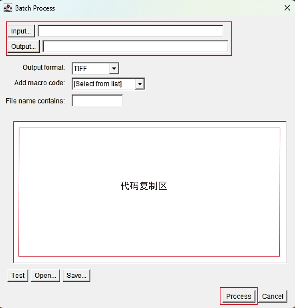
图1 Batch Process操作界面介绍
Figure 1 Introduction to Batch Process operation interface
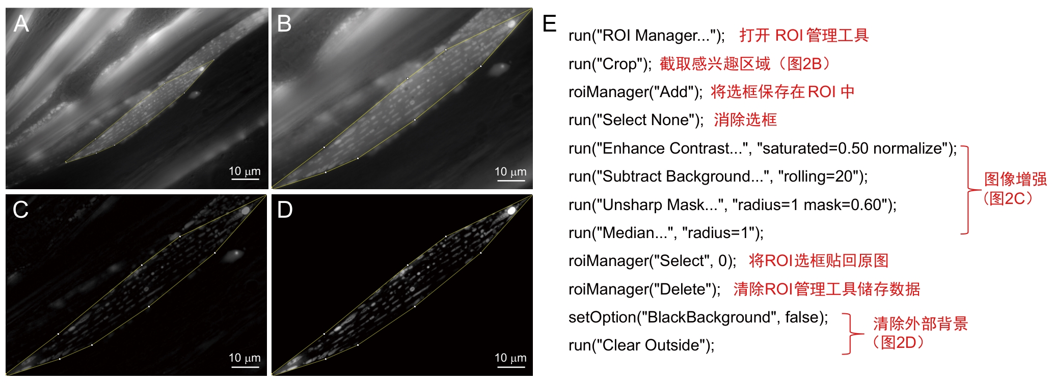
图2 前处理步骤及注释
Figure 2 Preprocessing procedures and annotations
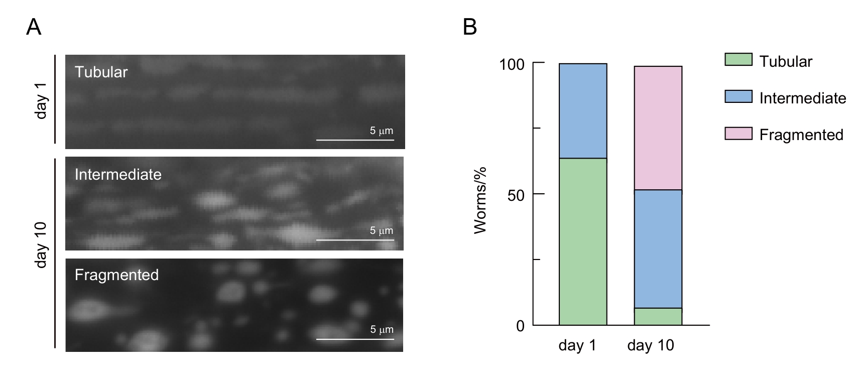
图3 线虫肌肉细胞线粒体形态分类
Figure 3 Classification of mitochondrial morphology in C. elegans muscle cells
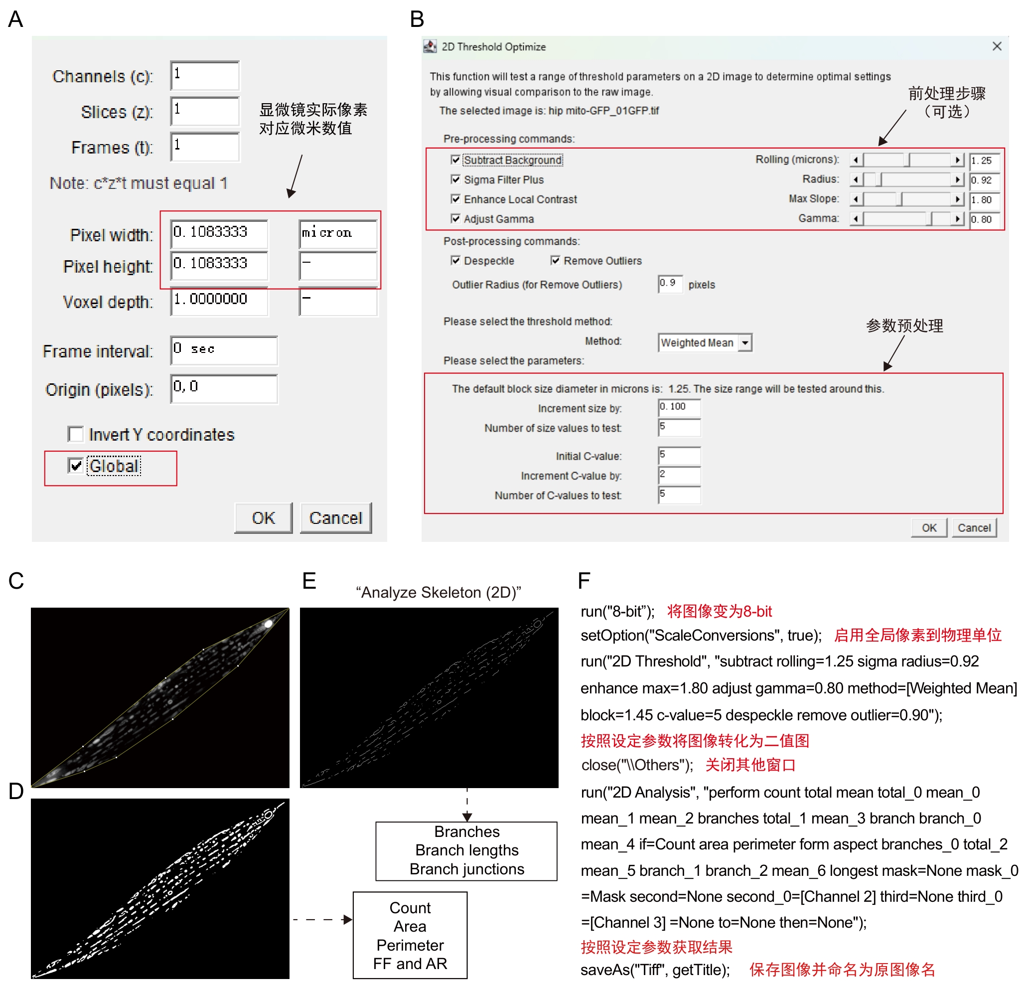
图4 Mitochondria Analyzer插件步骤及注释
Figure 4 Procedures and annotations of Mitochondria Analyzer plugin
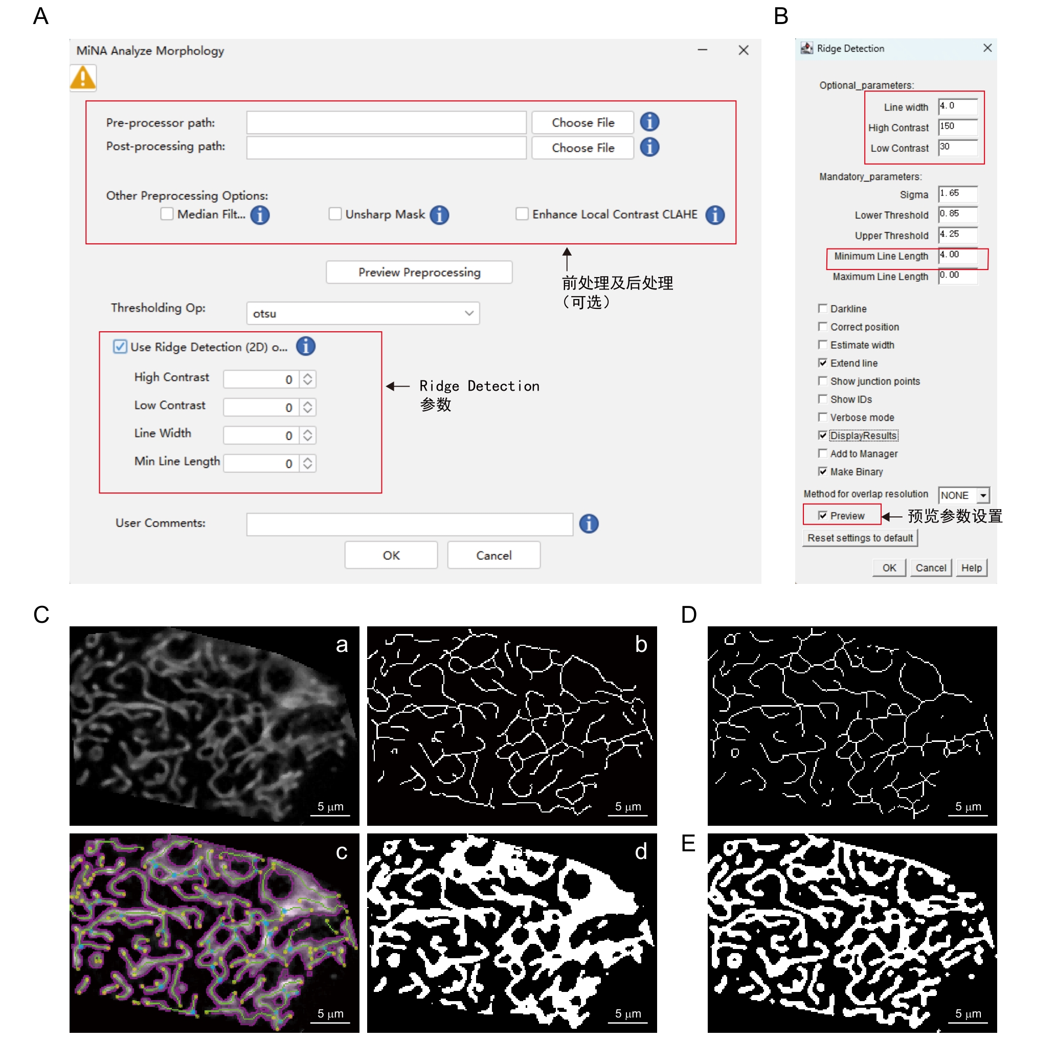
图5 MiNA - Mitochondrial Network Analysis插件步骤及注释
Figure 5 Procedures and annotations of MiNA - Mitochondrial Network Analysis plugin
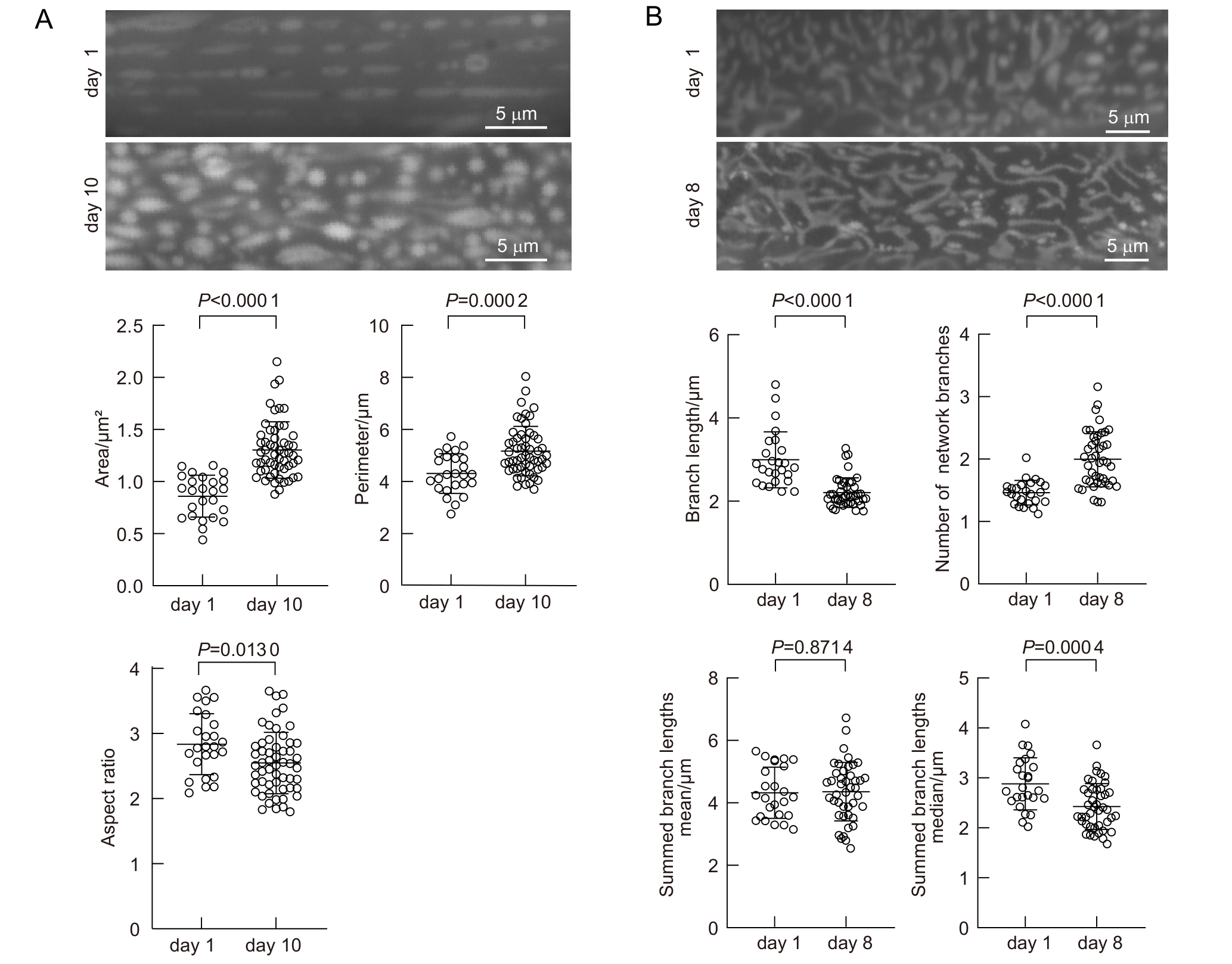
图6 老年线虫线粒体形态发生改变
Figure 6 Age-dependent mitochondrial morphology changes in C. elegans
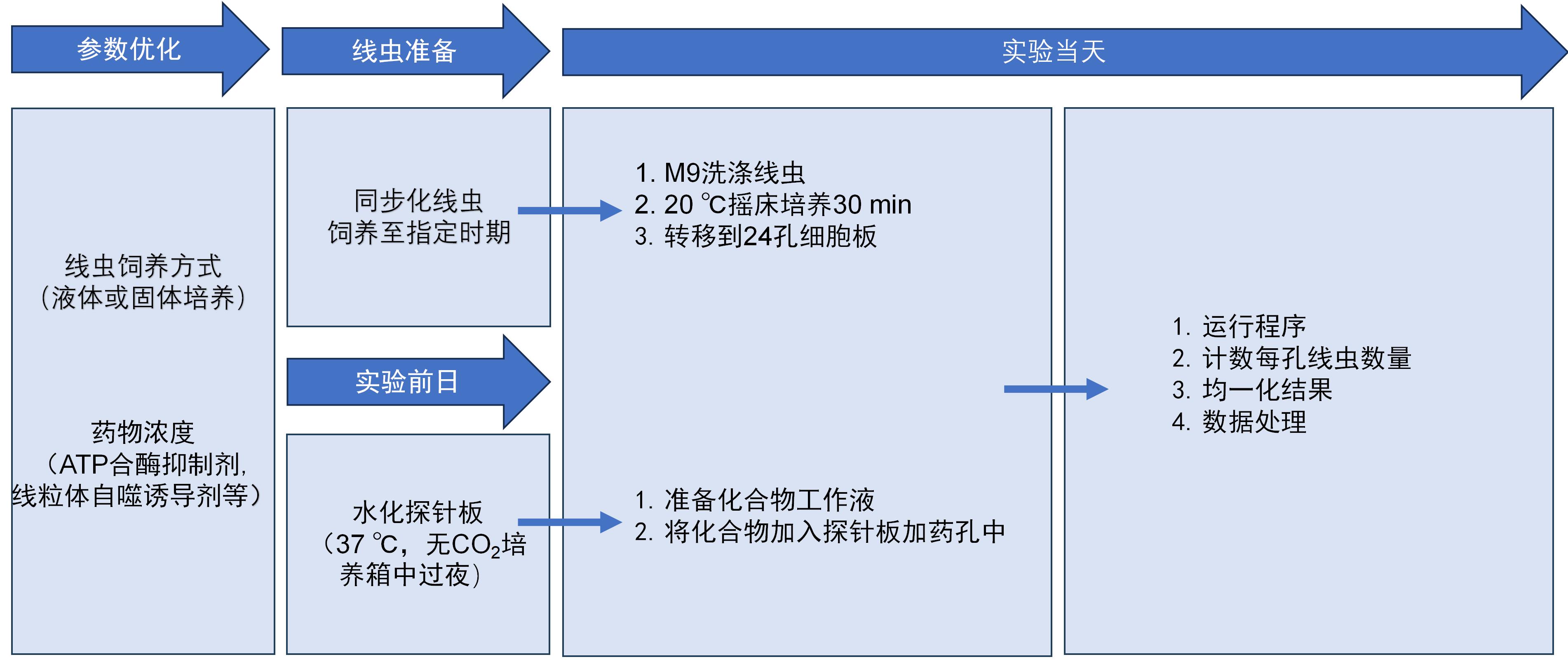
图7 Seahorse工作流程
Figure 7 Seahorse workflow
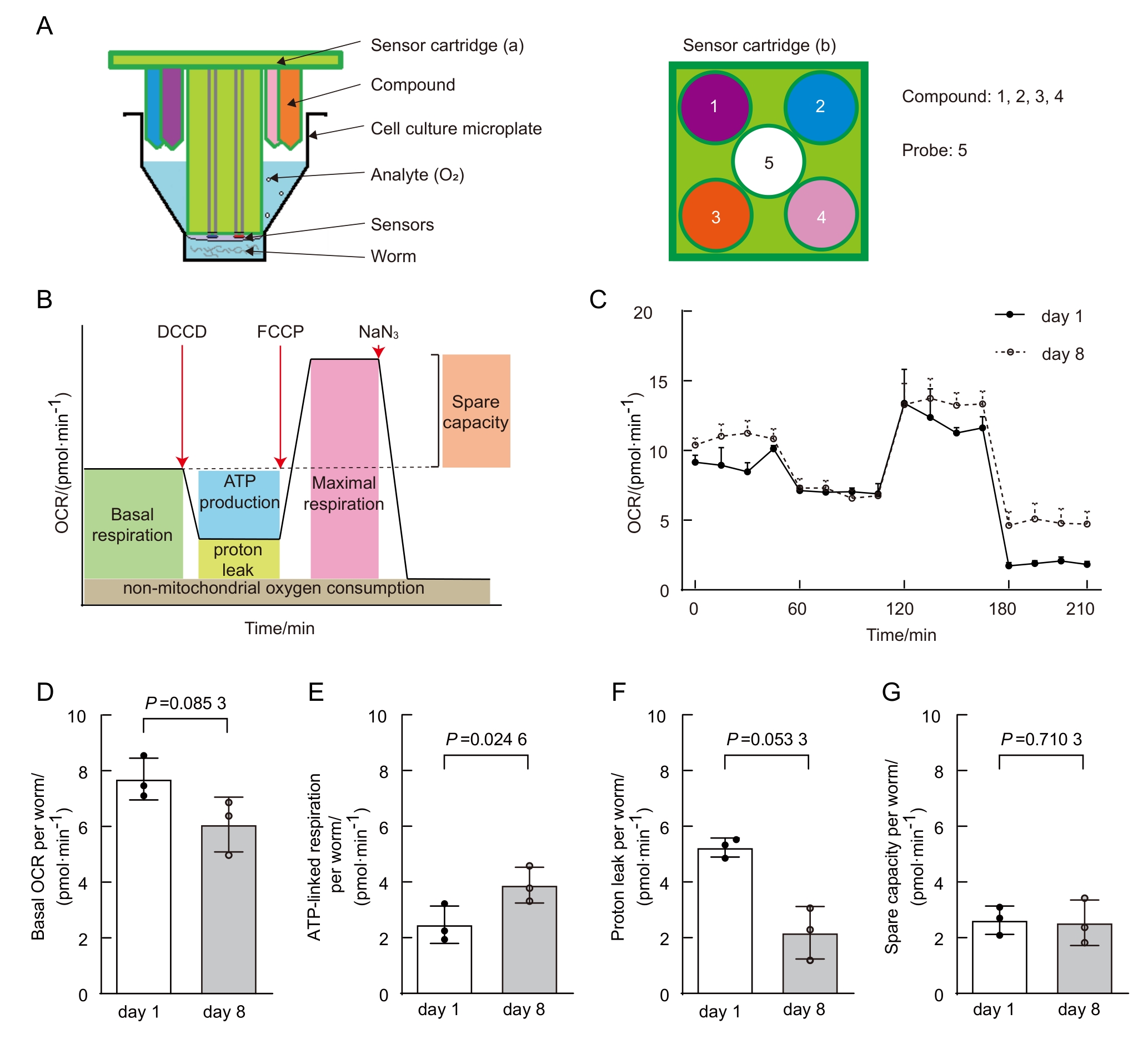
图8 年老线虫线粒体代谢发生改变(数据来自本实验室已发表文献[
Figure 8 Age-dependent mitochondrial function changes in C.elegans (data from published paper of our lab [
| [1] | MUKHERJEE I, GHOSH M, MEINECKE M. MICOS and the mitochondrial inner membrane morphology–when things get out of shape[J]. FEBS Lett, 2021, 595(8):1159-1183. DOI:10.1002/1873-3468.14089 . |
| [2] | OKAMOTO K, SHAW J M. Mitochondrial morphology and dynamics in yeast and multicellular eukaryotes[J]. Annu Rev Genet, 2005, 39:503-536. DOI:10.1146/annurev.genet.38. 072902.093019 . |
| [3] | LÓPEZ-OTÍN C, BLASCO M A, PARTRIDGE L, et al. Hallmarks of aging: an expanding universe[J]. Cell, 2023, 186(2):243-278. DOI:10.1016/j.cell.2022.11.001 . |
| [4] | YAN C J, DUANMU X Y, ZENG L, et al. Mitochondrial DNA: distribution, mutations, and elimination[J]. Cells, 2019, 8(4):379. DOI:10.3390/cells8040379 . |
| [5] | KLIONSKY D J, ABDELMOHSEN K, ABE A, et al. Guidelines for the use and interpretation of assays for monitoring autophagy (3rd edition)[J]. Autophagy, 2016, 12(1):1-222. DOI:10.1080/15548627.2015.1100356 . |
| [6] | MACK H I D, HEIMBUCHER T, MURPHY C T. The nematode Caenorhabditis elegans as a model for aging research[J]. Drug Discov Today Dis Models, 2018, 27:3-13. DOI:10.1016/j.ddmod.2018.11.001 . |
| [7] | JI T, ZHANG X L, XIN Z L, et al. Does perturbation in the mitochondrial protein folding pave the way for neurodegeneration diseases?[J]. Ageing Res Rev, 2020, 57:100997. DOI:10.1016/j.arr.2019.100997 . |
| [8] | SOHRABI S, MOR D E, KALETSKY R, et al. High-throughput behavioral screen in C. elegans reveals Parkinson's disease drug candidates[J]. Commun Biol, 2021, 4(1):203. DOI:10.1038/s42003-021-01731-z . |
| [9] | YE S W, SONG S D, LIU X J, et al. A small-molecule screen identifies novel aging modulators by targeting 5-HT/DA signaling pathway[J]. Aging Cell, 2025, 24(3): 1-13. DOI:10.1111/acel.14411 . |
| [10] | PANDA M, FAKITSA M, MARKAKI M, et al. Caenorhabditis elegans as an emerging high throughput chronotherapeutic drug screening platform for human neurodegenerative disorders[J]. Adv Drug Deliv Rev, 2025, 224:115655. DOI:10.1016/j.addr.2025.115655 . |
| [11] | YOO I, AHN I, LEE J, et al. Extracellular flux assay (Seahorse assay): Diverse applications in metabolic research across biological disciplines[J]. Mol Cells, 2024, 47(8):100095. DOI:10.1016/j.mocell.2024.100095 . |
| [12] | STIERNAGLE T. Maintenance of C. elegans [J]. WormBook, 2006:1-11. DOI:10.1895/wormbook.1.101.1 . |
| [13] | MALLICK A, RANAWADE A, VAN DEN BERG W, et al. Axin-mediated regulation of lifespan and muscle health in C. elegans requires AMPK-FOXO signaling[J]. iScience, 2020, 23(12):101843. DOI:10.1016/j.isci.2020.101843 . |
| [14] | REGMI S G, ROLLAND S G, CONRADT B. Age-dependent changes in mitochondrial morphology and volume are not predictors of lifespan[J]. Aging (Albany NY), 2014, 6(2):118-30. DOI: 10.18632/aging.100639 . |
| [15] | XIA Q, LI P L, CASAS-MARTINEZ J C, et al. Peroxiredoxin 2 regulates DAF-16/FOXO mediated mitochondrial remodelling in response to exercise that is disrupted in ageing[J]. Mol Metab, 2024, 88:102003. DOI: 10.1016/j.molmet.2024.102003 . |
| [16] | CHAUDHRY A, SHI R, LUCIANI D S. A pipeline for multidimensional confocal analysis of mitochondrial morphology, function, and dynamics in pancreatic β-cells[J]. Am J Physiol Endocrinol Metab, 2020, 318(2):E87-E101. DOI:10.1152/ajpendo.00457.2019 . |
| [17] | Mitochondria Analyzer[Z/OL]. [2025-07-16]. . |
| [18] | VALENTE A J, MADDALENA L A, ROBB E L, et al. A simple ImageJ macro tool for analyzing mitochondrial network morphology in mammalian cell culture[J]. Acta Histochem, 2017, 119(3):315-326. DOI:10.1016/j.acthis.2017.03.001 . |
| [19] | MiNA - Mitochondrial Network Analysis[Z/OL]. [2025-07-16]. . |
| [20] | NG L F, GRUBER J. Measurement of respiration rate in live Caenorhabditis elegans [J]. Bio Protoc, 2019, 9(10):1-12. DOI:10.21769/BioProtoc.3243 . |
| [21] | HAROON S, VERMULST M. Oxygen consumption measurements in Caenorhabditis elegans using the seahorse XF24[J]. Bio Protoc, 2019, 9(13):e3288. DOI:10.21769/BioProtoc.3288 . |
| [22] | SONG M J, DONG S M, ZHANG X F, et al. A moderate static magnetic field promotes C. elegans longevity through cytochrome P450s[J]. Sci Rep, 2022, 12:16108. DOI:10.1038/s41598-022-20647-0 . |
| [23] | DILLIN A, HSU A L, ARANTES-OLIVEIRA N, et al. Rates of behavior and aging specified by mitochondrial function during development[J]. Science, 2002, 298(5602):2398-2401. DOI: 10.1126/science/1077780 . |
| [24] | AMORIM J A, COPPOTELLI G, ROLO A P, et al. Mitochondrial and metabolic dysfunction in ageing and age-related diseases[J]. Nat Rev Endocrinol, 2022, 18(4):243-258. DOI:10.1038/s41574-021-00626-7 . |
| [25] | SUN N, YOULE R J, FINKEL T. The mitochondrial basis of aging[J]. Mol Cell, 2016, 61(5):654-666. DOI:10.1016/j.molcel.2016.01.028 . |
| [26] | BAR-ZIV R, BOLAS T, DILLIN A. Systemic effects of mitochondrial stress[J]. EMBO Rep, 2020, 21(6):e50094. DOI:10.15252/embr.202050094 . |
| [27] | REGMI S G, ROLLAND S G, CONRADT B. Age-dependent changes in mitochondrial morphology and volume are not predictors of lifespan[J]. Aging, 2014, 6(2):118-130. DOI:10.18632/aging.100639 . |
| [28] | SON H G, ALTINTAS O, KIM E J E, et al. Age-dependent changes and biomarkers of aging in Caenorhabditis elegans [J]. Aging Cell, 2019, 18(2):e12853. DOI:10.1111/acel.12853 . |
| [29] | MAGLIONI S, MELLO D F, SCHIAVI A, et al. Mitochondrial bioenergetic changes during development as an indicator of C. elegans health-span[J]. Aging, 2019, 11(16):6535-6554. DOI:10.18632/aging.102208 . |
| [30] | MITRA K, WUNDER C, ROYSAM B, et al. A hyperfused mitochondrial state achieved at G1–S regulates cyclin E buildup and entry into S phase[J]. Proc Natl Acad Sci U S A, 2009, 106(29):11960-11965. DOI:10.1073/pnas.0904875106 . |
| [31] | DAGDA R K, CHERRA S J, KULICH S M, et al. Loss of PINK1 function promotes mitophagy through effects on oxidative stress and mitochondrial fission[J]. J Biol Chem, 2009, 284(20):13843-13855. DOI:10.1074/jbc.M808515200 . |
| [1] | 孙涵, 郭芃, 俞昕何, 张隽巧, 尧莹, 杨文. 秀丽隐杆线虫作为退行性疾病模型的分子通路研究进展[J]. 实验动物与比较医学, 2025, 45(6): 738-751. |
| [2] | 成慧, 方菲, 石嘉豪, 杨桦, 张梦杰, 杨平, 费俭. hil-1基因通过饮食限制通路调节秀丽隐杆线虫寿命[J]. 实验动物与比较医学, 2023, 43(3): 271-281. |
| [3] | 李晗, 张笑瑞, 张成芳. 间歇禁食法在改善奥氮平诱导小鼠代谢紊乱中的机制研究[J]. 实验动物与比较医学, 2023, 43(1): 3-10. |
| [4] | 李英娘, 戴玮, 盛健. 斑马鱼甲状腺肿大病例分析[J]. 实验动物与比较医学, 2019, 39(6): 462-466. |
| [5] | 沈艳, 徐汪洋, 朱后保. 氧化还原酶DHTKD1基因突变致病机制及小鼠模型研究进展[J]. 实验动物与比较医学, 2018, 38(6): 468-472. |
| [6] | 濮祥强, 王祥, 钱光辉, 马锦, 丁粤粤, 吕海涛. 小鼠免疫性冠状动脉炎内皮细胞线粒体的动态变化[J]. 实验动物与比较医学, 2018, 38(3): 169-175. |
| [7] | 常凯, 王裕, 庞文彪, 高继萍, 陈朝阳, 宋国华. 硒对氟致大鼠肾小管上皮细胞线粒体膜电位改变的拮抗作用[J]. 实验动物与比较医学, 2017, 37(3): 179-184. |
| [8] | 李媛, 张梅英. 自噬与帕金森疾病及相关性模型的研究进展[J]. 实验动物与比较医学, 2015, 35(4): 335-340. |
| [9] | 申幸娇, 岳秉飞, 马丽颖. 三个封闭群实验兔线粒体DNA D-loop区多态性分析[J]. 实验动物与比较医学, 2014, 34(1): 29-34. |
| [10] | 叶红梅, 钟春燕, 黄敏贤, 吕俊华. 荔枝核提取物对D-半乳糖诱发小鼠学习记忆损伤的保护机制[J]. 实验动物与比较医学, 2013, 33(4): 285-289. |
| [11] | 高骏, 倪丽菊, 孙凤萍, 王金祥, 胡建华, 高诚, 李凯, 肖君华, 周宇荀. 东方田鼠指名亚种的线粒体基因组序列分析及系统进化研究[J]. 实验动物与比较医学, 2013, 33(3): 167-173. |
| [12] | 张评浒, 陶元清, 江振洲, 王忠东, 范薇, 张陆勇. 喜马拉雅旱獭作为药物线粒体毒性替代模型的可行性分析[J]. 实验动物与比较医学, 2012, 32(5): 436-440. |
| [13] | 李洪涛1,吴清洪2,肖东2,袁进2,王万山2,张嘉宁2,顾为望2. 四种实验用小型猪mtDNA控制区5'端序列的比较研究[J]. 实验动物与比较医学, 2009, 29(4): 237-240. |
| [14] | 管敏强1,曹琼洁2,陈忠义2,阮冬芬2,楼哲丰2,金龙金2. 线粒体DNA序列分析封闭群小鼠遗传稳定性[J]. 实验动物与比较医学, 2009, 29(2): 113-116. |
| [15] | 谢建云1,2,冯洁1,柏熊3,胡建华1,高诚1. 四种群东方田鼠线粒体DNA[J]. 实验动物与比较医学, 2008, 28(5): 299-303. |
| 阅读次数 | ||||||
|
全文 |
|
|||||
|
摘要 |
|
|||||
