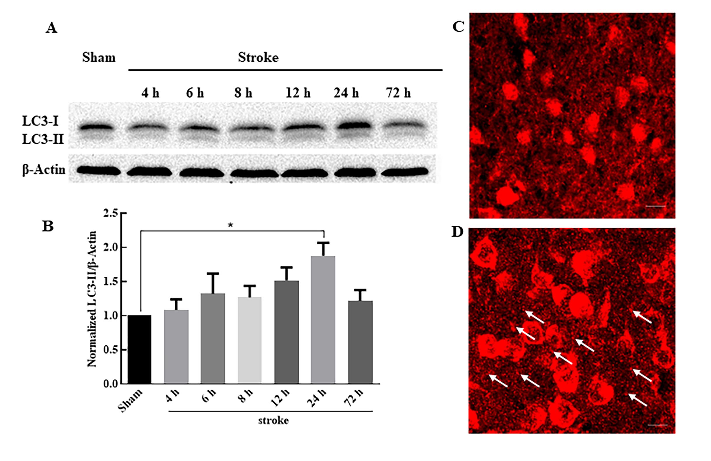
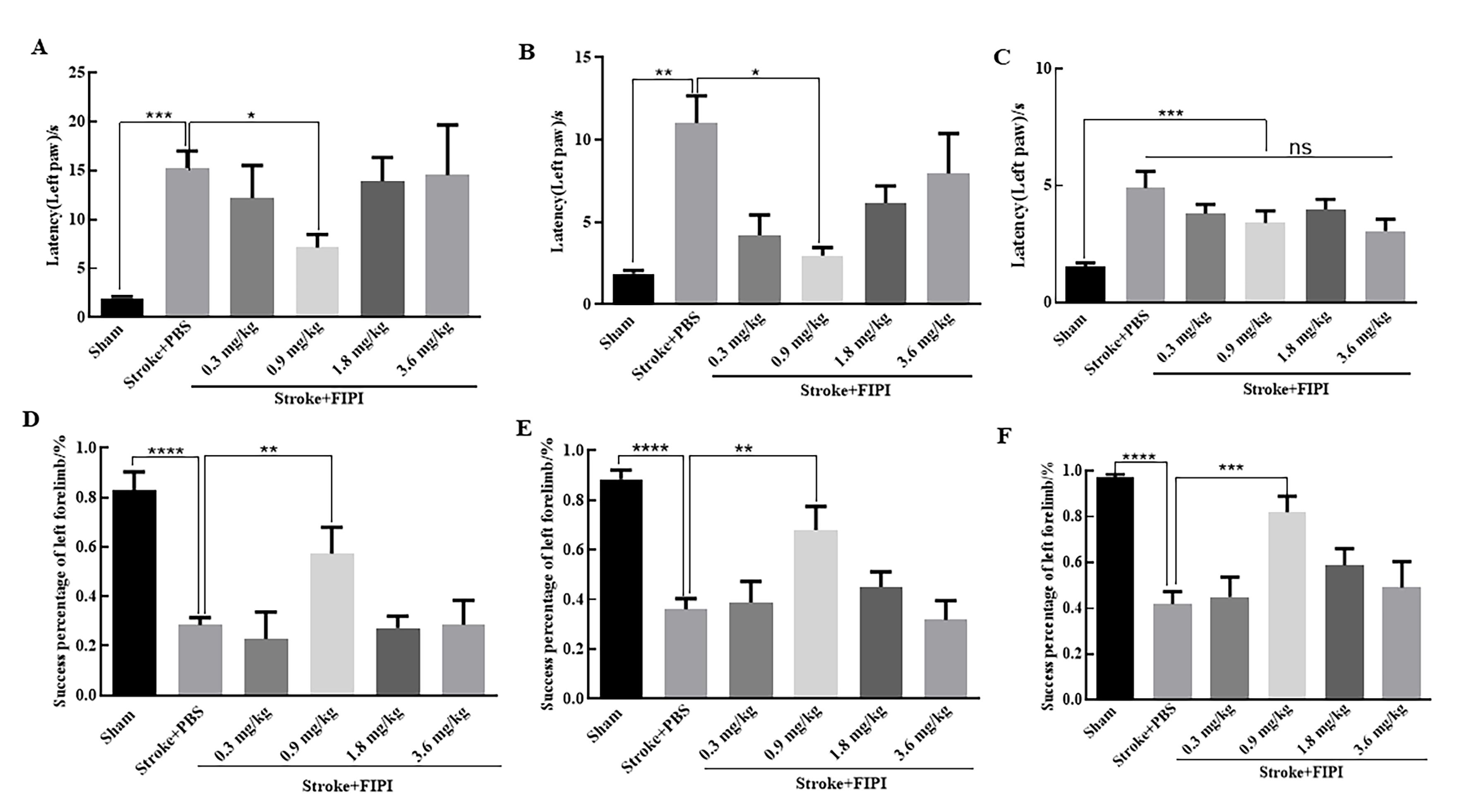
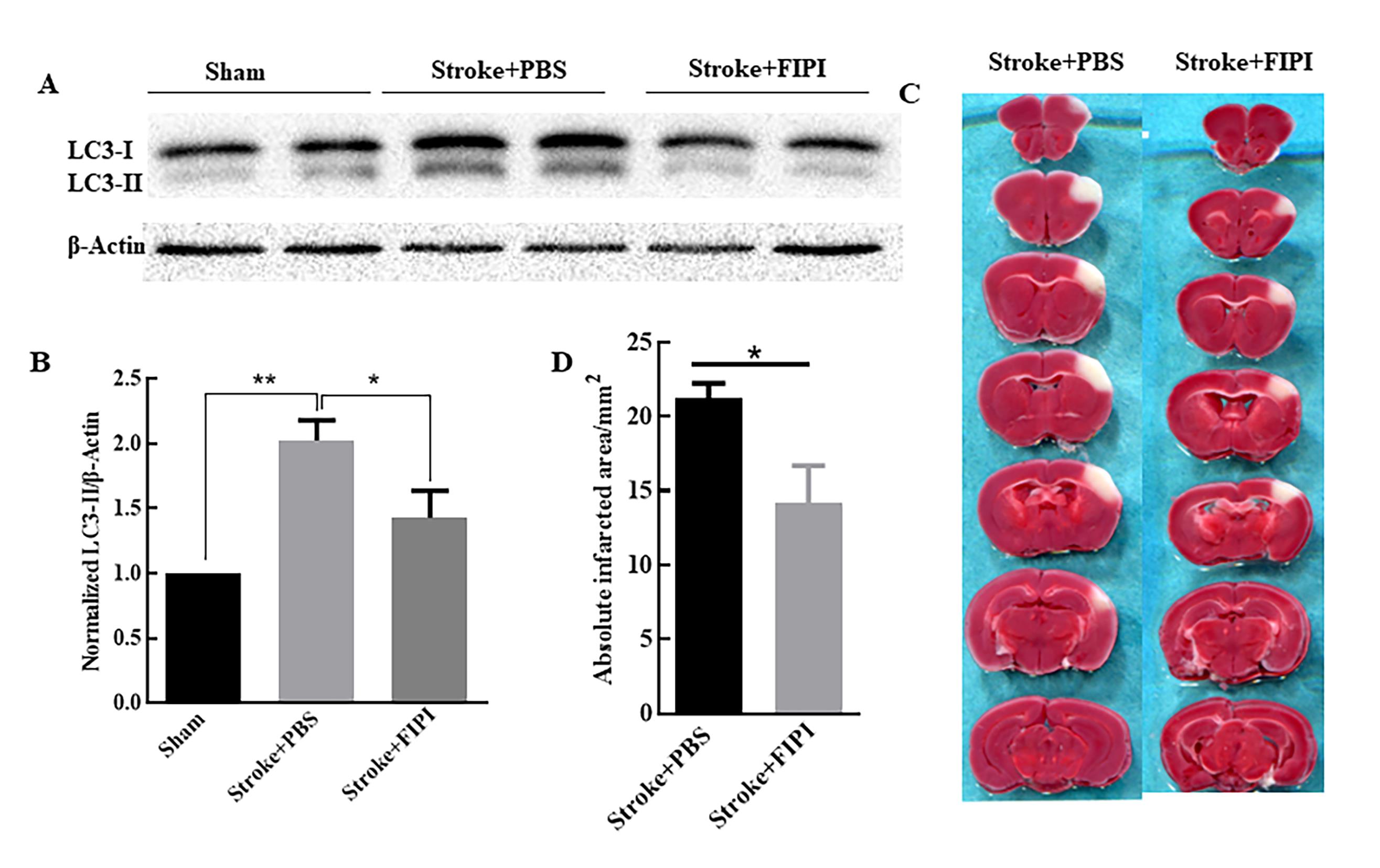
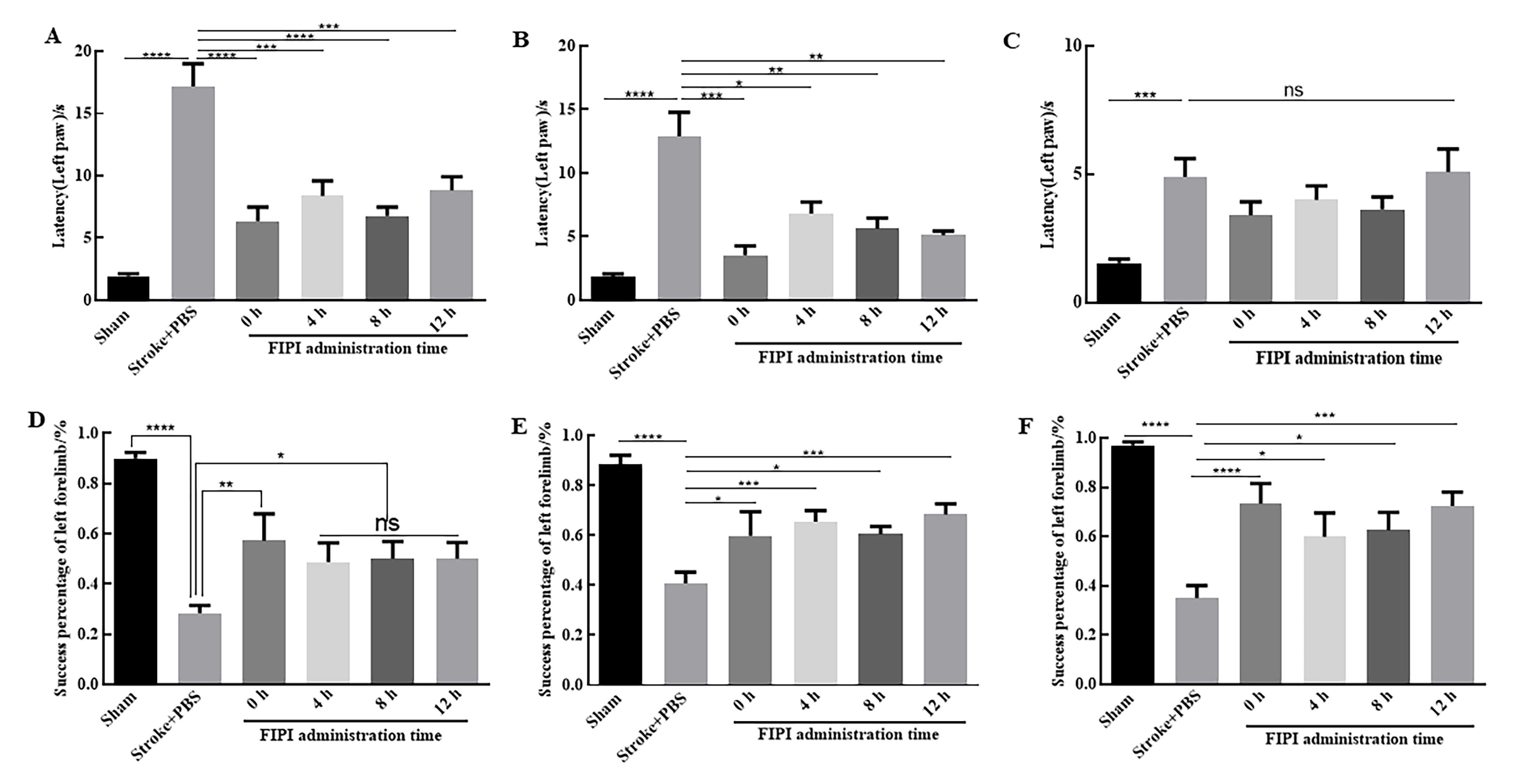
| 1 |
CAMPBELL B C V, DE SILVA D A, MACLEOD M R, et al. Ischaemic stroke[J]. Nat Rev Dis Primers, 2019, 5(1):70. DOI:10.1038/s41572-019-0118-8 .
|
| 2 |
WANG P, SHAO B Z, DENG Z Q, et al. Autophagy in ischemic stroke[J]. Prog Neurobiol, 2018, 163-164:98-117. DOI: 10.1016/j.pneurobio.2018.01.001 .
|
| 3 |
HWANG J Y, GERTNER M, PONTARELLI F, et al. Global ischemia induces lysosomal-mediated degradation of mTOR and activation of autophagy in hippocampal neurons destined to Die[J]. Cell Death Differ, 2017, 24(2):317-329. DOI:10.1038/cdd.2016.140 .
|
| 4 |
DALL'ARMI C, DEVEREAUX K A, DI PAOLO G. The role of lipids in the control of autophagy[J]. Curr Biol, 2013, 23(1): R33-R45. DOI:10.1016/j.cub.2012.10.041 .
|
| 5 |
PURI C, RENNA M, BENTO C F, et al. Diverse autophagosome membrane sources coalesce in recycling endosomes[J]. Cell, 2013, 154(6):1285-1299. DOI:10.1016/j.cell.2013.08.044 .
|
| 6 |
HUR J H, PARK S Y, DALL'ARMI C, et al. Phospholipase D1 deficiency in mice causes nonalcoholic fatty liver disease via an autophagy defect[J]. Sci Rep, 2016, 6:39170. DOI:10.1038/srep39170 .
|
| 7 |
DALL'ARMI C, HURTADO-LORENZO A, TIAN H, et al. The phospholipase D1 pathway modulates macroautophagy[J]. Nat Commun, 2010, 1:142. DOI:10.1038/ncomms1144 .
|
| 8 |
YOON M S. Vps34 and PLD1 take center stage in nutrient signaling: their dual roles in regulating autophagy[J]. Cell Commun Signal, 2015, 13:44. DOI:10.1186/s12964-015-0122-x .
|
| 9 |
ZHU Y B, KANG K, ZHANG Y, et al. PLD1 negatively regulates dendritic branching[J]. J Neurosci, 2012, 32(23):7960-7969. DOI:10.1523/jneurosci.5378-11.2012 .
|
| 10 |
ZHU Y B, GAO W, ZHANG Y, et al. Astrocyte-derived phosphatidic acid promotes dendritic branching[J]. Sci Rep, 2016, 6:21096. DOI:10.1038/srep21096 .
|
| 11 |
陶少鑫, 朱彦兵, 于山平, 等. 小鼠缺血性脑卒中后磷脂酶D1在自噬和神经损伤中的作用[J]. 临床和实验医学杂志, 2019, 18(8): 790-794. DOI:10.3969/j.issn.1671-4695.2019.08.002 .
|
|
TAO S X, ZHU Y B, YU S P, et al. Role of phospholipase D1 in autophagy and neuronal damage after focal ischemic stroke in mice[J]. J Clin Exp Med, 2019, 18(8): 790-794. DOI:10.3969/j.issn.1671-4695.2019.08.002 .
|
| 12 |
KLIER M, GOWERT N S, JÄCKEL S, et al. Phospholipase D1 is a regulator of platelet-mediated inflammation[J]. Cell Signal, 2017, 38:171-181. DOI:10.1016/j.cellsig.2017.07.007 .
|
| 13 |
SU W, YEKU O, OLEPU S, et al. 5-Fluoro-2-indolyl des-chlorohalopemide (FIPI), a phospholipase D pharmacological inhibitor that alters cell spreading and inhibits chemotaxis[J]. Mol Pharmacol, 2009, 75(3):437-446. DOI:10.1124/mol.108. 053298 .
|
| 14 |
HENKELS K M, MUPPANI N R, GOMEZ-CAMBRONERO J. PLD-specific small-molecule inhibitors decrease tumor-associated macrophages and neutrophils infiltration in breast tumors and lung and liver metastases[J]. PLoS One, 2016, 11(11): e0166553. DOI:10.1371/journal.pone.0166553 .
|
| 15 |
MAO L L, LI P Y, ZHU W, et al. Regulatory T cells ameliorate tissue plasminogen activator-induced brain haemorrhage after stroke[J]. Brain, 2017, 140(7):1914-1931. DOI:10.1093/brain/awx111 .
|
| 16 |
ANDREJEVA G, GOWAN S, LIN G, et al. De novo phosphatidylcholine synthesis is required for autopha-gosome membrane formation and maintenance during autophagy[J]. Autophagy, 2020, 16(6):1044-1060. DOI:10.1080/15548627.2019.1659608 .
|
| 17 |
CAI M, HE J, XIONG J, et al. Phospholipase D1-regulated autophagy supplies free fatty acids to counter nutrient stress in cancer cells[J]. Cell Death Dis, 2016, 7(11): e2448. DOI:10.1038/cddis.2016.355 .
|
| 18 |
TIAN F, DEGUCHI K, YAMASHITA T, et al. In vivo imaging of autophagy in a mouse stroke model[J]. Autophagy, 2010, 6(8):1107-1114. DOI:10.4161/auto.6.8.13427 .
|
| 19 |
MARCHESAN D, RUTBERG M, ANDERSSON L, et al. A phospholipase D-dependent process forms lipid droplets containing caveolin, adipocyte differentiation-related protein, and vimentin in a cell-free system[J]. J Biol Chem, 2003, 278(29):27293-27300. DOI:10.1074/jbc.m301430200 .
|
| 20 |
NGUYEN T B, OLZMANN J A. Lipid droplets and lipotoxicity during autophagy[J]. Autophagy, 2017, 13(11):2002-2003. DOI:10.1080/15548627.2017.1359451 .
|
| 21 |
MCDERMOTT M I, WANG Y, WAKELAM M J O, et al. Mammalian phospholipase D: function, and therapeutics[J]. Prog Lipid Res, 2020, 78:101018. DOI:10.1016/j.plipres. 2019.101018 .
|
| 22 |
BAE E J, LEE H J, JANG Y H, et al. Phospholipase D1 regulates autophagic flux and clearance of α-synuclein aggregates[J]. Cell Death Differ, 2014, 21(7):1132-1141. DOI:10.1038/cdd. 2014.30 .
|
| 23 |
OLIVEIRA T G, CHAN R B, TIAN H, et al. Phospholipase d2 ablation ameliorates Alzheimer's disease-linked synaptic dysfunction and cognitive deficits[J]. J Neurosci, 2010, 30(49):16419-16428. DOI:10.1523/jneurosci.3317-10.2010 .
|
| 24 |
KIM S H, PARK M Y, YUN N J, et al. Targeting PLD2 in adipocytes augments adaptive thermogenesis by improving mitochondrial quality and quantity in mice[J]. J Exp Med, 2022, 219(2): e20211523. DOI:10.1084/jem.20211523 .
|
| 25 |
YUZHU W, DAN T, CHANGWEI W, et al. Propofol attenuates α-synuclein aggregation and neuronal damage in a mouse model of ischemic stroke [J]. Neurosci Bull, 2020, 36(3):289-298. DOI:10.1007/s12264-019-00426-0
|


 ), 白帆3,4, 陶少鑫1,2, 潘雨花蕾1,2, 王欢1, 赵羽商1, 王崧1, 于艳3,4(
 )(
)(