



• •
蒙飞彪1, 王林仪1,2, 张文强1, 唐瑞欣1, 黄雪映1, 方美荣1, 杨威1,2, 郭健敏1,2( )(
)( )
)
出版日期:2026-07-28
作者简介:蒙飞彪(1987—),男,本科,研究方向:药物药理毒理研究。E-mail:mengfeibiao@lewwin.com.cn;基金资助:
MENG Feibiao1, WANG Linyi1,2, ZHANG Wenqiang1, TANG Ruixin1, HUANG Xueying1, FANG Meirong1, YANG Wei1,2, GUO Jianmin1,2()()
Published:2026-07-28
Correspondence to:
GUO Jianmin (ORCID: 0000-0003-3319-5580), E-mail: guojianmin@lewwin.com.cn.摘要:
目的 采用5%盐酸普萘洛尔乳剂诱导豚鼠耳部银屑病,建立一种稳定的局部银屑病动物模型,并验证其在药效评价中的应用价值。 方法 29只6~8周龄的普通级雄性Hartley豚鼠,按体重随机分组,筛选9只豚鼠作为正常组,其余动物为造模组,造模组涂抹5%盐酸普萘洛尔乳剂于右耳背部,每日4次,连续4天,以耳厚度差>0.05 mm及银屑病样皮损面积和疾病严重程度指数(PASI)评分>3分为成模标准;成模动物以右耳耳厚度差值为主指标,以PASI评分、体重为副指标均衡随机分组,分为模型组、1.6 mg/kg 甲氨蝶呤组,连续给药10 d。试验期间观察皮损、体重、耳厚度差值及PASI评分变化,末期取材检测耳重量、脾脏指数、耳组织病理变化以及肥大细胞浸润程度指标。 结果 造模组动物造模2 d即出现红肿以及增厚的银屑病样皮损现象,并伴有明显搔抓行为,于4~9 d为模型的高峰期,耳部皮肤显著红肿、增厚、鳞屑增多;模型组的PASI评分、耳厚度差值、耳肿胀、脾脏指数、组织病理变化以及肥大细胞浸润均显著高于正常组;甲氨蝶呤均能显著改善上述检测指标。 结论 成功建立了一种稳定、可重复的盐酸普萘洛尔诱导豚鼠耳部银屑病模型,适用于局部银屑病的药效作用评价。
中图分类号:
蒙飞彪,王林仪,张文强,等.盐酸普萘洛尔诱导豚鼠耳部银屑病模型的建立与验证[J]. 实验动物与比较医学.. DOI: 10.12300/j.issn.1674-5817.2026.011.
MENG Feibiao,WANG Linyi,ZHANG Wenqiang,et al. Establishment and Validation of a Guinea Pig Model of Ear Psoriasis Induced by Propranolol Hydrochloride[J]. Laboratory Animal and Comparative Medicine. DOI: 10.12300/j.issn.1674-5817.2026.011.
比例 Ratio | 组分 Component | 重量(g) Weight (g) | 百分比(%) Percentage (%) |
|---|---|---|---|
| 油相Oil phase | 硬脂酸Stearic acid | 1.4 | 14 |
| 液体石蜡Liquid paraffin | 2.4 | 24 | |
| 水相Aqueous phase | 三乙醇胺Triethanolamine | 0.3 | 3 |
| 甘油Glycerol | 0.9 | 9 | |
| 蒸馏水Distilled water | 4.5 | 45 | |
| 药物Medicine | 盐酸普萘洛尔Propranolol hydrochloride | 0.5 | 5 |
| 总量Total quantity | 10 | 100 | |
表1 5%盐酸普萘洛尔乳剂配方
Table 1 5% Propranolol Hydrochloride Formula
比例 Ratio | 组分 Component | 重量(g) Weight (g) | 百分比(%) Percentage (%) |
|---|---|---|---|
| 油相Oil phase | 硬脂酸Stearic acid | 1.4 | 14 |
| 液体石蜡Liquid paraffin | 2.4 | 24 | |
| 水相Aqueous phase | 三乙醇胺Triethanolamine | 0.3 | 3 |
| 甘油Glycerol | 0.9 | 9 | |
| 蒸馏水Distilled water | 4.5 | 45 | |
| 药物Medicine | 盐酸普萘洛尔Propranolol hydrochloride | 0.5 | 5 |
| 总量Total quantity | 10 | 100 | |
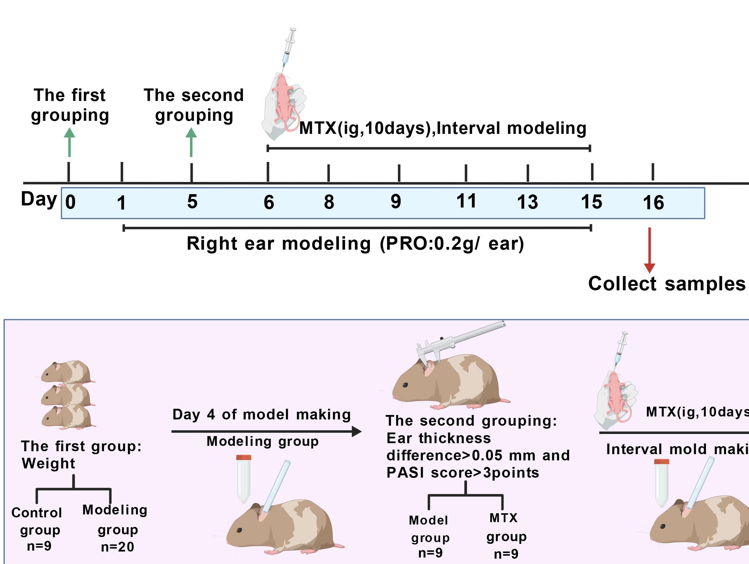
图1 实验流程示意图
Figure 1 Schematic diagram of the experimental process
皮肤泛红程度 Degree of skin redness | 鳞屑 Scale | 增厚程度 Degree of thickening | 严重程度 Different degrees of severity | 转换评分 Conversion score |
|---|---|---|---|---|
无泛红现象 No redness at all | 耳部表面无可见鳞屑 | 未见增厚 | 无症状 | 0 |
耳部皮肤呈淡红色 The skin on the ear is a light red color. | 部分皮损表面完全或覆有鳞屑,以细微鳞屑为主 | 略有增厚,但尚属轻微范围 | 轻度 | 1 |
耳部皮肤呈红色 The skin on the ear is red. | 大多数皮损表面完全或不完全覆有鳞屑,鳞屑呈片状 | 呈中等程度增厚,变化较为显著 | 中度 | 2 |
耳部皮肤呈深红色 The skin on the ear is a deep red color | 几乎所有皮损表面覆有鳞屑,较厚成层 | 明显增厚,变化突出 | 重度 | 3 |
耳部皮肤红色极深 The skin on the ear is extremely red | 全部皮损表面均覆有鳞屑,鳞屑很厚成层 | 异常增厚,已达严重程度 | 极重度 | 4 |
表2 PASI评分细则
Table 2 PASI Scoring Criteria
皮肤泛红程度 Degree of skin redness | 鳞屑 Scale | 增厚程度 Degree of thickening | 严重程度 Different degrees of severity | 转换评分 Conversion score |
|---|---|---|---|---|
无泛红现象 No redness at all | 耳部表面无可见鳞屑 | 未见增厚 | 无症状 | 0 |
耳部皮肤呈淡红色 The skin on the ear is a light red color. | 部分皮损表面完全或覆有鳞屑,以细微鳞屑为主 | 略有增厚,但尚属轻微范围 | 轻度 | 1 |
耳部皮肤呈红色 The skin on the ear is red. | 大多数皮损表面完全或不完全覆有鳞屑,鳞屑呈片状 | 呈中等程度增厚,变化较为显著 | 中度 | 2 |
耳部皮肤呈深红色 The skin on the ear is a deep red color | 几乎所有皮损表面覆有鳞屑,较厚成层 | 明显增厚,变化突出 | 重度 | 3 |
耳部皮肤红色极深 The skin on the ear is extremely red | 全部皮损表面均覆有鳞屑,鳞屑很厚成层 | 异常增厚,已达严重程度 | 极重度 | 4 |
病变程度 Degree of lesion | 组织学改变 Histological changes | 评分(分) Score(points) |
|---|---|---|
| 正常Normal | 未观察到组织学改变或组织结构基本正常 | 0 |
| 轻微Slight | 病理损伤范围零星可见,不构成区域性 | 0.5 |
| 轻度Mild | 灶性、多灶性或散在分布的损伤,损伤范围在100倍镜下可见1/3-1个视野 | 1 |
| 中度Moderate | 灶性、多灶性或散在分布的损伤,损伤范围在100倍镜下可见1-3个视野 | 2 |
| 重度Severe | 灶性、多灶性或散在分布的损伤,损伤范围在100倍镜下可见3个视野以上 | 3 |
表3 病理诊断病变程度判读以及半定量评分标准[
Table 3 Criteria for Pathological Diagnosis, Degree of Lesion, and Semi-quantitative Scoring System
病变程度 Degree of lesion | 组织学改变 Histological changes | 评分(分) Score(points) |
|---|---|---|
| 正常Normal | 未观察到组织学改变或组织结构基本正常 | 0 |
| 轻微Slight | 病理损伤范围零星可见,不构成区域性 | 0.5 |
| 轻度Mild | 灶性、多灶性或散在分布的损伤,损伤范围在100倍镜下可见1/3-1个视野 | 1 |
| 中度Moderate | 灶性、多灶性或散在分布的损伤,损伤范围在100倍镜下可见1-3个视野 | 2 |
| 重度Severe | 灶性、多灶性或散在分布的损伤,损伤范围在100倍镜下可见3个视野以上 | 3 |
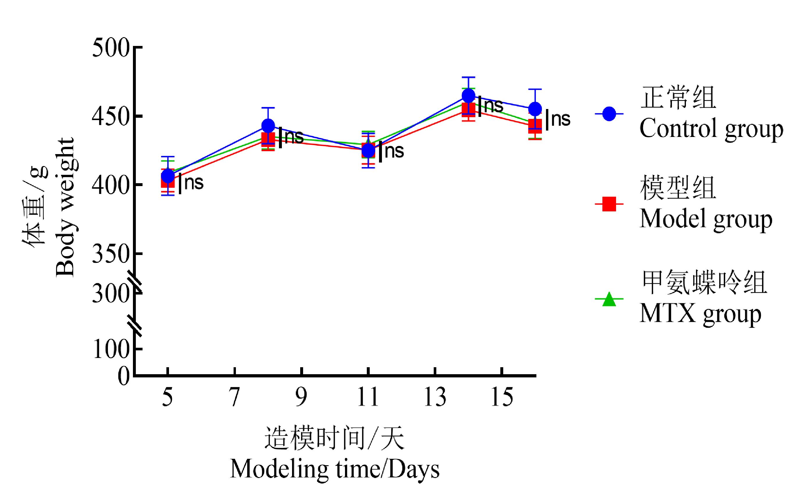
图2 豚鼠耳部银屑病模型体重变化趋势(ˉn = 9)
Figure 2 Body weight change trend of the guinea pig ear psoriasis model (n = 9)
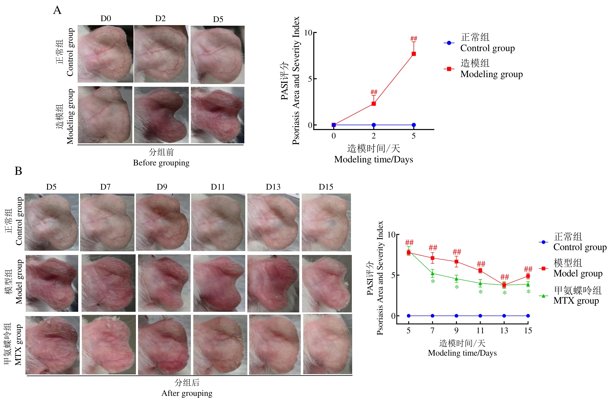
图3 豚鼠耳部造模部位皮肤观察和PASI评分变化趋势(n = 9)
Figure 3 Observation of the skin at the ear modeling site in guinea pigs and the trend of PASI score changes(n = 9)
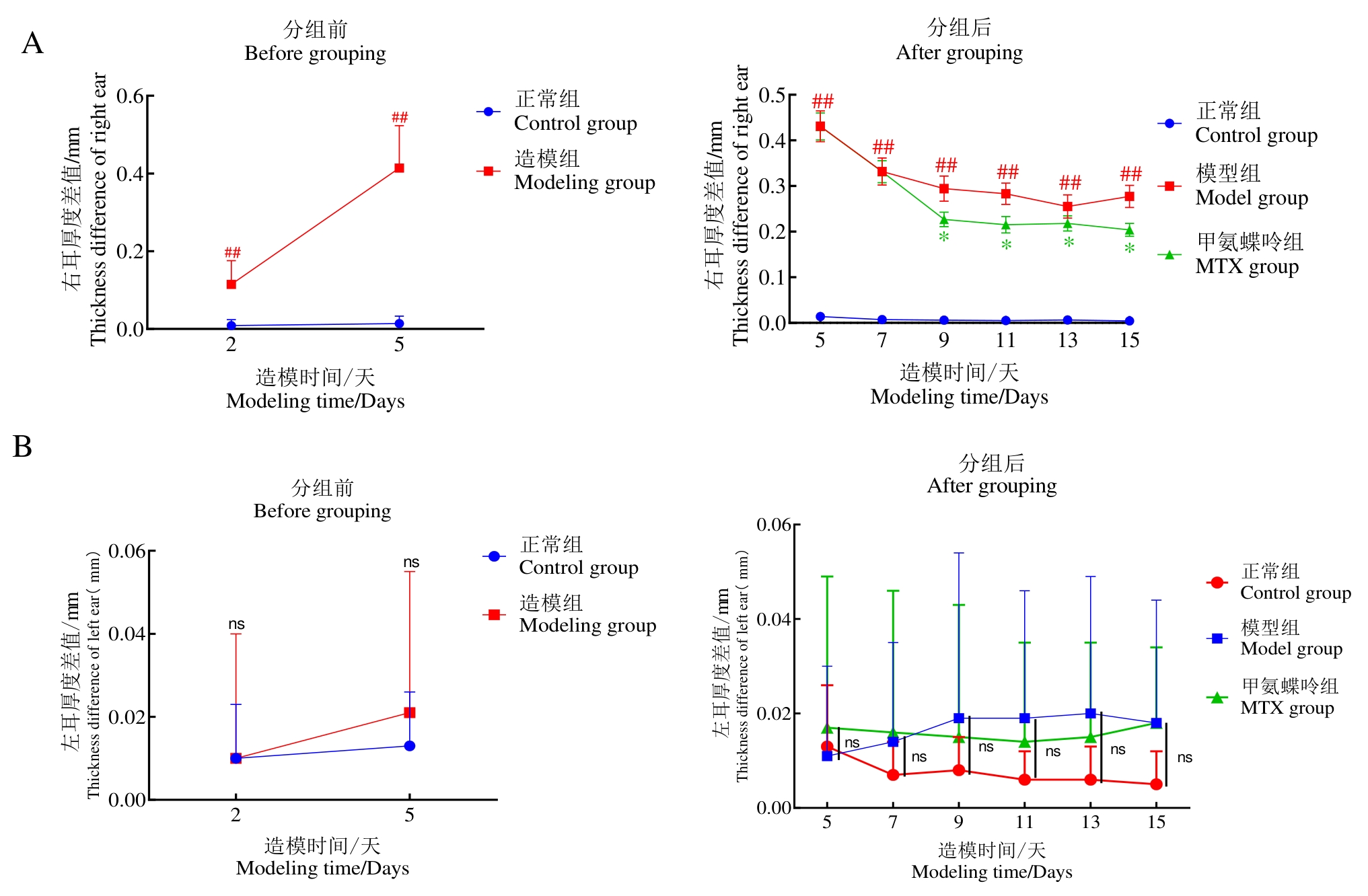
图4 豚鼠耳部银屑病模型实验期间耳厚度差变化
Figure 4 Changes in ear thickness during the experiment of the guinea pig ear psoriasis model
组别 Groups | 左耳片(g) Left ear(g) | 右耳片(g) Right ear(g) | 耳肿胀度(g) Ear swelling degree (g) |
|---|---|---|---|
正常组 control group | 0.0695±0.004 | 0.0669±0.0050 | 0.0004±0.0024 |
模型组 model group | 0.0713±0.002 | 0.0896±0.0052## | 0.0187±0.0040## |
甲氨蝶呤组 MTX group | 0.0702±0.004 | 0.0826±0.0056* | 0.0123±0.0051** |
表4 豚鼠耳部银屑病模型耳肿胀度 (
Table 4 The ear swelling of the guinea pig psoriasis model
组别 Groups | 左耳片(g) Left ear(g) | 右耳片(g) Right ear(g) | 耳肿胀度(g) Ear swelling degree (g) |
|---|---|---|---|
正常组 control group | 0.0695±0.004 | 0.0669±0.0050 | 0.0004±0.0024 |
模型组 model group | 0.0713±0.002 | 0.0896±0.0052## | 0.0187±0.0040## |
甲氨蝶呤组 MTX group | 0.0702±0.004 | 0.0826±0.0056* | 0.0123±0.0051** |
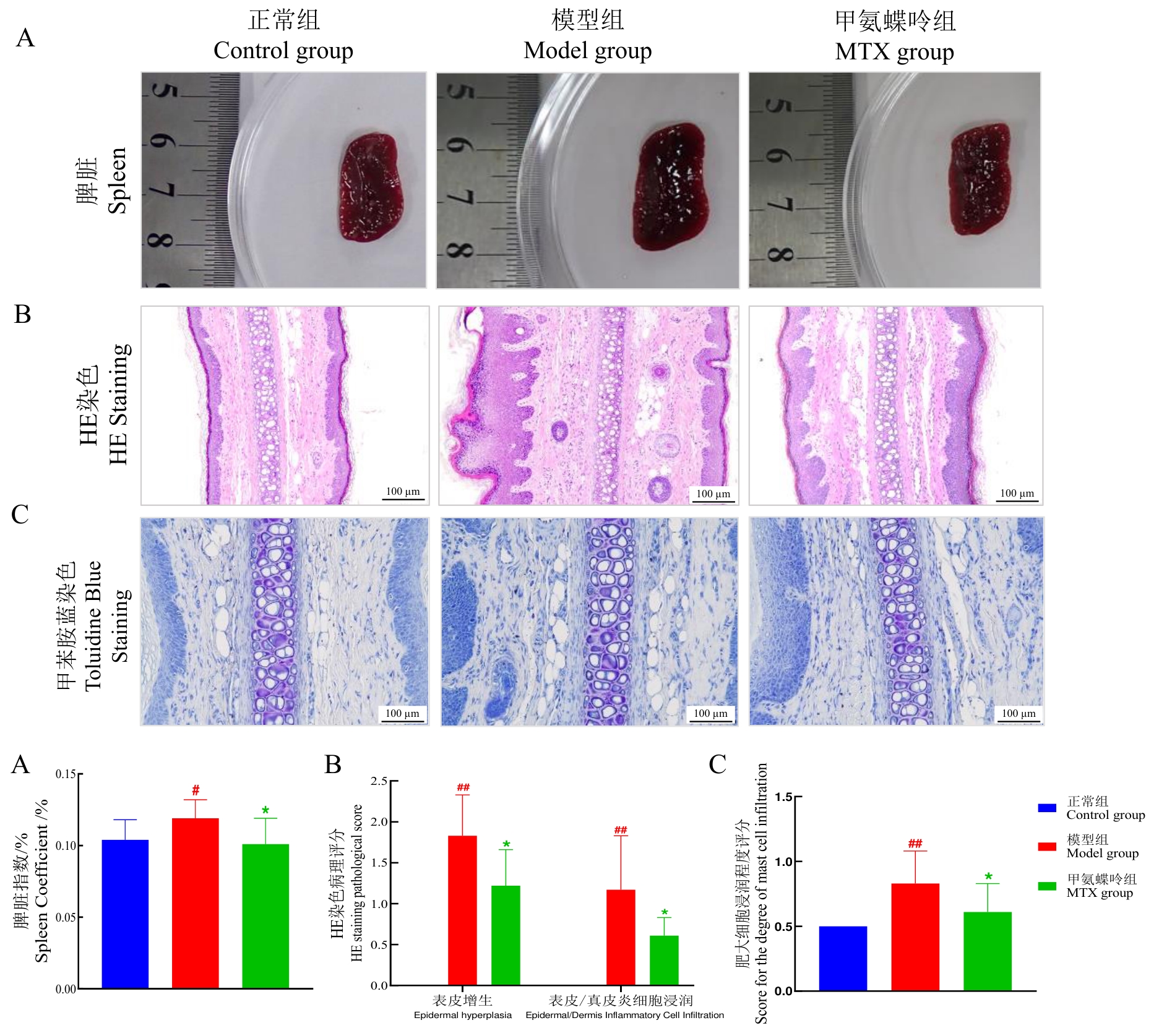
图5 豚鼠耳部银屑病模型脾脏指数、耳组织组织病理改变与肥大细胞浸润
Figure 5 Spleen index, histopathological changes of ear tissue, and mast cell infiltration in the murine ear psoriasis model
| [1] | Geng R S Q, Sibbald R G. Cutaneous psoriasis: clinical aspects and treatments[J]. Adv Skin Wound Care, 2025, 38(4): 176-182. DOI: 10.1097/ASW.0000000000000243 . |
| [2] | Helm M F, Clebak K T, Critchfield L, et al. Psoriasis and papulosquamous disorders[J]. Prim Care, 2025, 52(3): 567-591. DOI: 10.1016/j.pop.2025.05.007 . |
| [3] | Chan J J. Psoriasis: an update on topical and systemic therapies[J]. Aust Prescr, 2025, 48(3): 87-92. DOI: 10.18773/austprescr.2025.026 . |
| [4] | Parab S, Doshi G. The experimental animal models in psoriasis research: a comprehensive review[J]. Int Immunopharmacol, 2023, 117: 109897. DOI: 10.1016/j.intimp.2023.109897 . |
| [5] | Tatu A L, Elisei A M, Chioncel V, et al. Immunologic adverse reactions of β-blockers and the skin[J]. Exp Ther Med, 2019, 18(2): 955-959. DOI: 10.3892/etm.2019.7504 . |
| [6] | Kim G K, del Rosso J Q. Drug-provoked psoriasis: is it drug induced or drug aggravated?: understanding pathophysiology and clinical relevance[J]. J Clin Aesthet Dermatol, 2010, 3(1): 32-38. |
| [7] | 张萌萌, 刘树民, 孙慧娟, 等. 盐酸普萘洛尔致银屑病样大鼠模型的剂量、时间及性别探讨[J]. 海南医科大学学报, 2025, 31(5): 351-359, 372. DOI: 10.13210/j.cnki.jhmu.20240830.004 . |
| Zhang Mengmeng, Liu Shumin, Sun Huijuan, et al. Exploration of dose, time and sex in a psoriasis-like rat model caused by propranolol hydrochloride[J]. J Hainan Med Univ, 2025, 31(5): 351-359, 372. DOI: 10.13210/j.cnki.jhmu.20240830.004 . | |
| [8] | Shackelford C, Long G, Wolf J, et al. Qualitative and quantitative analysis of nonneoplastic lesions in toxicology studies[J]. Toxicol Pathol, 2002, 30(1): 93-96. DOI: 10.1080/01926230252824761 . |
| [9] | Tampa M, Mitran M I, Mitran C I, et al. Psoriasis: what is new in markers of disease severity?[J]. Medicina (Kaunas), 2024, 60(2): 337. DOI: 10.3390/medicina60020337 . |
| [10] | Kshirsagar S J, Adhav P S, Laddha U D, et al. Navigating psoriasis: From immune mechanisms to natural healing approaches[J]. Int Immunopharmacol, 2025, 144: 113626. DOI: 10.1016/j.intimp.2024.113626 . |
| [11] | 李慧贤, 胡丽, 郑焱, 等. 基于全球疾病负担(GBD)大数据的中国银屑病流行病学负担分析[J]. 中国皮肤性病学杂志, 2021, 35(4): 386-392. DOI: 10.13735/j.cjdv.1001-7089.202003197 . |
| Li Huixian, Hu Li, Zheng Yan, et al. Analysis of the epidemiological burden of psoriasis in China based on the big data of global burden of disease study (GBD)[J]. Chin J Dermatovenereology, 2021, 35(4): 386-392. DOI: 10.13735/j.cjdv.1001-7089.202003197 . | |
| [12] | Shirley S N, Watson A E, Yusuf N. Pathogenesis of inflammation in skin disease: from molecular mechanisms to pathology[J]. Int J Mol Sci, 2024, 25(18): 10152. DOI: 10.3390/ijms251810152 . |
| [13] | Lin Z C, Yang S C, Tran T T P, et al. Animal models of psoriasis for novel drug discovery: a literature update[J]. Expert Opin Drug Discov, 2025, 20(9): 1193-1208. DOI: 10.1080/17460441.2025.2528959 . |
| [14] | Xi Ruofan, Nie Yiwen, Lu Hanzhi, et al. Phytochemicals from Stellera chamaejasme alleviate psoriasis by modulating the immune microenvironment via the JAK2/PI3K/AKT pathway[J]. Int Immunopharmacol, 2025, 165: 115540. DOI: 10.1016/j.intimp.2025.115540 . |
| [15] | 牛凡琪, 李炳男, 何世烨, 等. 桃红四物汤调控MAPK/NF-κB信号通路治疗大鼠银屑病的作用机理[J]. 中国皮肤性病学杂志, 2025, 39(1): 17-29. DOI: 10.13735/j.cjdv.1001-7089.202406010 . |
| Niu Fanqi, Li Bingnan, He Shiye, et al. Mechanism of Taohong Siwu decoction regulating MAPK/NF-κB signaling pathway in the treatment of psoriasis in rats[J]. Chin J Dermatovenereology, 2025, 39(1): 17-29. DOI: 10.13735/j.cjdv.1001-7089.202406010 . | |
| [16] | Vinardell M P. Methodological shortcomings in the reports of the imiquimod psoriatic model[J]. Exp Dermatol, 2022, 31(3): 299-303. DOI: 10.1111/exd.14479 . |
| [17] | 李倾, 屠佳妮, 胡佳, 等. 咪喹莫特诱导银屑病动物模型的研究进展[J]. 中国实验动物学报, 2025, 33(8): 1222-1234. DOI: 10.3969/j.issn.1005-4847.2025.08.011 . |
| Li Qing, Tu Jiani, Hu Jia, et al. Research progress on animal models of imiquimod-induced psoriasis[J]. Acta Lab Anim Sci Sin, 2025, 33(8): 1222-1234. DOI: 10.3969/j.issn.1005-4847.2025.08.011 . | |
| [18] | Awad V M, Sakhamuru S, Kambampati S, et al. Mechanisms of beta-blocker induced psoriasis, and psoriasis de novo at the cellular level[J]. Cureus, 2020, 12(7): e8964. DOI: 10.7759/cureus.8964 . |
| [19] | 伍长娟, 沈娟. 盐酸普萘洛尔造模周期对豚鼠银屑病模型的影响[J]. 承德医学院学报, 2015, 32(5): 374-376. DOI: CNKI:SUN:CDYX.0.2015-05-004 . |
| Wu Changjuan, Shen Juan. Effects of propranolol hydrochloride modeling period on guinea pig models of psoriasis[J]. J Chengde Med Coll, 2015, 32(5): 374-376. DOI: CNKI:SUN:CDYX.0.2015-05-004 . | |
| [20] | Zhao Fang, Wang Yujie, Zuo Haojie, et al. Cyclin-Dependent kinase 9 (CDK9) inhibitor Atuveciclib ameliorates Imiquimod-Induced Psoriasis-Like dermatitis in mice by inhibiting various inflammation factors via STAT3 signaling pathway[J]. Int Immunopharmacol, 2024, 129: 111652. DOI: 10.1016/j.intimp.2024.111652 . |
| [21] | Sbidian E, Chaimani A, Garcia-Doval I, et al. Systemic pharmacological treatments for chronic plaque psoriasis: a network meta-analysis[J]. Cochrane Database Syst Rev, 2022, 5(5): CD011535. DOI: 10.1002/14651858.CD011535.pub5 . |
| [22] | 杨怡冰, 乔媛媛, 李灿哲, 等. 血热证与血瘀证银屑病大鼠模型的建立及证候差异评价[J]. 世界科学技术-中医药现代化, 2025, 27(8): 2360-2372. DOI: 10.11842/wst.20240724001 . |
| Yang Yibing, Qiao Yuanyuan, Li Canzhe, et al. Establishment and evaluation of psoriasis rat model with blood-heat syndrome and blood-stasis syndrome[J]. World Sci Technol Mod Tradit Chin Med, 2025, 27(8): 2360-2372. DOI: 10.11842/wst.20240724001 . | |
| [23] | Bai Linkun, Su Yazhen, Ning Zongdi, et al. Challenges and opportunities in animal models of psoriatic arthritis[J]. Inflamm Res, 2023, 72(6): 1291-1301. DOI: 10.1007/s00011-023-01752-w . |
| [24] | 高祥, 刘宁, 葛文萍, 等. 三转基因银屑病小鼠模型建立与表型分析[J]. 中国比较医学杂志, 2015, 25(7): 11-15. DOI: 10.3969/j.issn.1671-7856.2015.07.003 . |
| Gao Xiang, Liu Ning, Ge Wenping, et al. Establishment of the psoriasis transgenic mouse model and analysis of the phenotype[J]. Chin J Comp Med, 2015, 25(7): 11-15. DOI: 10.3969/j.issn.1671-7856.2015.07.003 . | |
| [25] | 泰刚, 李洋, 杨志敏, 等. 银屑病(血热证)豚鼠复合模型的建立及评价研究[J]. 药物评价研究, 2020, 43(11): 2176-2180. DOI: 10.7501/j.issn.1674-6376.2020.11.005 . |
| Tai Gang, Li Yang, Yang Zhimin, et al. Establishment and evaluation of a compound model of psoriasis (blood heat syndrome) in guinea pigs[J]. Drug Eval Res, 2020, 43(11): 2176-2180. DOI: 10.7501/j.issn.1674-6376.2020.11.005 . |
| [1] | 何苗, 张旭兴, 吴刚, 周毅, 饶豪, 胡翔宇. 基于中西医临床病证特点的腰椎管狭窄症动物模型分析[J]. 实验动物与比较医学, 2026, 46(4): 541-552. |
| [2] | 潘林琴, 邓向亮, 罗云霞. 缺血性脑卒中动物模型的中西医整合转化研究进展[J]. 实验动物与比较医学, 2026, 46(3): 344-356. |
| [3] | 中国研究型医院学会医学动物实验专家委员会, 中国医药生物技术协会再生医学分会, 韩发彬, 陈琳, 陈志国, 卢明, 李英俊. 帕金森病药物和干细胞治疗临床前动物实验模型选择指南(2026年版)[J]. 实验动物与比较医学, 2026, 46(2): 153-177. |
| [4] | 李慧. 生物语言学动物模型研究进展[J]. 实验动物与比较医学, 2026, 46(2): 297-305. |
| [5] | 杨琳, 金梦, 吴汗青, 李顺, 周晓辉. 痘苗病毒天坛株感染AG6小鼠脑病模型的建立与初步分析[J]. 实验动物与比较医学, 2026, 46(1): 3-10. |
| [6] | 廖望钥, 雷爽, 李璇, 郭闵, 周若然. 注意缺陷多动障碍动物模型文献分析及效度评价[J]. 实验动物与比较医学, 2026, 46(1): 66-80. |
| [7] | 陈子宜, 孙红燕, 康品方, 武文娟. 肺动脉高压动物模型构建方法及新型技术研究进展[J]. 实验动物与比较医学, 2026, 46(1): 81-93. |
| [8] | 杨韵蓉, 吴文郁, 谭跃, 严国锋, 李垚, 卢今. 脑卒中动物模型构建与评价方法评述[J]. 实验动物与比较医学, 2026, 46(1): 94-106. |
| [9] | 刘松, 莫倩茹, 王瑾, 崔颖, 田铃. 黑水虻的主要生物学特性及其作为模式生物的开发应用前景[J]. 实验动物与比较医学, 2025, 45(6): 803-809. |
| [10] | 雷林蓓, 万小娟, 谢婧, 刘雨新, 邹节新, 谢宪兵. 无菌蜜蜂在生物医学研究中的应用、优势与挑战[J]. 实验动物与比较医学, 2025, 45(6): 784-793. |
| [11] | 王笑铭, 孟晨晨, 范鹿, 李艳阳, 张军平, 吕仕超. 中医证候类动物模型构建方法概述[J]. 实验动物与比较医学, 2025, 45(5): 596-610. |
| [12] | 高超奇, 祝志波, 孙显东. 大鼠血管重构模型的应用进展与分类分析[J]. 实验动物与比较医学, 2025, 45(5): 542-550. |
| [13] | 刘子琪, 李云英, 李钦, 李元涵, 何芳雁, 温伟波. 脾胃虚寒型胃溃疡动物模型研究进展[J]. 实验动物与比较医学, 2025, 45(5): 574-585. |
| [14] | 刘洋, 程来洋, 郭中坤. 中医病症结合的围绝经期综合征动物模型研究进展[J]. 实验动物与比较医学, 2025, 45(5): 586-595. |
| [15] | 中国研究型医院学会医学动物实验专家委员会, 中国研究型医院学会神经再生与组织器官损伤修复专业委员会, 中国解剖学会工程解剖学分会. 椎间盘退行性病变的临床前研究动物模型选择指南(2025年版)[J]. 实验动物与比较医学, 2025, 45(5): 524-541. |
| 阅读次数 | ||||||
|
全文 |
|
|||||
|
摘要 |
|
|||||
