
Laboratory Animal and Comparative Medicine ›› 2023, Vol. 43 ›› Issue (6): 595-603.DOI: 10.12300/j.issn.1674-5817.2023.093
• Animal Experimental Techniques and Methods • Previous Articles Next Articles
Xin LIU1,2,3, Shaobo SHI1,2,3, Cui ZHANG1,2,3, Bo YANG1,2,3, Chuan QU1,2,3( )(
)( )
)
Received:2023-06-30
Revised:2023-10-18
Online:2023-12-25
Published:2023-12-25
Correspondence to:
Chuan QU
CLC Number:
Xin LIU,Shaobo SHI,Cui ZHANG,et al. Construction and Evaluation of End-to-side Anastomosis Model of Autologous Arteriovenous Fistula in Mice[J]. Laboratory Animal and Comparative Medicine, 2023, 43(6): 595-603. DOI: 10.12300/j.issn.1674-5817.2023.093.
Add to citation manager EndNote|Ris|BibTeX
URL: https://www.slarc.org.cn/dwyx/EN/10.12300/j.issn.1674-5817.2023.093
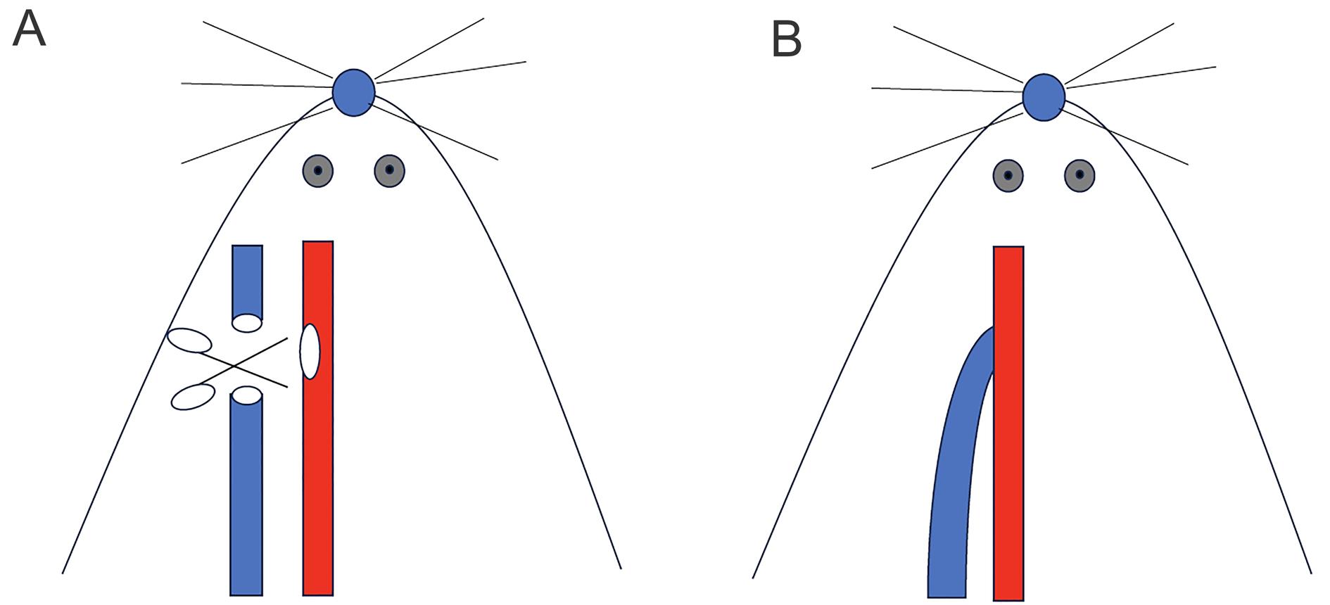
Figure 1 Operation diagram of autologous arteriovenous fistula in mice
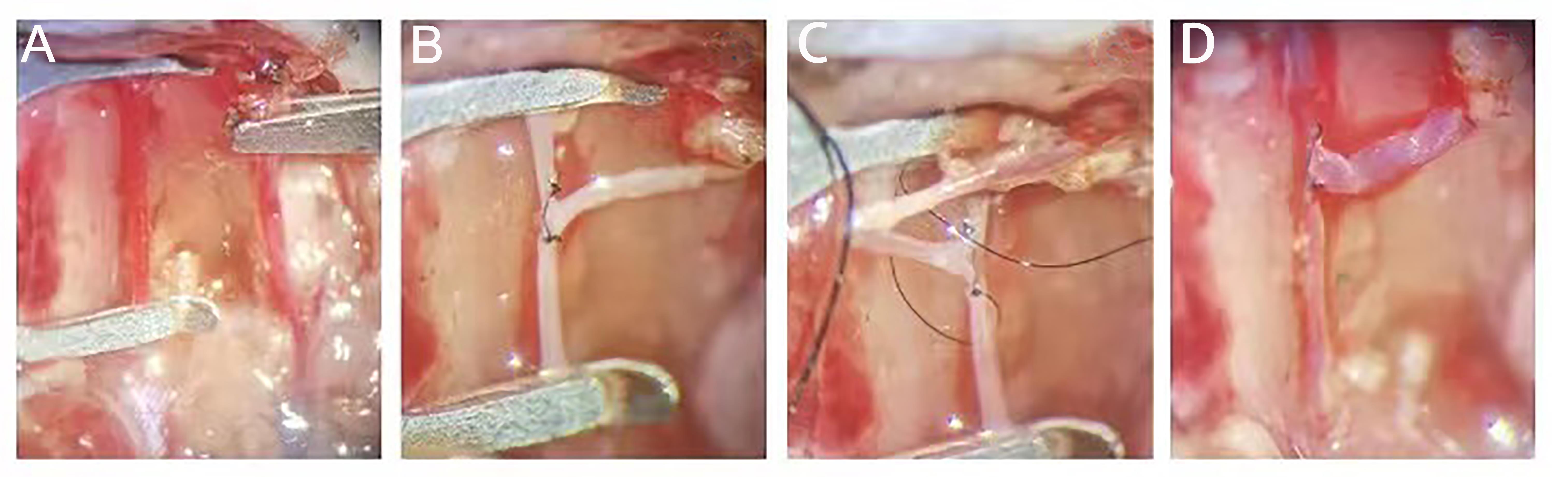
Figure 2 Procedures of end-to-side anastomosis of external jugular vein and common carotid artery in mice
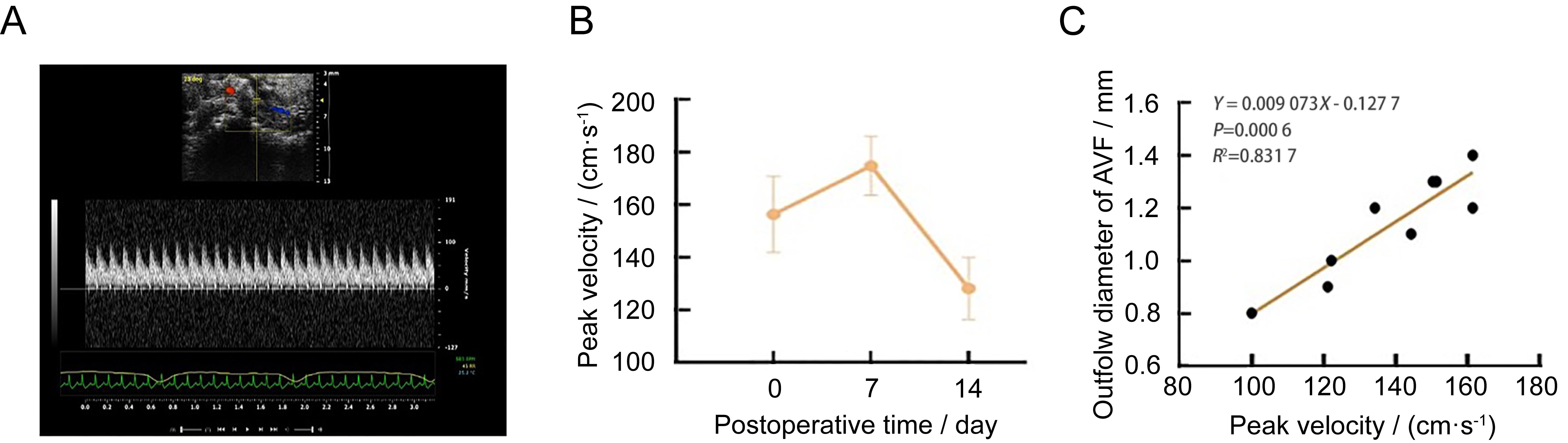
Figure 3 Noninvasive Doppler ultrasound assessment in mice autologous arteriovenous fistula (AVF) model
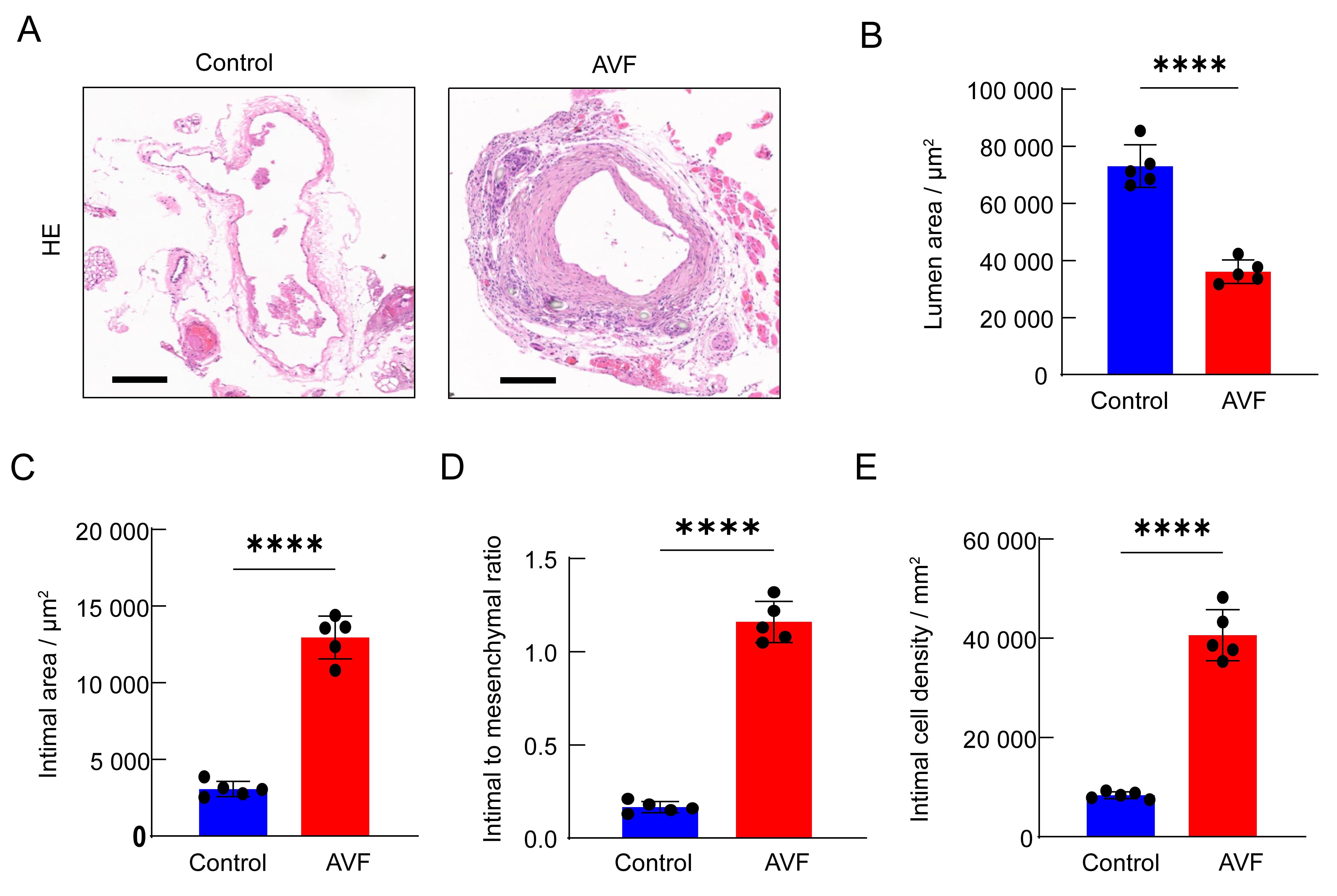
Figure 4 Analysis of vascular intima in mice autologous arteriovenous fistula (AVF) model
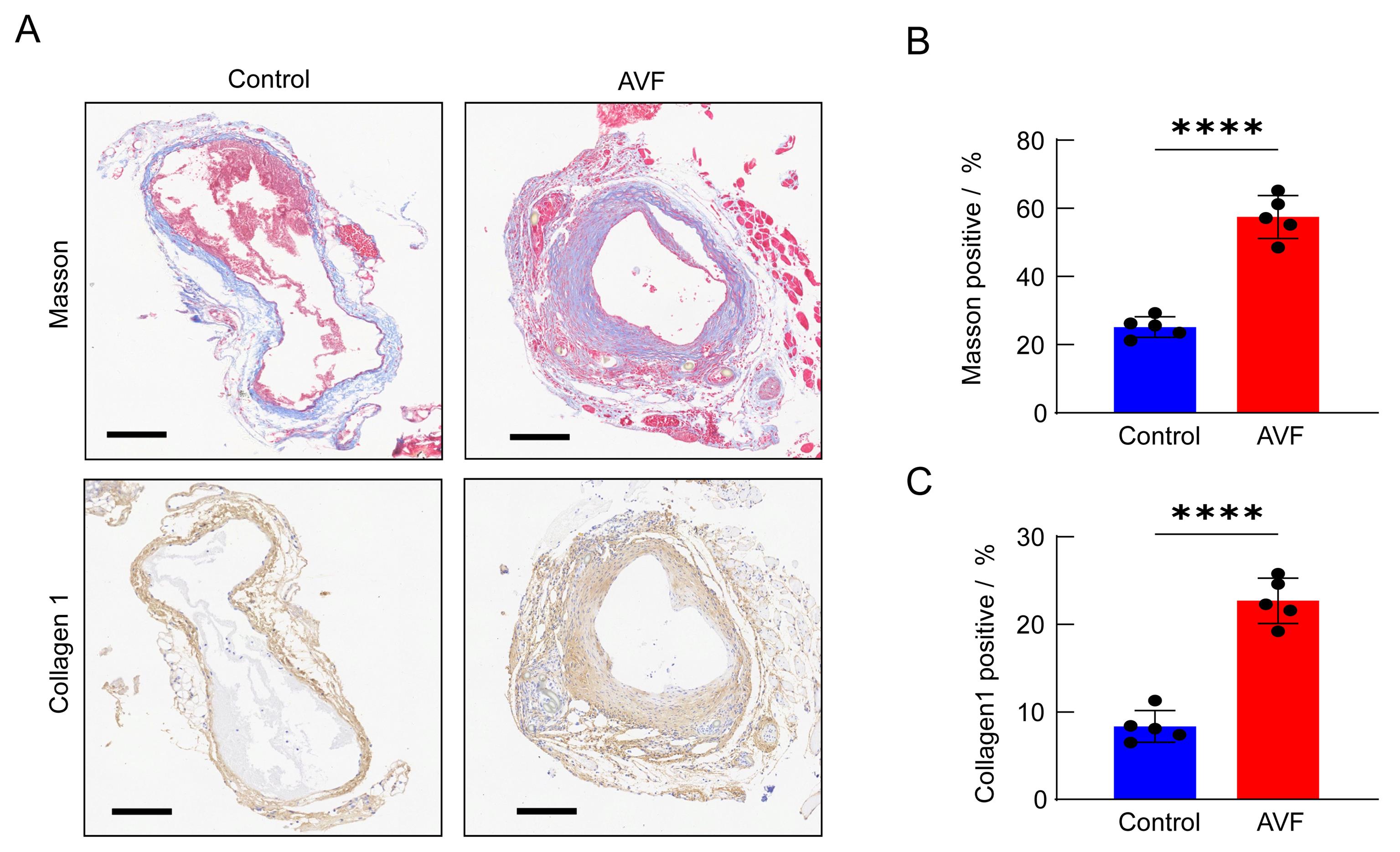
Figure 5 Analysis of collagen deposition in mice autologous arteriovenous fistula (AVF) model
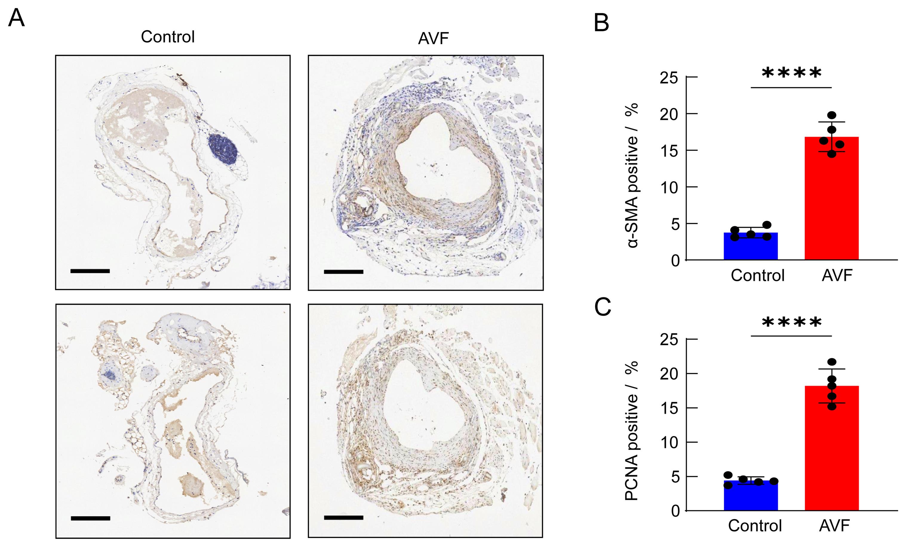
Figure 6 α-SMA and PCNA analysis in mice autologous arteriovenous fistula (AVF) model
| 1 | LIYANAGE T, NINOMIYA T, JHA V, et al. Worldwide access to treatment for end-stage kidney disease: a systematic review[J]. Lancet, 2015, 385(9981):1975-1982. DOI:10.1016/S0140-6736(14)61601-9 . |
| 2 | ROOIJENS P P G M, TORDOIR J H M, STIJNEN T, et al. Radiocephalic wrist arteriovenous fistula for hemodialysis: meta-analysis indicates a high primary failure rate[J]. Eur J Vasc Endovasc Surg, 2004, 28(6):583-589. DOI: 10.1016/j.ejvs.2004.08.014 . |
| 3 | HUIJBREGTS H J T, BOTS M L, WITTENS C H A, et al. Hemodialysis arteriovenous fistula patency revisited: results of a prospective, multicenter initiative[J]. Clin J Am Soc Nephrol, 2008, 3(3):714-719. DOI: 10.2215/CJN.02950707 . |
| 4 | AL-JAISHI A A, OLIVER M J, THOMAS S M, et al. Patency rates of the arteriovenous fistula for hemodialysis: a systematic review and meta-analysis[J]. Am J Kidney Dis, 2014, 63(3):464-478. DOI: 10.1053/j.ajkd.2013.08.023 . |
| 5 | WASSE H, HUANG R, NAQVI N, et al. Inflammation, oxidation and venous neointimal hyperplasia precede vascular injury from AVF creation in CKD patients[J]. J Vasc Access, 2012, 13(2):168-174. DOI: 10.5301/jva.5000024 . |
| 6 | ROY-CHAUDHURY P, WANG Y, KRISHNAMOORTHY M, et al. Cellular phenotypes in human stenotic lesions from haemodialysis vascular access[J]. Nephrol Dial Transplant, 2009, 24(9):2786-2791. DOI: 10.1093/ndt/gfn708 . |
| 7 | ROY-CHAUDHURY P, AREND L, ZHANG J H, et al. Neointimal hyperplasia in early arteriovenous fistula failure[J]. Am J Kidney Dis, 2007, 50(5):782-790. DOI: 10.1053/j.ajkd. 2007. 07. 019 . |
| 8 | YANG B X, SHERGILL U, FU A A, et al. The mouse arteriovenous fistula model[J]. J Vasc Interv Radiol, 2009, 20(7):946-950. DOI: 10.1016/j.jvir.2009.03.044 . |
| 9 | YAMAMOTO K, PROTACK C D, TSUNEKI M, et al. The mouse aortocaval fistula recapitulates human arteriovenous fistula maturation[J]. Am J Physiol Heart Circ Physiol, 2013, 305(12): H1718-H1725. DOI: 10.1152/ajpheart.00590.2013 . |
| 10 | LIANG M, WANG Y, LIANG A L, et al. Migration of smooth muscle cells from the arterial anastomosis of arteriovenous fistulas requires Notch activation to form neointima[J]. Kidney Int, 2015, 88(3):490-502. DOI: 10.1038/ki.2015.73 . |
| 11 | MISRA S, KILARI S, YANG B X, et al. Anti human CX3CR1 VHH molecule attenuates venous neointimal hyperplasia of arteriovenous fistula in mouse model[J]. J Am Soc Nephrol, 2021, 32(7):1630-1648. DOI: 10.1681/ASN.2020101458 . |
| 12 | CAI C Q, KILARI S, ZHAO C L, et al. Therapeutic effect of adipose derived mesenchymal stem cell transplantation in reducing restenosis in a murine angioplasty model[J]. J Am Soc Nephrol, 2020, 31(8):1781-1795. DOI: 10.1681/ASN. 2019101042 . |
| 13 | ROY-CHAUDHURY P, KHAN R, CAMPOS B, et al. Pathogenetic role for early focal macrophage infiltration in a pig model of arteriovenous fistula (AVF) stenosis[J]. J Vasc Access, 2014, 15(1):25-28. DOI: 10.5301/jva.5000151 . |
| 14 | CHAN J S, CAMPOS B, WANG Y, et al. Proliferation patterns in a pig model of AV fistula stenosis: can we translate biology into novel therapies?[J]. Semin Dial, 2014, 27(6):626-632. DOI: 10.1111/sdi.12240 . |
| 15 | KOKOZIDOU M, KATSARGYRIS A, VERHOEVEN E L G, et al. Vascular access animal models used in research[J]. Ann Anat Anat Anz, 2019, 225:65-75. DOI: 10.1016/j.aanat.2019.06.002 . |
| 16 | FLORESCU M C, FOSTER K W, SACKS A R, et al. Sheep model of hemodialysis arteriovenous fistula using superficial veins[J]. Semin Dial, 2015, 28(6):687-691. DOI: 10.1111/sdi.12407 . |
| 17 | CASTIER Y, LEHOUX S, HU Y, et al. Characterization of neointima lesions associated with arteriovenous fistulas in a mouse model[J]. Kidney Int, 2006, 70(2):315-320. DOI: 10.1038/sj.ki.5001569 . |
| 18 | CAI C Q, ZHAO C L, KILARI S, et al. Experimental murine arteriovenous fistula model to study restenosis after transluminal angioplasty[J]. Lab Anim, 2020, 49(11):320-334. DOI: 10.1038/s41684-020-00659-x . |
| [1] | PAN Linqin, DENG Xiangliang, LUO Yunxia. Advances in Integrative Translational Research on Animal Models of Ischemic Stroke in Traditional Chinese and Western Medicine [J]. Laboratory Animal and Comparative Medicine, 2026, 46(3): 344-356. |
| [2] | WANG Juan, XU Jiahui, TIAN Yunyuan, ZHANG Mengmeng, LI Min, WANG Siwang, LI Yao. Comparison and Behavioral Observation of Two Female Mice Models of Ulcerative Colitis [J]. Laboratory Animal and Comparative Medicine, 2026, 46(3): 332-343. |
| [3] | BU Yu, HOU Jinting, LI Yuanyuan, SHA Jingtao, XIE Chenlu, WANG Wengang, SUN Xingwei. A Review and Evaluation of Integrated Disease and Syndrome Animal Models for Hemorrhoids in Traditional Chinese and Western Medicine [J]. Laboratory Animal and Comparative Medicine, 2026, 46(3): 357-366. |
| [4] | LI Longxue, WAN Chongfan, ZHANG Qi, LEI Ruting, WANG Xiaoyue, CHENG Leyan, LAI Qi, LIU Ronghua, LIU Xuan, XU Tielong. Molecular Mechanisms of Qingfei Paidu Decoction in the Prevention and Treatment of Acute Lung Injury in Mice Based on miRNA Sequencing [J]. Laboratory Animal and Comparative Medicine, 2026, 46(3): 311-320. |
| [5] | AI Xiufeng, ZHANG Lizong, FANG Mingsun, LÜ Dongying, CHEN Chu, CAI Zhaowei, WANG Dejun. Analysis of Differences in the Intestinal Flora of Rats and Mice after Drinking Chlorinated Water Based on 16S rRNA Sequencing [J]. Laboratory Animal and Comparative Medicine, 2026, 46(3): 437-445. |
| [6] | Committee of Experts on Medical Animal Experiments, Chinese Research Hospital Association , Committee of Regenerative Medicine Branch, Chinese Medicinal Biotech Association , HAN Fabin, CHEN Lin, CHEN Zhiguo, LU Ming, LI Yingjun. Guidelines for Selecting Preclinical Animal Models for Drugs and Stem Cell Therapies for Parkinson Disease (2026 Edition) [J]. Laboratory Animal and Comparative Medicine, 2026, 46(2): 153-177. |
| [7] | LI Hui. Advances in Animal Models for Biolinguistic Research [J]. Laboratory Animal and Comparative Medicine, 2026, 46(2): 297-305. |
| [8] | RONG Wenshuang, NIU Yuanfei, LIU Meiting, YANG Mengyuan, CUI Shuang, MA Lina, FU Yao, WANG Lianmei, CAO Junling. Influence of Antigen Type on the Establishment of an Induced Sjögren Syndrome Mouse Model [J]. Laboratory Animal and Comparative Medicine, 2026, 46(2): 178-190. |
| [9] | WU Xianwen, LIU Lili, CHEN Ye, XU Guoheng. Optimization of Cage-Changing Intervals and Wood Shavings Usage for Mice During the Growth Phase in Breeding Systems [J]. Laboratory Animal and Comparative Medicine, 2026, 46(2): 251-260. |
| [10] | LIU Sai, FU Bin, LI Sidi, CHEN Zhida, ZHANG Yue, GUO Zhongkun, WANG Yongan, WANG Kezhou. Adra2a Regulates LPS-Induced Inflammation in Hepatocytes of Lbp-/- Mice via the MAPK Signaling Pathway [J]. Laboratory Animal and Comparative Medicine, 2026, 46(2): 212-221. |
| [11] | ZHAO He, ZHANG Tao, LI Li, XIAO Yuzhou, AN Xuefang, ZHANG Fan. Enhancement Effect of Porcine Inhibin Polyclonal Antibody on Superovulation in C57BL/6J Mice [J]. Laboratory Animal and Comparative Medicine, 2026, 46(2): 271-278. |
| [12] | CUI Changwan, LU Yiping, YU Miao, WANG Shuang, WU Si, SUN Zhengrong. Inhibitory Effect of Solute Carrier Family 7 Member 5 Inhibitor JPH203 on Renal Fibrosis Induced by Unilateral Ureteral Obstruction in Mice [J]. Laboratory Animal and Comparative Medicine, 2026, 46(2): 205-211. |
| [13] | YANG Lin, JIN Meng, WU Hanqing, LI Shun, ZHOU Xiaohui. Establishment and Preliminary Analysis of an AG6 Mouse Encephalopathy Model Induced by Vaccinia Virus Tiantan Strain Infection [J]. Laboratory Animal and Comparative Medicine, 2026, 46(1): 3-10. |
| [14] | LIAO Wangyue, LEI Shuang, LI Xuan, GUO Min, ZHOU Ruoran. Literature Analysis and Validity Assessment for Animal Models of Attention Deficit and Hyperactive Disorder [J]. Laboratory Animal and Comparative Medicine, 2026, 46(1): 66-80. |
| [15] | LIU Chang, XIANG Xuesong, HE Huihuang, CHEN Xiaoqing, QIU Wenhong. Establishment and Evaluation of an Oxidative Stress Model of Atopic Dermatitis Induced by 2,4-dinitrofluorobenzene [J]. Laboratory Animal and Comparative Medicine, 2026, 46(1): 46-54. |
| Viewed | ||||||
|
Full text |
|
|||||
|
Abstract |
|
|||||