
Laboratory Animal and Comparative Medicine ›› 2026, Vol. 46 ›› Issue (4): 476-486.DOI: 10.12300/j.issn.1674-5817.2025.181
• Animal Models of Human Diseases • Previous Articles
DU Hongzhi1( ), FAN Jin2,3, WEI Xiaolu2, WANG Lianmei2, LIU Yan2, CHEN Weiya2, CHEN Tengfei2, GAO Yunhang2, SONG Ling2, ZHANG Guangping2()(
 ), HOU Hongping2()
Received:2025-11-07
Revised:2026-04-23
Online:2026-08-25
Published:2026-08-22
Correspondence to:
ZHANG Guangping, HOU Hongping
CLC Number:
DU Hongzhi,FAN Jin,WEI Xiaolu,et al. Multimodal Assessment of a Hypoxic Pulmonary Hypertension Mouse Model[J]. Laboratory Animal and Comparative Medicine, 2026, 46(4): 476-486. DOI: 10.12300/j.issn.1674-5817.2025.181.
Add to citation manager EndNote|Ris|BibTeX
URL: https://www.slarc.org.cn/dwyx/EN/10.12300/j.issn.1674-5817.2025.181
评价维度 Evaluation dimensions | 评分标准/分 Scoring criteria/points | |||
|---|---|---|---|---|
| 0 | 1 | 2 | 3 | |
精神状态 Mental status | 精神饱满,反应灵敏,受刺激(如轻触)后迅速回避 | 精神稍萎靡,反应较迟钝,受刺激后缓慢回避 | 精神萎靡,嗜睡,刺激后无明显回避反应 | 精神极度萎靡,处于濒死状态,对刺激无反应 |
毛发状态 Hair condition | 毛发光洁顺滑,紧贴体表,无杂乱、脱毛 | 毛发略粗糙,少量杂乱,无脱毛 | 毛发干枯、杂乱,局部蓬松,伴少量脱毛 | 毛发严重干枯、蓬乱,大面积脱毛或结块 |
活动度 Activity level | 活动频繁,自主探索活跃,笼内穿梭自如 | 活动减少,多静坐,偶尔穿梭走动 | 活动极少,仅在摄食饮水时短暂活动,大部分时间蜷缩一隅 | 几乎无自主活动,蜷缩不动 |
呼吸状态 Respiratory status | 呼吸平稳,胸腹起伏均匀,无异常呼吸音 | 呼吸频率轻度加快,胸腹起伏略明显,无张口呼吸 | 呼吸急促,胸腹起伏剧烈,伴轻微张口呼吸或喘鸣 | 严重呼吸困难,张口呼吸,喘鸣明显,甚至出现发绀(口唇/肢端青紫) |
饮食与体况 Diet and body condition | 摄食、饮水正常,体重稳定增长,体形匀称 | 摄食、饮水量轻度下降,体重增长缓慢或无明显变化 | 摄食、饮水量显著下降,体重下降>5%,体型偏瘦 | 几乎不进食、饮水,体重下降>10%,体型极度消瘦,皮肤弹性差 |
Table 1 General condition scoring scale
评价维度 Evaluation dimensions | 评分标准/分 Scoring criteria/points | |||
|---|---|---|---|---|
| 0 | 1 | 2 | 3 | |
精神状态 Mental status | 精神饱满,反应灵敏,受刺激(如轻触)后迅速回避 | 精神稍萎靡,反应较迟钝,受刺激后缓慢回避 | 精神萎靡,嗜睡,刺激后无明显回避反应 | 精神极度萎靡,处于濒死状态,对刺激无反应 |
毛发状态 Hair condition | 毛发光洁顺滑,紧贴体表,无杂乱、脱毛 | 毛发略粗糙,少量杂乱,无脱毛 | 毛发干枯、杂乱,局部蓬松,伴少量脱毛 | 毛发严重干枯、蓬乱,大面积脱毛或结块 |
活动度 Activity level | 活动频繁,自主探索活跃,笼内穿梭自如 | 活动减少,多静坐,偶尔穿梭走动 | 活动极少,仅在摄食饮水时短暂活动,大部分时间蜷缩一隅 | 几乎无自主活动,蜷缩不动 |
呼吸状态 Respiratory status | 呼吸平稳,胸腹起伏均匀,无异常呼吸音 | 呼吸频率轻度加快,胸腹起伏略明显,无张口呼吸 | 呼吸急促,胸腹起伏剧烈,伴轻微张口呼吸或喘鸣 | 严重呼吸困难,张口呼吸,喘鸣明显,甚至出现发绀(口唇/肢端青紫) |
饮食与体况 Diet and body condition | 摄食、饮水正常,体重稳定增长,体形匀称 | 摄食、饮水量轻度下降,体重增长缓慢或无明显变化 | 摄食、饮水量显著下降,体重下降>5%,体型偏瘦 | 几乎不进食、饮水,体重下降>10%,体型极度消瘦,皮肤弹性差 |
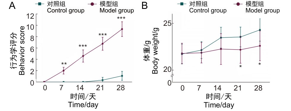
Figure 1 General condition scores (A) and body weight changes (B) of mice in the control and model groups
指标 Parameters | 对照组 Control group | 模型组 Model group |
|---|---|---|
| RVWT δ/mm | 0.35±0.02 | 0.50±0.10** |
| RVAPD l/mm | 1.23±0.13 | 1.59±0.09*** |
| MPAD l/mm | 1.45±0.10 | 1.69±0.10*** |
| Vpa /(mm·s-1) | 694.33±41.70 | 579.48±41.43*** |
| RVTD l/mm | 2.39±0.11 | 3.07±0.15*** |
| RVLD l/mm | 3.29±0.29 | 3.87±0.30** |
| TAPSV /(mm·s-1) | 63.84±9.02 | 38.90±8.90*** |
| TAPSE l/mm | 1.17±0.11 | 0.69±0.24*** |
Table 2 Echocardiograms parameters in mice of the control and model groups
指标 Parameters | 对照组 Control group | 模型组 Model group |
|---|---|---|
| RVWT δ/mm | 0.35±0.02 | 0.50±0.10** |
| RVAPD l/mm | 1.23±0.13 | 1.59±0.09*** |
| MPAD l/mm | 1.45±0.10 | 1.69±0.10*** |
| Vpa /(mm·s-1) | 694.33±41.70 | 579.48±41.43*** |
| RVTD l/mm | 2.39±0.11 | 3.07±0.15*** |
| RVLD l/mm | 3.29±0.29 | 3.87±0.30** |
| TAPSV /(mm·s-1) | 63.84±9.02 | 38.90±8.90*** |
| TAPSE l/mm | 1.17±0.11 | 0.69±0.24*** |
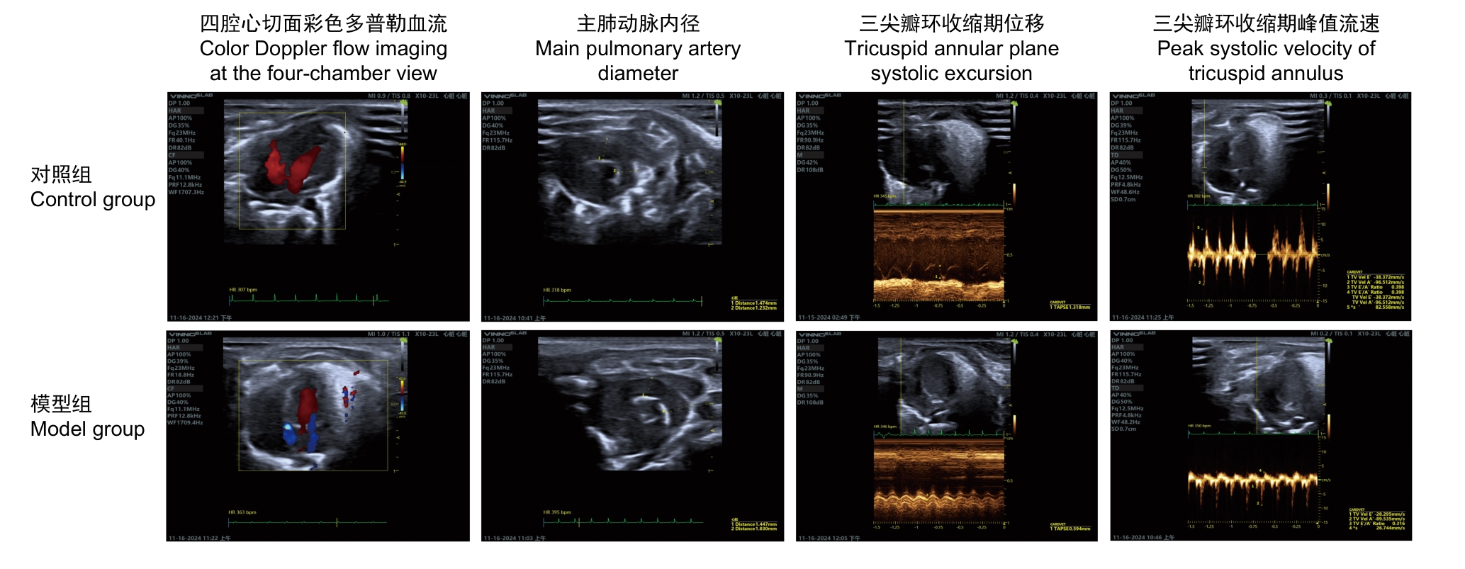
Figure 2 Echocardiograms of control and model mice
检测指标 Measured parameters | 对照组 Control group | 模型组 Model group | |
|---|---|---|---|
右心室收缩压 Right ventricular systolic pressure | RVSP p/mmHg | 4.76±0.95 | 9.46±0.75*** |
血气指标 Blood gas indicators | pH | 7.037±0.089 | 6.899±0.045** |
| PCO2p/mmHg | 67.9±4.5 | 57.6±11.6 | |
| PO2p/mmHg | 108.0±4.0 | 85.3±4.1*** | |
| SO2φ/% | 94.2±1.6 | 85.5±3.8*** | |
| HCO3-c/(mmol·L-1) | 18.0±4.0 | 10.8±1.7** | |
| TCO2 c/(mmol·L-1) | 20.0±3.8 | 12.0±2.1** | |
| a/A | 1 | 1 | |
| Lac c/(mmol·L-1) | 6.59±0.63 | 9.51±1.13*** | |
| WBC Ccell /(109·L-1) | 5.12±0.26 | 7.11±0.81*** | |
血液学指标 Hematological indicators | RBC Ccell /(1012·L-1) | 10.46±0.29 | 11.31±0.40** |
| PCV φ/% | 47.03±0.75 | 49.57±1.30** | |
| HGB ρ/(g·L-1) | 157.33±1.37 | 169.00±3.46*** | |
| PLT Ccell /(109·L-1) | 535.67±276.84 | 1 229.67±90.20*** | |
| RDW-SD/fL | 23.4±0.5 | 26.1±0.2*** | |
| RDW-CV/% | 19.3±0.3 | 19.9±0.3** | |
| RDW φ/fL | 7.53±0.51 | 8.17±0.40* | |
| NEUT Ccell /(109·L-1) | 0.83±0.07 | 1.46±0.32** | |
| LYMPH Ccell /(109·L-1) | 5.39±1.21 | 3.84±0.44* | |
| RET Ccell /(109·L-1) | 3.62±0.27 | 2.06±0.31*** | |
Table 3 Results of blood gas and hematological tests in mice of the control and model groups
检测指标 Measured parameters | 对照组 Control group | 模型组 Model group | |
|---|---|---|---|
右心室收缩压 Right ventricular systolic pressure | RVSP p/mmHg | 4.76±0.95 | 9.46±0.75*** |
血气指标 Blood gas indicators | pH | 7.037±0.089 | 6.899±0.045** |
| PCO2p/mmHg | 67.9±4.5 | 57.6±11.6 | |
| PO2p/mmHg | 108.0±4.0 | 85.3±4.1*** | |
| SO2φ/% | 94.2±1.6 | 85.5±3.8*** | |
| HCO3-c/(mmol·L-1) | 18.0±4.0 | 10.8±1.7** | |
| TCO2 c/(mmol·L-1) | 20.0±3.8 | 12.0±2.1** | |
| a/A | 1 | 1 | |
| Lac c/(mmol·L-1) | 6.59±0.63 | 9.51±1.13*** | |
| WBC Ccell /(109·L-1) | 5.12±0.26 | 7.11±0.81*** | |
血液学指标 Hematological indicators | RBC Ccell /(1012·L-1) | 10.46±0.29 | 11.31±0.40** |
| PCV φ/% | 47.03±0.75 | 49.57±1.30** | |
| HGB ρ/(g·L-1) | 157.33±1.37 | 169.00±3.46*** | |
| PLT Ccell /(109·L-1) | 535.67±276.84 | 1 229.67±90.20*** | |
| RDW-SD/fL | 23.4±0.5 | 26.1±0.2*** | |
| RDW-CV/% | 19.3±0.3 | 19.9±0.3** | |
| RDW φ/fL | 7.53±0.51 | 8.17±0.40* | |
| NEUT Ccell /(109·L-1) | 0.83±0.07 | 1.46±0.32** | |
| LYMPH Ccell /(109·L-1) | 5.39±1.21 | 3.84±0.44* | |
| RET Ccell /(109·L-1) | 3.62±0.27 | 2.06±0.31*** | |
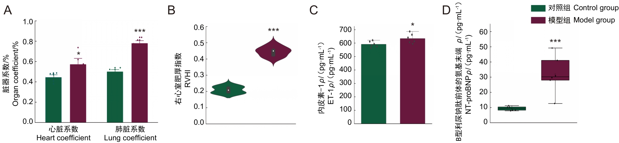
Figure 3 Organ coefficient (A), right ventricular hypertrophy index (B) and endothelin-1 and N-terminal pro-B-type natriuretic peptide (C-D) in lung tissue of control and model mice
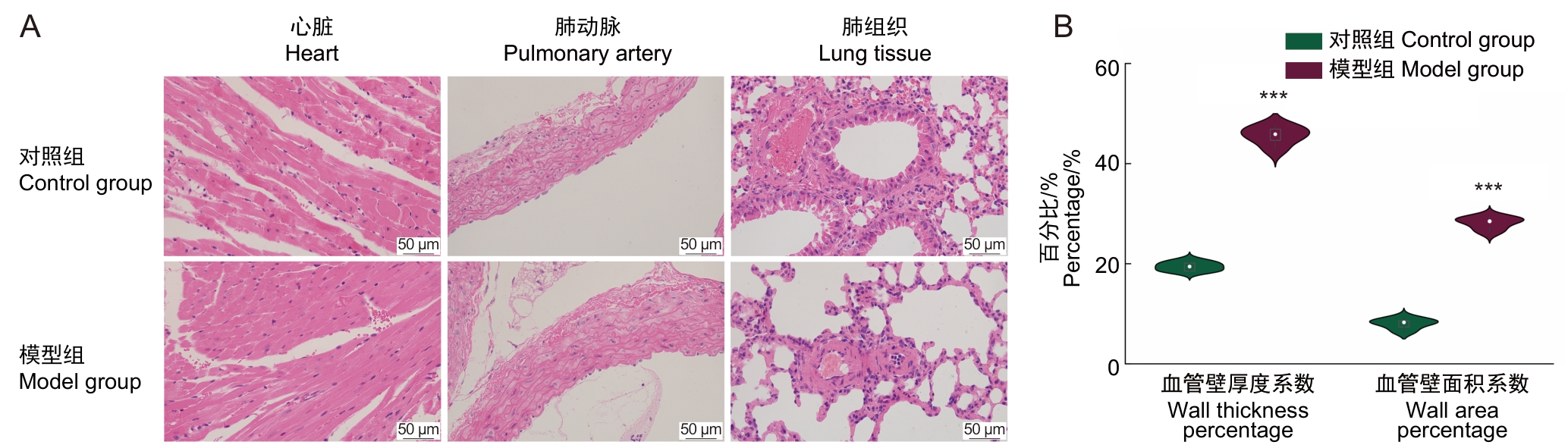
Figure 4 Histopathological observations and pulmonary vascular parameters in mice of the control and model groups

Figure 5 Heatmap of correlation analysis among various indicators in the model group mice
| [1] | POCH D, MANDEL J. Pulmonary hypertension[J]. Ann Intern Med, 2021, 174(4): ITC49-ITC64. DOI: 10.7326/aitc202104200 . |
| [2] | MAI A S, LIM O Z H, HO Y J, et al. Prevalence, risk factors and intervention for depression and anxiety in pulmonary hypertension: a systematic review and meta-analysis[J]. Front Med, 2022, 9: 765461. DOI: 10.3389/fmed.2022.765461 . |
| [3] | KOVACS G, BARTOLOME S, DENTON C P, et al. Definition, classification and diagnosis of pulmonary hypertension[J]. Eur Respir J, 2024, 64(4): 2401324. DOI: 10.1183/13993003. 01324-2024 . |
| [4] | 田婷, 徐磊. 活性氧在缺氧性肺动脉高压发病机制中的作用[J]. 心脏杂志, 2025, 37(1): 99-103. DOI: 10.12125/j.chj.202302064 . |
| TIAN T, XU L. Role of reactive oxygen species in pathogenesis of hypoxic pulmonary hypertension[J]. Chin Heart J, 2025, 37(1): 99-103. DOI: 10.12125/j.chj.202302064 . | |
| [5] | 张学佳, 李天骐, 李平, 等. 缺氧诱导BMPR2突变大鼠左心室损伤[J]. 中国分子心脏病学杂志, 2024, 24(3): 6128-6135. DOI: 10.16563/j.cnki.1671-6272.2024.06.010 . |
| ZHANG X J, LI T Q, LI P, et al. Hypoxia induced left ventricular injury in BMPR2 mutant rats[J]. Mol Cardiol China, 2024, 24(3): 6128-6135. DOI: 10.16563/j.cnki.1671-6272.2024.06.010 . | |
| [6] | WAN J J, YI J, WANG F Y, et al. Expression and regulation of HIF-1a in hypoxic pulmonary hypertension: Focus on pathological mechanism and Pharmacological Treatment[J]. Int J Med Sci, 2024, 21(1): 45-60. DOI: 10.7150/ijms.88216 . |
| [7] | YE Y, XU Q Y, WUREN T N. Inflammation and immunity in the pathogenesis of hypoxic pulmonary hypertension[J]. Front Immunol, 2023, 14: 1162556. DOI: 10.3389/fimmu.2023.1162556 . |
| [8] | GELZINIS T A. Pulmonary hypertension in 2021: part Ⅰ—definition, classification, pathophysiology, and presentation[J]. J Cardiothorac Vasc Anesth, 2022, 36(6): 1552-1564. DOI: 10.1053/j.jvca.2021.06.036 . |
| [9] | AHMAD K, KHANGOORA V, NATHAN S D. Lung disease-related pulmonary hypertension[J]. Cardiol Clin, 2022, 40(1): 77-88. DOI: 10.1016/j.ccl.2021.08.005 . |
| [10] | HUMBERT M, KOVACS G, HOEPER M M, et al. 2022 ESC/ERS Guidelines for the diagnosis and treatment of pulmonary hypertension[J]. Eur Heart J, 2022, 43(38): 3618-3731. DOI: 10.1093/eurheartj/ehac237 . |
| [11] | 肖家旺, 王建铭, 庚靖淞, 等. 肺动脉高压患者右心房压与超声心动图参数的相关性[J]. 心脏杂志, 2025, 37(3): 304-309. DOI: 10.12125/j.chj.202402019 . |
| XIAO J W, WANG J M, GENG J S, et al. Correlation between right atrial pressure and echocardiographic parameters and its influencing factors in patients with pulmonary arterial hypertension[J]. Chin Heart J, 2025, 37(3): 304-309. DOI: 10.12125/j.chj.202402019 . | |
| [12] | 吴道雄, 李妍锦, 王玉明, 等. 肺动脉高压早期诊断的生物标志物最新研究进展[J]. 现代检验医学杂志, 2025, 40(1): 208-212. DOI: 10.3969/j.issn.1671-7414.2025.01.040 . |
| WU D X, LI Y J, WANG Y M, et al. Recent progress in biomarkers for the early diagnosis of pulmonary hypertension[J]. J Mod Lab Med, 2025, 40(1): 208-212. DOI: 10.3969/j.issn.1671-7414.2025.01.040 . | |
| [13] | 谢锦程, 肖梦媛, 邓少东, 等. 肺动脉高压实验模型的研究进展[J]. 中国比较医学杂志, 2023, 33(9): 79-89. DOI: 10.3969/j.issn.1671-7856.2023.09.010 . |
| XIE J C, XIAO M Y, DENG S D, et al. Research progress in experimental models of pulmonary arterial hypertension[J]. Chin J Comp Med, 2023, 33(9): 79-89. DOI: 10.3969/j.issn.1671-7856.2023.09.010 . | |
| [14] | 叶芳, 叶玲燕, 赵素玉, 等. 肺动脉高压患者肝中静脉血流频谱参数、三尖瓣环收缩期位移与右心功能的相关性分析[J]. 临床超声医学杂志, 2022, 24(9): 691-695. DOI: 10.3969/j.issn.1008-6978.2022.09.012 . |
| YE F, YE L Y, ZHAO S Y, et al. Correlation analysis of middle hepatic vein blood flow spectrum parameters, tricuspid annular plane systolic excursion and right ventricular function in patients with pulmonary hypertension[J]. J Clin Ultrasound Med, 2022, 24(9): 691-695. DOI: 10.3969/j.issn.1008-6978.2022.09.012 . | |
| [15] | 胡慧敏, 李一丹, 郭迪晨, 等. 超声心动图评估西那卡塞对动脉性肺动脉高压大鼠右心功能影响的实验研究[J]. 中华超声影像学杂志, 2023, 32(1): 79-85. |
| HU H M, LI Y D, GUO D C, et al. Assessment of the effect of cinacalcet on right ventricular function in rats with pulmonary arterial hypertension by echocardiography: an experimental study[J]. Chin J Ultrason, 2023, 32(1): 79-85. | |
| [16] | 孙行行, 吴文汇, 袁平, 等. 三尖瓣环收缩期位移评价先天性心脏病相关肺动脉高压疾病严重程度的价值[J]. 同济大学学报(医学版), 2018, 39(4): 56-60, 65. DOI: 10.16118/j.1008-0392.2018.04.011 . |
| SUN X X, WU W H, YUAN P, et al. Tricuspid annular plane systolic excursion for evaluation of severity of pulmonary arterial hypertension in patients with congenital heart disease[J]. J Tongji Univ (Med Sci), 2018, 39(4): 56-60, 65. DOI: 10.16118/j.1008-0392.2018.04.011 . | |
| [17] | MAUERMANN E, VANDENHEUVEL M, FRANÇOIS K, et al. Right ventricular systolic assessment by transesophageal versus transthoracic echocardiography: displacement, velocity, and myocardial deformation[J]. J Cardiothorac Vasc Anesth, 2020, 34(8): 2152-2161. DOI: 10.1053/j.jvca.2020.03.009 . |
| [18] | YUCHI Y, SUZUKI R, TESHIMA T, et al. Utility of tricuspid annular plane systolic excursion normalized by right ventricular size indices in dogs with postcapillary pulmonary hypertension[J]. J Vet Intern Med, 2021, 35(1): 107-119. DOI: 10.1111/jvim.15984 . |
| [19] | 郭丽敏, 武晓峰, 张淑莹, 等. 超声心动图评估野百合碱诱导的大鼠肺动脉高压[J]. 中华超声影像学杂志, 2019, 28(7): 629-636. DOI: 10.3760/cma.j.issn.1004-4477.2019.07.011 . |
| GUO L M, WU X F, ZHANG S Y, et al. Echocardiographic evaluation of monocrotaline induced pulmonary arterial hypertension in rats[J]. Chin J Ultrason, 2019, 28(7): 629-636. DOI: 10.3760/cma.j.issn.1004-4477.2019.07.011 . | |
| [20] | PAKHOMOV N V, KOSTYUNINA D S, MACORI G, et al. High-soluble-fiber diet attenuates hypoxia-induced vascular remodeling and the development of hypoxic pulmonary hypertension[J]. Hypertension, 2023, 80(11): 2372-2385. DOI: 10.1161/hypertensionaha.123.20914 . |
| [21] | BAČÁKOVÁ L, SEDLÁŘ A, MUSÍLKOVÁ J, et al. Mechanisms controlling the behavior of vascular smooth muscle cells in hypoxic pulmonary hypertension[J]. Physiol Res, 2024: S569-S596. DOI: 10.33549/physiolres.935394 . |
| [22] | 陈迪, 袁天翊, 陈俞材, 等. DL0805-2防治野百合碱诱导的大鼠肺动脉高压的药效评价[J]. 药学学报, 2021, 56(1): 208-216. DOI: 10.16438/j.0513-4870.2020-0981 . |
| CHEN D, YUAN T Y, CHEN Y C, et al. Evaluation of the preventive effect of DL0805-2 against monocrotaline induced rat pulmonary arterial hypertension[J]. Acta Pharm Sin, 2021, 56(1): 208-216. DOI: 10.16438/j.0513-4870.2020-0981 . | |
| [23] | 周英, 王乐, 李明霞. 缺氧性肺动脉高压新生儿血清HIF-1α、ET-1及Ca2+变化及意义[J]. 中国当代儿科杂志, 2011, 13(3): 181-184. |
| ZHOU Y, WANG L, LI M X. Serum levels of HIF-1α, ET-1 and Ca2+ in neonates with hypoxic pulmonary hypertension[J]. Chin J Contemp Pediatr, 2011, 13(3): 181-184. | |
| [24] | ZHANG T T, CUI B, DAI D Z, et al. Pharmacological efficacy of CPU 86017 on hypoxic pulmonary hypertension in rats: mediated by direct inhibition of calcium channels and antioxidant action, but indirect effects on the ET-1 pathway[J]. J Cardiovasc Pharmacol, 2005, 46(6): 727-734. DOI: 10.1097/01.fjc.0000184470.58047.79 . |
| [25] | MELLOR A, BOOS C, HOLDSWORTH D, et al. Cardiac biomarkers at high altitude[J]. High Alt Med Biol, 2014, 15(4): 452-458. DOI: 10.1089/ham.2014.1035 . |
| [26] | MENDIOLA E A, SILVA GONÇALVES BOS D DA, LEICHTER D M, et al. Right ventricular architectural remodeling and functional adaptation in pulmonary hypertension[J]. Circ Heart Fail, 2023, 16(2): e009768. DOI: 10.1161/circheartfailure. 122.009768 . |
| [1] | WANG Juan, XU Jiahui, TIAN Yunyuan, ZHANG Mengmeng, LI Min, WANG Siwang, LI Yao. Comparison and Behavioral Observation of Two Female Mice Models of Ulcerative Colitis [J]. Laboratory Animal and Comparative Medicine, 2026, 46(3): 332-343. |
| [2] | LI Longxue, WAN Chongfan, ZHANG Qi, LEI Ruting, WANG Xiaoyue, CHENG Leyan, LAI Qi, LIU Ronghua, LIU Xuan, XU Tielong. Molecular Mechanisms of Qingfei Paidu Decoction in the Prevention and Treatment of Acute Lung Injury in Mice Based on miRNA Sequencing [J]. Laboratory Animal and Comparative Medicine, 2026, 46(3): 311-320. |
| [3] | AI Xiufeng, ZHANG Lizong, FANG Mingsun, LÜ Dongying, CHEN Chu, CAI Zhaowei, WANG Dejun. Analysis of Differences in the Intestinal Flora of Rats and Mice after Drinking Chlorinated Water Based on 16S rRNA Sequencing [J]. Laboratory Animal and Comparative Medicine, 2026, 46(3): 437-445. |
| [4] | RONG Wenshuang, NIU Yuanfei, LIU Meiting, YANG Mengyuan, CUI Shuang, MA Lina, FU Yao, WANG Lianmei, CAO Junling. Influence of Antigen Type on the Establishment of an Induced Sjögren Syndrome Mouse Model [J]. Laboratory Animal and Comparative Medicine, 2026, 46(2): 178-190. |
| [5] | LIU Sai, FU Bin, LI Sidi, CHEN Zhida, ZHANG Yue, GUO Zhongkun, WANG Yongan, WANG Kezhou. Adra2a Regulates LPS-Induced Inflammation in Hepatocytes of Lbp-/- Mice via the MAPK Signaling Pathway [J]. Laboratory Animal and Comparative Medicine, 2026, 46(2): 212-221. |
| [6] | ZHAO He, ZHANG Tao, LI Li, XIAO Yuzhou, AN Xuefang, ZHANG Fan. Enhancement Effect of Porcine Inhibin Polyclonal Antibody on Superovulation in C57BL/6J Mice [J]. Laboratory Animal and Comparative Medicine, 2026, 46(2): 271-278. |
| [7] | CUI Changwan, LU Yiping, YU Miao, WANG Shuang, WU Si, SUN Zhengrong. Inhibitory Effect of Solute Carrier Family 7 Member 5 Inhibitor JPH203 on Renal Fibrosis Induced by Unilateral Ureteral Obstruction in Mice [J]. Laboratory Animal and Comparative Medicine, 2026, 46(2): 205-211. |
| [8] | WU Xianwen, LIU Lili, CHEN Ye, XU Guoheng. Optimization of Cage-Changing Intervals and Wood Shavings Usage for Mice During the Growth Phase in Breeding Systems [J]. Laboratory Animal and Comparative Medicine, 2026, 46(2): 251-260. |
| [9] | XU Bo, CHEN Tairen, FANG Qian, WU Ji. Research on Spatiotemporal Gene Expression Profiles and Repair Mechanisms of Spinal Cord Compression and Hemisection Spinal Cord Injury Mouse Models [J]. Laboratory Animal and Comparative Medicine, 2026, 46(1): 32-45. |
| [10] | XU Yingtao, WANG Mengmeng, LIN Ping, CHI Haitao, WANG Yi, BAI Ying. Exosomes Treat Ischemic Stroke by Regulation of Ferroptosis Through the NRF2/SLC7A11/GPX4 Pathway in Mice [J]. Laboratory Animal and Comparative Medicine, 2026, 46(1): 20-31. |
| [11] | YANG Lin, JIN Meng, WU Hanqing, LI Shun, ZHOU Xiaohui. Establishment and Preliminary Analysis of an AG6 Mouse Encephalopathy Model Induced by Vaccinia Virus Tiantan Strain Infection [J]. Laboratory Animal and Comparative Medicine, 2026, 46(1): 3-10. |
| [12] | LIU Chang, XIANG Xuesong, HE Huihuang, CHEN Xiaoqing, QIU Wenhong. Establishment and Evaluation of an Oxidative Stress Model of Atopic Dermatitis Induced by 2,4-dinitrofluorobenzene [J]. Laboratory Animal and Comparative Medicine, 2026, 46(1): 46-54. |
| [13] | NIE Yongqiang, WANG Zhaoxia. Exploration and Practice of Building a One-Stop Service Platform for Gene-Edited Mice in University Animal Centers: A Case Study of Shanghai Jiao Tong University [J]. Laboratory Animal and Comparative Medicine, 2025, 45(5): 642-648. |
| [14] | WANG Chao, LI Shun, REN Xiaonan, YANG Hua, CHEN Lixiang, XU Chunhua, ZHOU Xiaohui. Aging Inhibits Memory Immune Response of CD8+ T Cells in Lungs of C57BL/6J Mice Against Influenza A (H1N1) Virus [J]. Laboratory Animal and Comparative Medicine, 2025, 45(5): 515-523. |
| [15] | LIU Yueqin, XUE Weiguo, WANG Shuyou, SHEN Yaohua, JIA Shuyong, WANG Guangjun, SONG Xiaojing. Observation of Digestive Tract Tissue Morphology in Mice Using Probe-Based Confocal Laser Endomicroscopy [J]. Laboratory Animal and Comparative Medicine, 2025, 45(4): 457-465. |
| Viewed | ||||||
|
Full text |
|
|||||
|
Abstract |
|
|||||
