
Laboratory Animal and Comparative Medicine ›› 2026, Vol. 46 ›› Issue (2): 178-190.DOI: 10.12300/j.issn.1674-5817.2025.175
• Animal Models of Human Diseases • Previous Articles Next Articles
RONG Wenshuang1,2,3, NIU Yuanfei4, LIU Meiting2,3, YANG Mengyuan5, CUI Shuang2,3, MA Lina6, FU Yao1, WANG Lianmei2,3( )(
)( ), CAO Junling1,6()()
Received:2025-10-24
Revised:2026-02-05
Online:2026-04-25
Published:2026-04-20
Contact:
WANG Lianmei, CAO Junling
CLC Number:
RONG Wenshuang,NIU Yuanfei,LIU Meiting,et al. Influence of Antigen Type on the Establishment of an Induced Sjögren Syndrome Mouse Model[J]. Laboratory Animal and Comparative Medicine, 2026, 46(2): 178-190. DOI: 10.12300/j.issn.1674-5817.2025.175.
Add to citation manager EndNote|Ris|BibTeX
URL: https://www.slarc.org.cn/dwyx/EN/10.12300/j.issn.1674-5817.2025.175
引物名称 Primer name | 引物序列 Primer sequence |
|---|---|
| m-SSA-F | 5'-GCTGGTCTCACAAAGATCTCCTCC-3' |
| m-SSA-R | 5'-TCTCAGCCTCCACAGAAAGCG-3' |
| m-SSB-F | 5'-ATGACTGCTTTGGAGGCCAAAATC-3' |
| m-SSB-R | 5'-TGTCAGCCGGTTTAGCCTGTTGAA-3' |
| m-β-Actin-F | 5'-AGAGGGAAATCGTGCGTGAC-3' |
| m-β-Actin-R | 5'-CAATAGTGATGACCTGGCCGT-3' |
Table 1 PCR primer sequences
引物名称 Primer name | 引物序列 Primer sequence |
|---|---|
| m-SSA-F | 5'-GCTGGTCTCACAAAGATCTCCTCC-3' |
| m-SSA-R | 5'-TCTCAGCCTCCACAGAAAGCG-3' |
| m-SSB-F | 5'-ATGACTGCTTTGGAGGCCAAAATC-3' |
| m-SSB-R | 5'-TGTCAGCCGGTTTAGCCTGTTGAA-3' |
| m-β-Actin-F | 5'-AGAGGGAAATCGTGCGTGAC-3' |
| m-β-Actin-R | 5'-CAATAGTGATGACCTGGCCGT-3' |
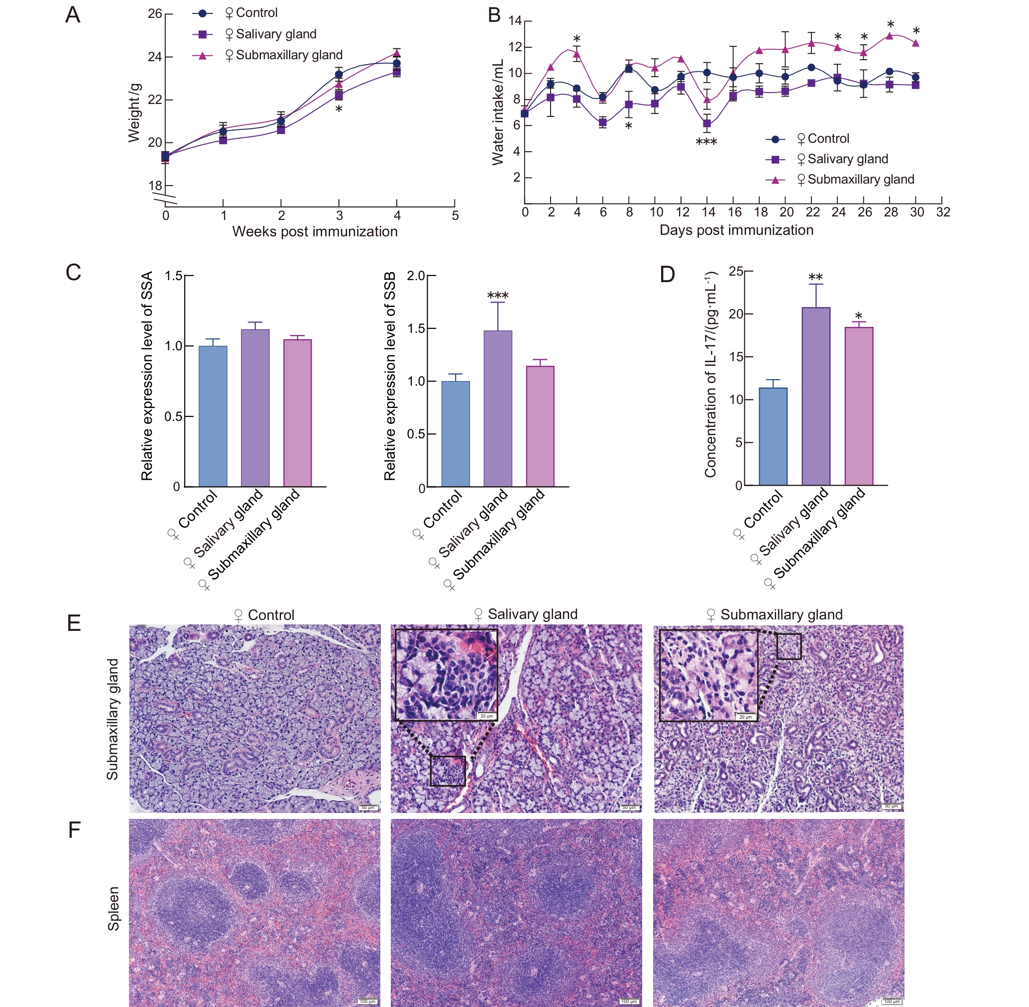
Figure 1 Sj?gren syndrome-related phenotypes in antigen-induced female C57BL/6J mice
组别 Group | 第0天唾液流量/ (mg·min-1) Day 0 salivary flow rate /(mg·min-1) | 第30天唾液流量/ (mg·min-1) Day 30 salivary flow rate /(mg·min-1) | 颌下腺指数 Submaxillary gland index | 胸腺指数 Thymus index | 脾指数 Spleen index |
|---|---|---|---|---|---|
PBS-佐剂雌性对照组 PBS-adjuvanted female control group | 6.60±1.11 | 14.77±1.04 | 0.28±0.03 | 0.26±0.04 | 0.44±0.04 |
唾液腺抗原雌性组 Salivary gland antigen female group | 6.28±1.10 | 13.10±1.05 | 0.29±0.03 | 0.20±0.02∗∗ | 0.51±0.13 |
颌下腺抗原雌性组 Submaxillary gland antigen female group | 6.14±1.16 | 10.52±1.06∗ | 0.30±0.03 | 0.24±0.04 | 0.50±0.06 |
Table 2 Salivary flow rate and organ indices in female C57BL/6J mice of each group
组别 Group | 第0天唾液流量/ (mg·min-1) Day 0 salivary flow rate /(mg·min-1) | 第30天唾液流量/ (mg·min-1) Day 30 salivary flow rate /(mg·min-1) | 颌下腺指数 Submaxillary gland index | 胸腺指数 Thymus index | 脾指数 Spleen index |
|---|---|---|---|---|---|
PBS-佐剂雌性对照组 PBS-adjuvanted female control group | 6.60±1.11 | 14.77±1.04 | 0.28±0.03 | 0.26±0.04 | 0.44±0.04 |
唾液腺抗原雌性组 Salivary gland antigen female group | 6.28±1.10 | 13.10±1.05 | 0.29±0.03 | 0.20±0.02∗∗ | 0.51±0.13 |
颌下腺抗原雌性组 Submaxillary gland antigen female group | 6.14±1.16 | 10.52±1.06∗ | 0.30±0.03 | 0.24±0.04 | 0.50±0.06 |
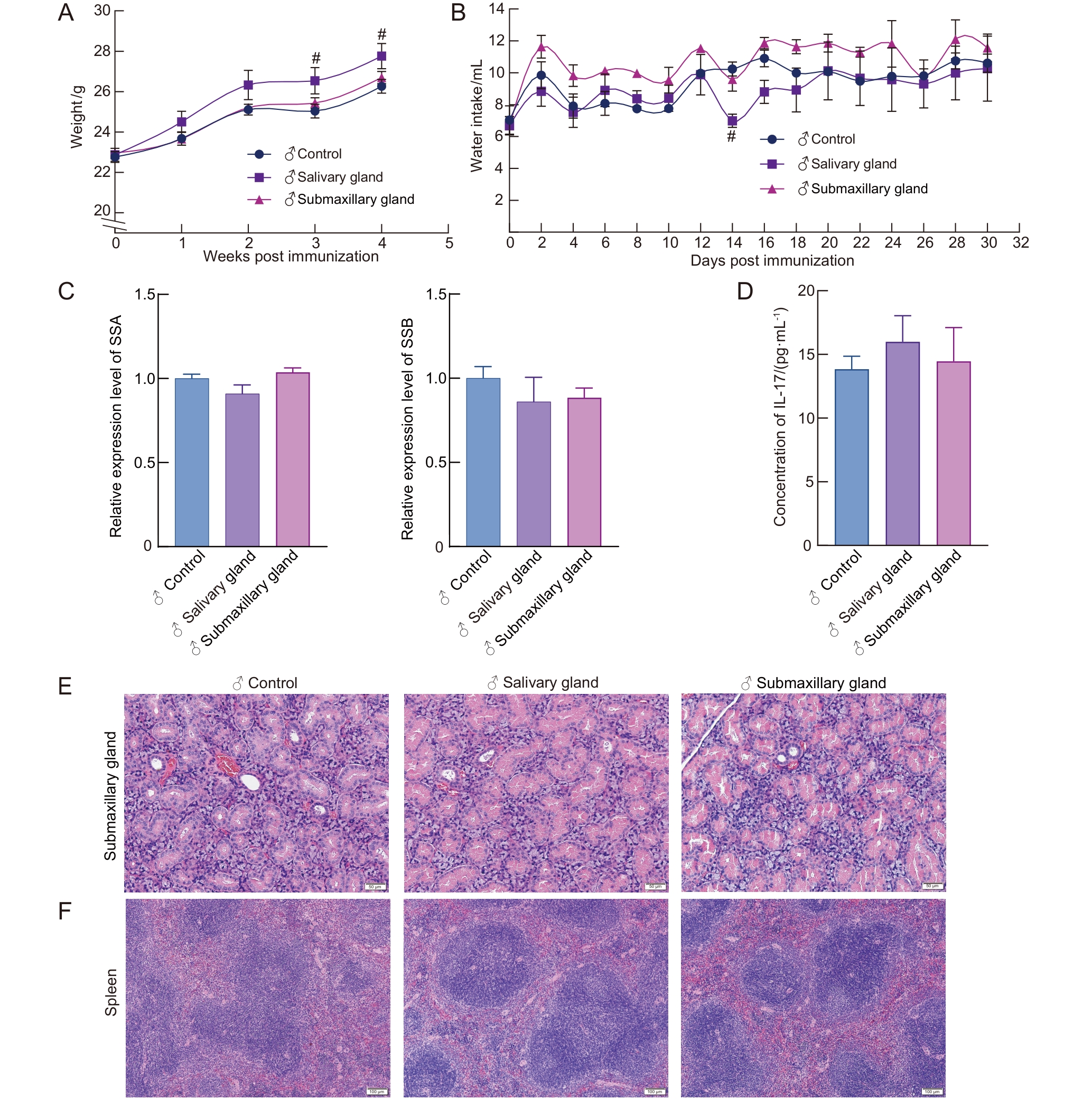
Figure 2 Sj?gren syndrome-related phenotypes in antigen-induced male C57BL/6J mice
组别 Group | 第0天唾液流量/ (mg·min-1) Day 0 salivary flow rate /(mg·min-1) | 第30天唾液流量/(mg·min-1) Day 30 salivary flow rate /(mg·min-1) | 颌下腺指数 Submaxillary gland index | 胸腺指数 Thymus index | 脾指数 Spleen index |
|---|---|---|---|---|---|
PBS-佐剂雄性对照组 PBS-adjuvanted male control group | 6.21±0.88 | 14.60±0.90 | 0.38±0.03 | 0.18±0.06 | 0.41±0.07 |
唾液腺抗原雄性组 Salivary gland antigen male group | 6.72±0.73 | 11.17±0.62 | 0.40±0.04 | 0.18±0.05 | 0.37±0.14 |
颌下腺抗原雄性组 Submaxillary gland antigen male group | 6.17±0.90 | 11.54±0.70 | 0.38±0.06 | 0.17±0.03 | 0.34±0.05 |
Table 3 Salivary flow rate and organ indices in male C57BL/6J mice of each group
组别 Group | 第0天唾液流量/ (mg·min-1) Day 0 salivary flow rate /(mg·min-1) | 第30天唾液流量/(mg·min-1) Day 30 salivary flow rate /(mg·min-1) | 颌下腺指数 Submaxillary gland index | 胸腺指数 Thymus index | 脾指数 Spleen index |
|---|---|---|---|---|---|
PBS-佐剂雄性对照组 PBS-adjuvanted male control group | 6.21±0.88 | 14.60±0.90 | 0.38±0.03 | 0.18±0.06 | 0.41±0.07 |
唾液腺抗原雄性组 Salivary gland antigen male group | 6.72±0.73 | 11.17±0.62 | 0.40±0.04 | 0.18±0.05 | 0.37±0.14 |
颌下腺抗原雄性组 Submaxillary gland antigen male group | 6.17±0.90 | 11.54±0.70 | 0.38±0.06 | 0.17±0.03 | 0.34±0.05 |
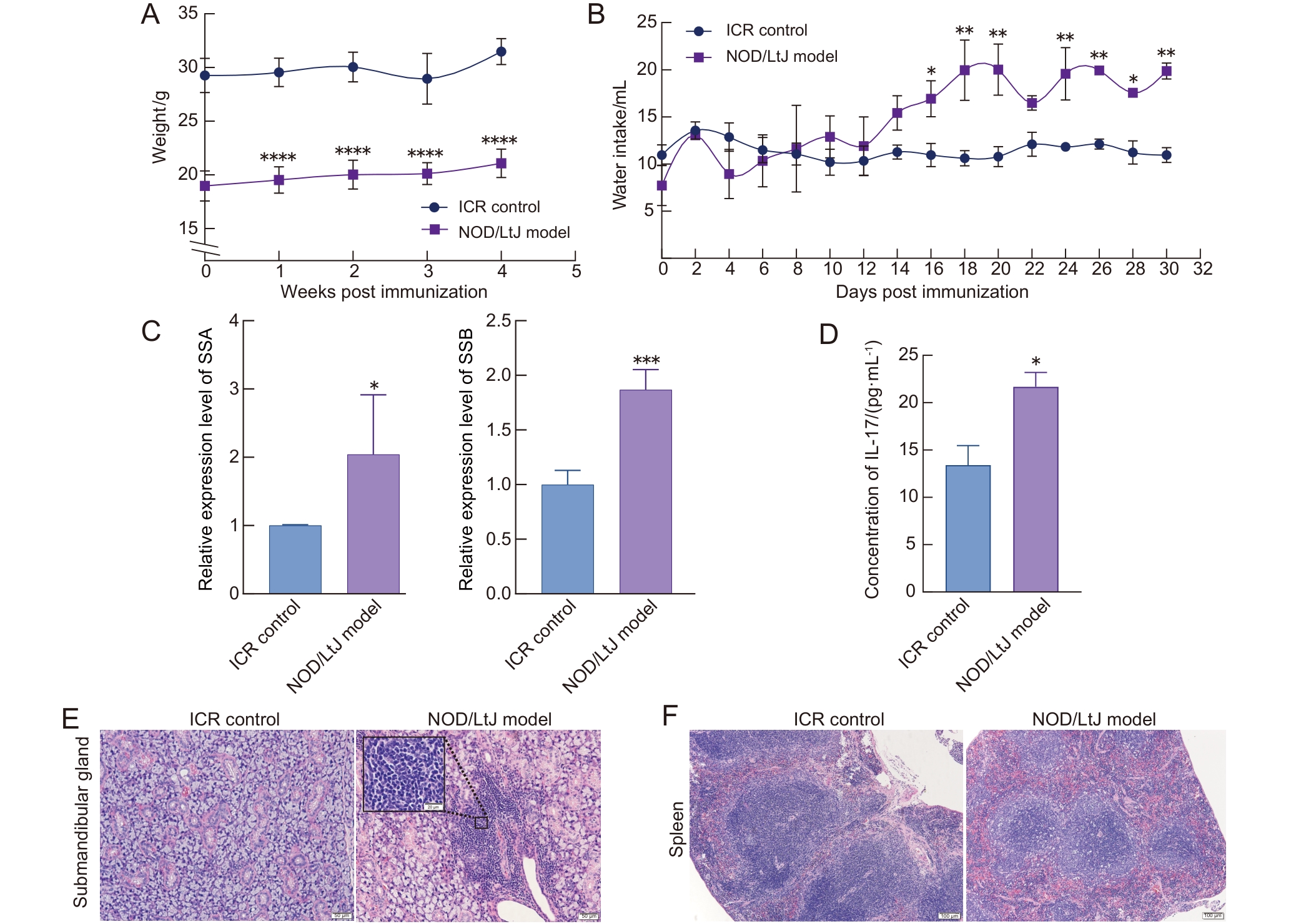
Figure 3 Sj?gren syndrome-related phenotypes in female NOD/LtJ model mice and ICR mice
组别 Group | 第0天唾液流量/(mg·min-1) Day 0 salivary flow rate /(mg·min-1) | 第30天唾液流量/(mg·min-1) Day 30 salivary flow rate / (mg·min-1) | 颌下腺指数 Submaxillary gland index | 胸腺指数 Thymus index | 脾指数 Spleen index |
|---|---|---|---|---|---|
| ICR mice | 8.37±0.46 | 15.85±1.25 | 0.39±0.01 | 0.13±0.03 | 0.39±0.08 |
| NOD/LtJ mice | 6.42±0.05∗ | 5.11±0.46∗∗ | 0.40±0.04 | 0.16±0.03∗ | 0.31±0.05 |
Table 4 Salivary flow rate and organ indices in female NOD/LtJ model mice and ICR mice
组别 Group | 第0天唾液流量/(mg·min-1) Day 0 salivary flow rate /(mg·min-1) | 第30天唾液流量/(mg·min-1) Day 30 salivary flow rate / (mg·min-1) | 颌下腺指数 Submaxillary gland index | 胸腺指数 Thymus index | 脾指数 Spleen index |
|---|---|---|---|---|---|
| ICR mice | 8.37±0.46 | 15.85±1.25 | 0.39±0.01 | 0.13±0.03 | 0.39±0.08 |
| NOD/LtJ mice | 6.42±0.05∗ | 5.11±0.46∗∗ | 0.40±0.04 | 0.16±0.03∗ | 0.31±0.05 |
| [1] | 刘维, 何东仪, 张文, 等. 干燥综合征中西医结合诊疗指南[J]. 中草药, 2025, 56(15): 5333-5346. DOI: 10.7501/j.issn.0253-2670.2025.15.001 . |
| LIU W, HE D Y, ZHANG W, et al. Guideline for diagnosis and treatment of Sjögren's syndrome with integrated traditional Chinese and Western medicine[J]. Chin Tradit Herb Drugs, 2025, 56(15): 5333-5346. DOI: 10.7501/j.issn.0253-2670.2025.15.001 . | |
| [2] | 张欢, 刘春红, 吴斌. 原发性干燥综合征的流行病学研究进展[J]. 现代预防医学, 2020, 47(16): 3056-3058, 3063. DOI: 10.20043/j.cnki.mpm.2020.16.041 . |
| ZHANG H, LIU C H, WU B. Advances in the epidemiology of primary Sjögren's syndrome[J]. Mod Prev Med, 2020, 47(16): 3056-3058, 3063. DOI: 10.20043/j.cnki.mpm.2020.16.041 . | |
| [3] | FAIRWEATHER D, BEETLER D J, MCCABE E J, et al. Mechanisms underlying sex differences in autoimmunity[J]. J Clin Investig, 2024, 134(18): e180076. DOI: 10.1172/jci180076 . |
| [4] | GAO Y Z, CHEN Y, ZHANG Z J, et al. Recent advances in mouse models of Sjögren's syndrome[J]. Front Immunol, 2020, 11: 1158. DOI: 10.3389/fimmu.2020.01158 . |
| [5] | JIANG T T, LIU X Q, WANG S M, et al. Paeoniflorin alleviated experimental Sjögren's syndrome by inhibiting NLRP3 inflammasome activation of submandibular gland cells via activating Nrf2/HO-1 pathway[J]. Free Radic Biol Med, 2025, 233: 355-364. DOI: 10.1016/j.freeradbiomed.2025.03.043 . |
| [6] | LIN X, RUI K, DENG J, et al. Th17 cells play a critical role in the development of experimental Sjögren's syndrome[J]. Ann Rheum Dis, 2015, 74(6): 1302-1310. DOI: 10.1136/annrheumdis-2013-204584 . |
| [7] | BAGAVANT H, DURSLEWICZ J, PYCLIK M, et al. Age-associated B cell infiltration in salivary glands represents a hallmark of Sjögren's-like disease in aging mice[J]. Geroscience, 2024, 46(6): 6085-6099. DOI: 10.1007/s11357-024-01159-3 . |
| [8] | XU J J, LIU O S, WANG D D, et al. In vivo generation of SSA/ro antigen-specific regulatory T cells improves experimental Sjögren's syndrome in mice[J]. Arthritis Rheumatol, 2022, 74(10): 1699-1705. DOI: 10.1002/art.42244 . |
| [9] | ARVIDSSON G, CZARNEWSKI P, JOHANSSON A, et al. Multimodal single-cell sequencing of B cells in primary Sjögren's syndrome[J]. Arthritis Rheumatol, 2024, 76(2): 255-267. DOI: 10.1002/art.42683 . |
| [10] | LEE A Y S, PUTTY T, LIN M W, et al. Isolated anti-Ro52 identifies a severe subset of Sjögren's syndrome patients[J]. Front Immunol, 2023, 14: 1115548. DOI: 10.3389/fimmu.2023. 1115548 . |
| [11] | WU Y, PENG L, FENG P Y, et al. Gut microbes consume host energy and reciprocally provide beneficial factors to sustain a symbiotic relationship with the host[J]. Sci Total Environ, 2023, 904: 166773. DOI: 10.1016/j.scitotenv.2023.166773 . |
| [12] | LUO Y, ZENG L T, WANG Y N, et al. Artemisinin derivatives modulate KEAP1-NRF2-xCT pathway to alleviate Sjögren's disease: insights from scRNA-seq and systems biology[J]. Front Immunol, 2025, 16: 1626230. DOI: 10.3389/fimmu. 2025. 1626230 . |
| [13] | 贵州省中西医结合学会. 2016ACR/EULAR共识: 原发性干燥综合征的最新分类标准[C/OL]//2019年贵州省中医、中西医结合风湿病学术会议论文集. (2019-04-26)[2025-10-23]. . |
| Guizhou Provincial Association of Integrated Traditional Chinese and Western Medicine. 2016ACR/EULAR consensus: updated classification criteria for primary Sjögren's syndrome[C/OL]//Proceedings of the 2019 Guizhou Provincial Conference on Rheumatology in Traditional Chinese Medicine and Integrated Traditional and Western Medicine. (2019-04-26)[2025-10-23]. . | |
| [14] | VAN GINKEL M S, NAKSHBANDI U, ARENDS S, et al. Increased diagnostic accuracy of the labial gland biopsy in primary Sjögren syndrome when multiple histopathological features are included[J]. Arthritis Rheumatol, 2024, 76(3): 421-428. DOI: 10.1002/art.42723 . |
| [15] | MATSUI K, SANO H. T helper 17 cells in primary Sjögren's syndrome[J]. J Clin Med, 2017, 6(7): 65. DOI: 10.3390/jcm 6070065 . |
| [16] | ZHAN Q P, ZHANG J N, LIN Y B, et al. Pathogenesis and treatment of Sjogren's syndrome: Review and update[J]. Front Immunol, 2023, 14: 1127417. DOI: 10.3389/fimmu. 2023. 1127417 . |
| [17] | FLOREZI G P, BARONE F P, PELISSARI C, et al. Salivary Th17-associated cytokines as potential biomarkers in primary Sjögren's disease[J]. Oral Surg Oral Med Oral Pathol Oral Radiol, 2025, 140(4): 428-435. DOI: 10.1016/j.oooo.2025.04.171 . |
| [18] | 郭朝焱. 病理诊断要点与应用[M]. 南昌: 江西科学技术出版社, 2023. |
| GUO C Y. Key points and applications of pathological diagnosis[M]. Nanchang: Jiangxi Science and Technology Press, 2023. | |
| [19] | YAO Y, MA J F, CHANG C, et al. Immunobiology of T cells in Sjögren's syndrome[J]. Clin Rev Allergy Immunol, 2021, 60(1): 111-131. DOI: 10.1007/s12016-020-08793-7 . |
| [20] | ROBERT P A, ARULRAJ T, MEYER-HERMANN M. Germinal centers are permissive to subdominant antibody responses[J]. Front Immunol, 2024, 14: 1238046. DOI: 10.3389/fimmu. 2023.1238046 . |
| [21] | PASUPULETI D, BAGWE P, FERGUSON A, et al. Evaluating nanoparticulate vaccine formulations for effective antigen presentation and T-cell proliferation using an in vitro overlay assay[J]. Vaccines, 2024, 12(9): 1049. DOI: 10.3390/vaccines 12091049 . |
| [22] | TURNER J D, TSAPPARELLI J, GILLIGAN L C, et al. Hormonal risk factors and androgen and glucocorticoid dysregulation in Sjogren's disease and non-Sjogren's sicca[J]. Rheumatology, 2026, 65: keaf546. DOI: 10.1093/rheumatology/keaf546 . |
| [23] | CHATZIS L G, GOULES A V, TZIOUFAS A G. Searching for the ″X factor″ in Sjögren's syndrome female predilection[J]. Clin Exp Rheumatol, 2021, 39(6): 206-214. DOI: 10.55563/clinexprheumatol/88dyrn . |
| [24] | 陈冬志, 赵会娟, 尹晓琳, 等. NOD/LtJ小鼠Ⅰ型糖尿病不同发病阶段细胞免疫状态研究[J]. 中国免疫学杂志, 2019, 35(5): 526-533. DOI: 10.3969/j.issn.1000-484X.2019.05.003 . |
| CHEN D Z, ZHAO H J, YIN X L, et al. Study on cellular immune status of NOD/LtJ mice at different stages of typeⅠdiabetes[J]. Chin J Immunol, 2019, 35(5): 526-533. DOI: 10.3969/j.issn.1000-484X.2019.05.003 . | |
| [25] | WONG F S, PEARSON J A, WEN L. Is the NOD mouse a good model for type 1 diabetes?[J]. Diabetologia, 2026, 69(1): 3-19. DOI: 10.1007/s00125-025-06579-0 . |
| [1] | WU Xianwen, LIU Lili, CHEN Ye, XU Guoheng. Optimization of Cage-Changing Intervals and Wood Shavings Usage for Mice During the Growth Phase in Breeding Systems [J]. Laboratory Animal and Comparative Medicine, 2026, 46(2): 251-260. |
| [2] | XU Yingtao, WANG Mengmeng, LIN Ping, CHI Haitao, WANG Yi, BAI Ying. Exosomes Treat Ischemic Stroke by Regulation of Ferroptosis Through the NRF2/SLC7A11/GPX4 Pathway in Mice [J]. Laboratory Animal and Comparative Medicine, 2026, 46(1): 20-31. |
| [3] | Yisu ZHANG, Xinru LIU, Ruojie WU, Rui LIU, Hong OUYANG, Xiaohong LI. Establishment and Evaluation of Mouse Model of Pregnancy Pain-depression Comorbidity Induced by Chronic Unpredictable Stress, Complete Freund's Adjuvant and Formalin [J]. Laboratory Animal and Comparative Medicine, 2024, 44(3): 259-269. |
| [4] | Min LIANG, Yang GUO, Jinjin WANG, Mengyan ZHU, Jun CHI, Yanjuan CHEN, Chengji WANG, Zhilan YU, Ruling SHEN. Construction of Dmd Gene Mutant Mice and Phenotype Verification in Muscle and Immune Systems [J]. Laboratory Animal and Comparative Medicine, 2024, 44(1): 42-51. |
| [5] | Han LI, Xiaorui ZHANG, Chengfang ZHANG. Mechanism of Intermittent Fasting in Improving Olanzapine-induced Metabolic Disorders in Mice [J]. Laboratory Animal and Comparative Medicine, 2023, 43(1): 3-10. |
| [6] | LI Zifa, ZHANG Hao, REN Meng, XU Kaiyong, HU Minghui, ZHOU Miaomiao, WANG Kezhou. Protective Effect of Quercetin on Lipid Metabolism Disorder in Mice Livers Caused by Cadmium [J]. Laboratory Animal and Comparative Medicine, 2021, 41(4): 305-312. |
| [7] | LEI Shan, LIU Qiang, HUANG Wei-jin, WANG You-chun. Influence of Strain, Gender and Hair Coat of Mice on Establishing Bioluminescent Imaging Pseudovirus Mouse Model [J]. Laboratory Animal and Comparative Medicine, 2019, 39(6): 423-428. |
| [8] | YANG Hua, ZHAO Ya-Juan, OU Qiang. Establishment of Nonalcoholic Fatty Liver Fibrosis Model and Expression of Inflammatory Factors in Mice [J]. Laboratory Animal and Comparative Medicine, 2017, 37(1): 20-24. |
| [9] | XIAO Nan1,ZHAO Wei2, BA Cai-feng2, SU Rong-jian1, SU Yu-hong1,3. Study on Gene Mutation and Muscles Subtypes’Expression in DMD Model Mice [J]. Laboratory Animal and Comparative Medicine, 2008, 28(6): 356-360. |
| Viewed | ||||||
|
Full text |
|
|||||
|
Abstract |
|
|||||