
Laboratory Animal and Comparative Medicine ›› 2025, Vol. 45 ›› Issue (5): 515-523.DOI: 10.12300/j.issn.1674-5817.2025.066
• Animal Models of Human Diseases • Next Articles
WANG Chao( ), LI Shun, REN Xiaonan, YANG Hua, CHEN Lixiang, XU Chunhua, ZHOU Xiaohui()(
 )
)
Received:2025-04-29
Revised:2025-06-05
Online:2025-10-25
Published:2025-10-23
Contact:
ZHOU Xiaohui
CLC Number:
WANG Chao,LI Shun,REN Xiaonan,et al. Aging Inhibits Memory Immune Response of CD8+ T Cells in Lungs of C57BL/6J Mice Against Influenza A (H1N1) Virus[J]. Laboratory Animal and Comparative Medicine, 2025, 45(5): 515-523. DOI: 10.12300/j.issn.1674-5817.2025.066.
Add to citation manager EndNote|Ris|BibTeX
URL: https://www.slarc.org.cn/dwyx/EN/10.12300/j.issn.1674-5817.2025.066
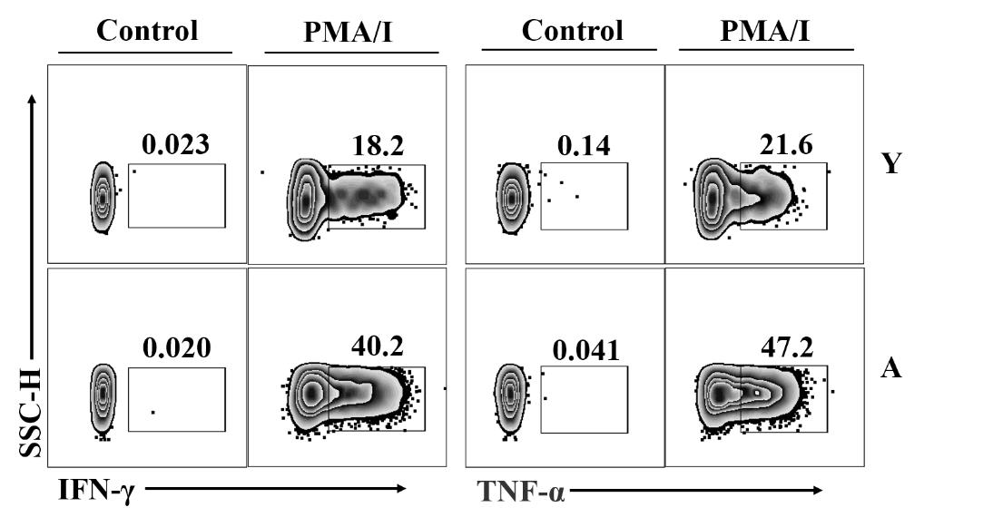
Figure 1 Detection of IFN-γ and TNF-α expression levels in CD8+CD44high T cell subset from lung tissues of aged and young mice after PMA/ionomycin stimulation by flow cytometryNote: Data gated on CD8+CD44high T cell subset; PMA, Phorbol 12-myristate 13-acetate; I, Ionomycin; Control, Unstimulated group; IFN-γ, Interferon-γ; TNF-α, Tumor necrosis factor-α; SSC-H, Side scatter-height; Y, Young group; A, Aged group.
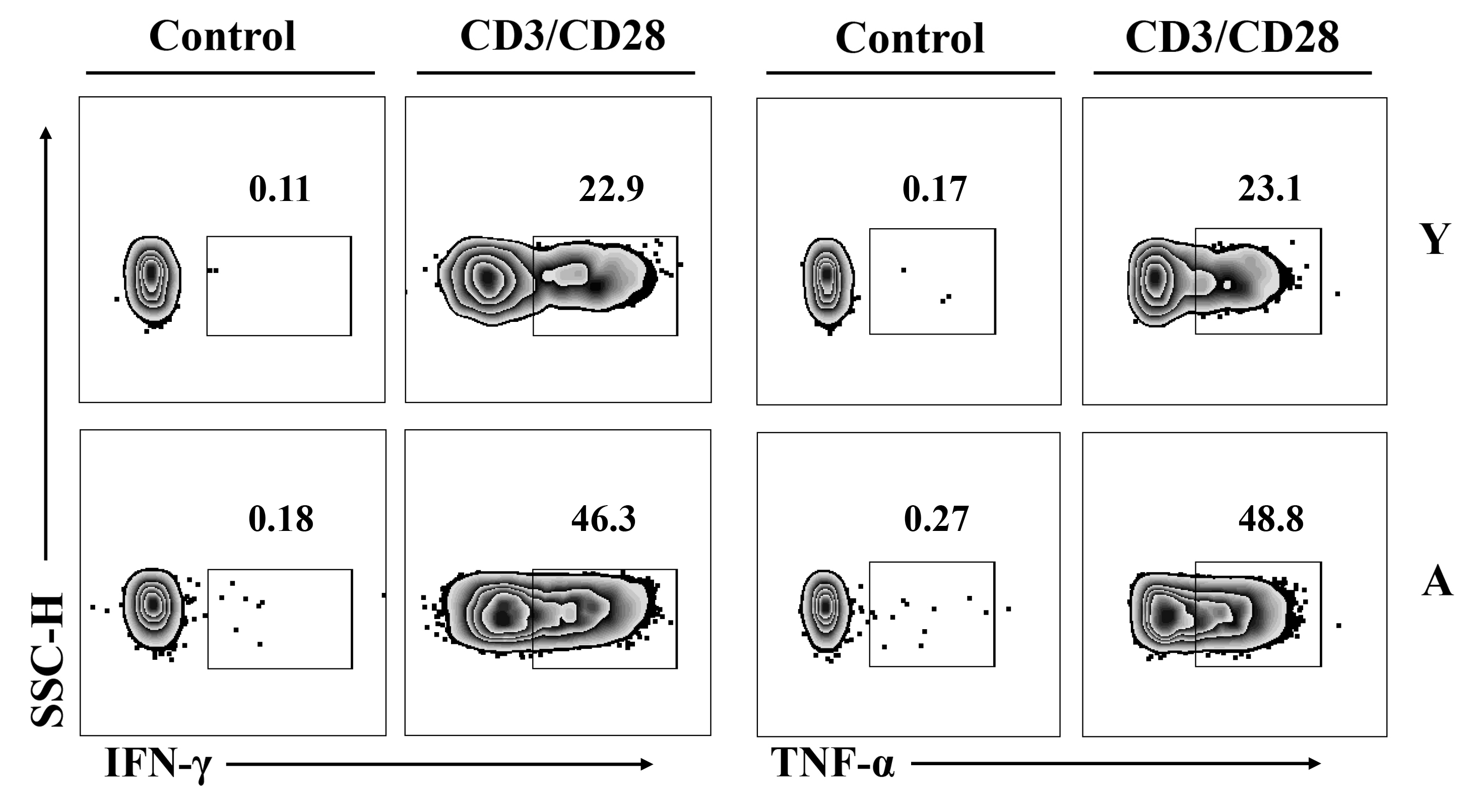
Figure 2 Detection of IFN-γ and TNF-α expression levels in CD8+CD44high T cell subset from lung tissues of aged and young mice after CD3/CD28 antibodies stimulation by flow cytometryNote: Data gated on CD8+CD44high T cell subset; CD3, Cluster of differentiation 3; CD28, Cluster of differentiation 28.
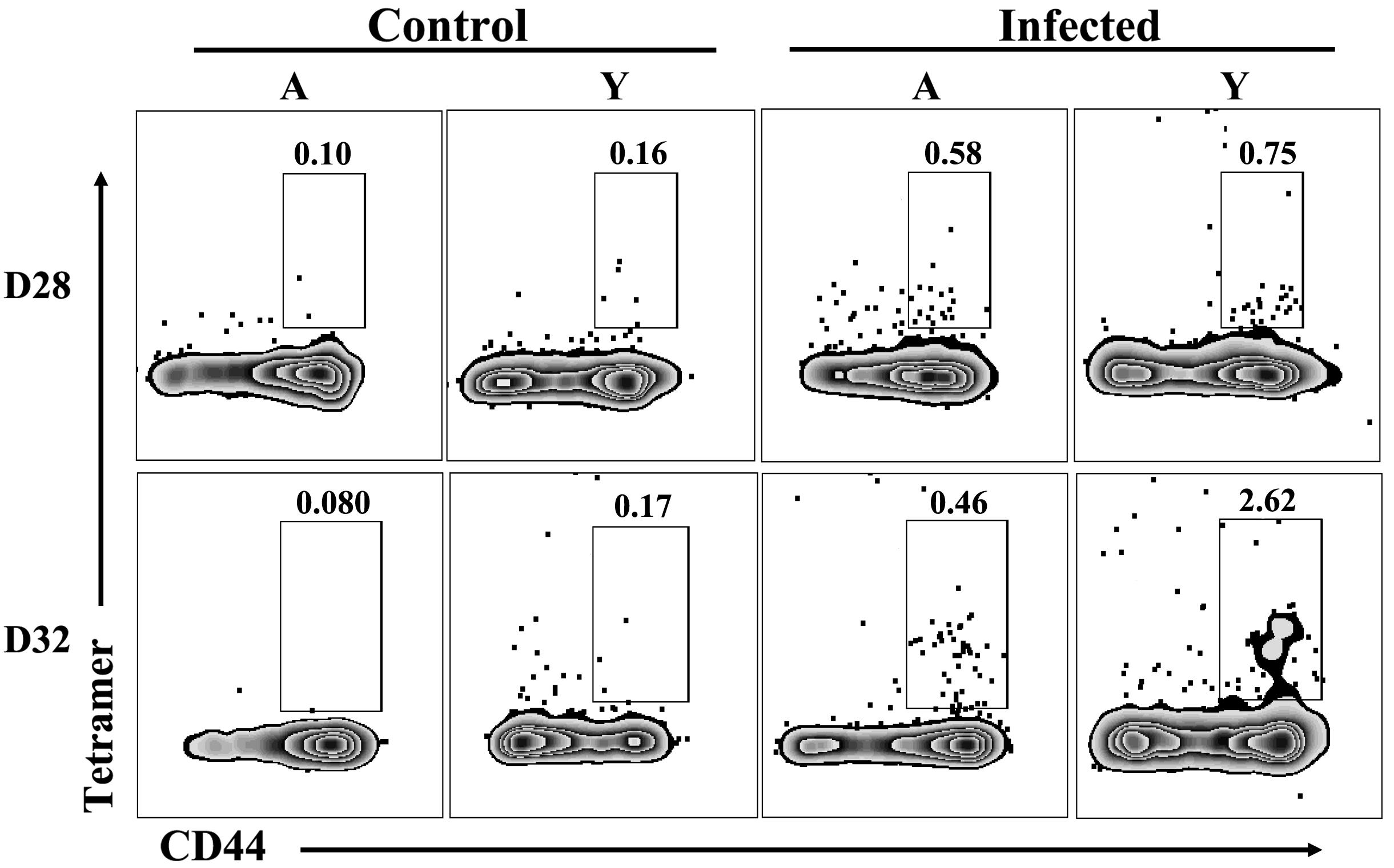
Figure 3 Proportions of virus-specific CD8+ T cells in lung tissues of aged and young mice on day 28 after first influenza virus infection and on day 4 (the 32nd day after first infection) after secondary infectionNote:Data gated on CD8+ T cell subset; D28, 28th day after the initial infection; D32, On the fourth day after the second infection, that is, on the 32nd day after the first infection; Control, The group not infected with influenza virus; Infected, The group infected with influenza virus; Tetramer, Influenza virus-specific MHC-Ⅰ tetramer; CD44, Cluster of differentiation 44.
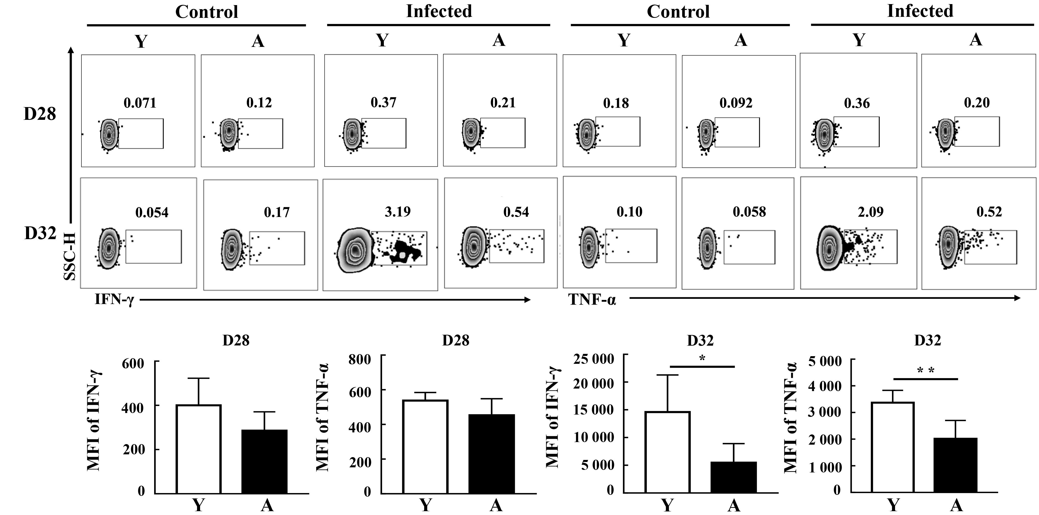
Figure 4 Expression levels and MFI of IFN-γ and TNF-α in viral peptide-stimulated CD8?CD44high T cells from lung tissues of aged and young mice on days 28 and 32 after influenza virus infection?Note: Data gated on CD8+CD44high T cell subset; *P<0.05, **P<0.01.
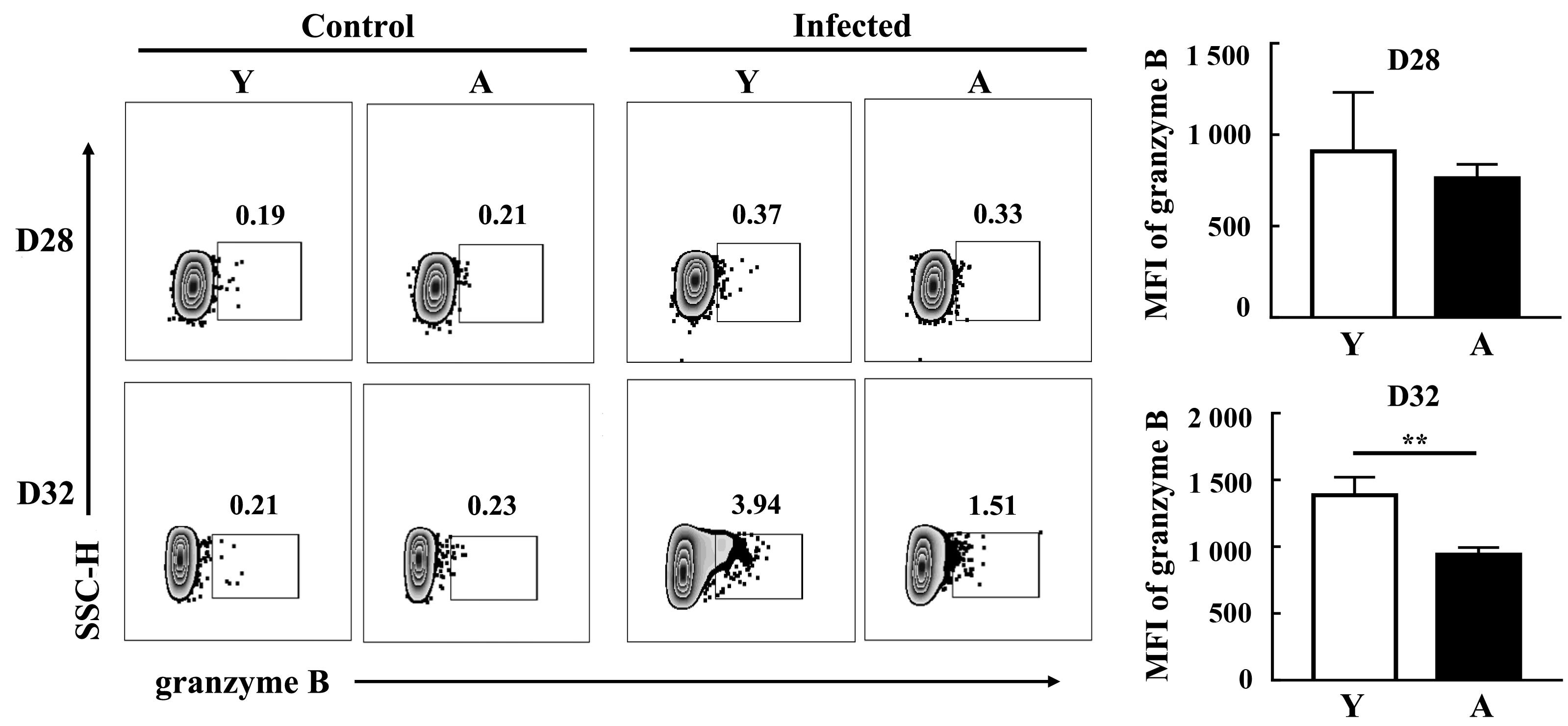
Figure 5 Expression levels and MFI of granzyme B in viral peptide-stimulated CD8?CD44high T cells from lung tissues of aged and young mice on days 28 and 32 after influenza virus infectionNote: Data gated on CD8+CD44high T cell subset; **P<0.01.
| [1] | KUMAR R, BURNS E A. Age-related decline in immunity: implications for vaccine responsiveness[J]. Expert Rev Vaccines, 2008, 7(4):467-479. DOI:10.1586/14760584.7.4.467 . |
| [2] | CIABATTINI A, NARDINI C, SANTORO F, et al. Vaccination in the elderly: The challenge of immune changes with aging[J]. Semin Immunol, 2018, 40:83-94. DOI:10.1016/j.smim. 2018.10.010 . |
| [3] | 丁相荣, 霍姝汭, 代解杰. 甲型流感病毒对人与实验动物神经系统影响的研究进展[J]. 实验动物与比较医学, 2023, 43(2):180-185. DOI:10.12300/j.issn.1674-5817.2022.142 . |
| DING X R, HUO S R, DAI J J. Research progress on influenza A virus and nervous system disease of human and experimental animals[J]. Lab Anim Comp Med, 2023, 43(2):180-185. DOI:10.12300/j.issn.1674-5817.2022.142 . | |
| [4] | KADYRZHANOVA G, TAMAI M, SARKAR S, et al. Aging impairs CD8 T cell responses in adoptive T-cell therapy against solid tumors[J]. Front Immunol, 2025, 16:1484303. DOI:10.3389/fimmu.2025.1484303 . |
| [5] | BORSA M, BARANDUN N, GRÄBNITZ F, et al. Asymmetric cell division shapes naive and virtual memory T-cell immunity during ageing[J]. Nat Commun, 2021, 12(1):2715. DOI:10.1038/s41467-021-22954-y . |
| [6] | HWANG J R, BYEON Y, KIM D, et al. Recent insights of T cell receptor-mediated signaling pathways for T cell activation and development[J]. Exp Mol Med, 2020, 52(5):750-761. DOI:10.1038/s12276-020-0435-8 . |
| [7] | CHEN Y, ZANDER R, KHATUN A, et al. Transcriptional and epigenetic regulation of effector and memory CD8 T cell differentiation[J]. Front Immunol, 2018, 9:2826. DOI:10.3389/fimmu.2018.02826 . |
| [8] | SHAN Q, HU S S, ZHU S Q, et al. Tcf1 preprograms the mobilization of glycolysis in central memory CD8+ T cells during recall responses[J]. Nat Immunol, 2022, 23(3):386-398. DOI: 10.1038/s41590-022-01131-3 . |
| [9] | CLAMBEY E T, WHITE J, KAPPLER J W, et al. Identification of two major types of age-associated CD8 clonal expansions with highly divergent properties[J]. Proc Natl Acad Sci USA, 2008, 105(35):12997-13002. DOI:10.1073/pnas.0805465105 . |
| [10] | YAGER E J, AHMED M, LANZER K, et al. Age-associated decline in T cell repertoire diversity leads to holes in the repertoire and impaired immunity to influenza virus[J]. J Exp Med, 2008, 205(3):711-723. DOI:10.1084/jem.20071140 . |
| [11] | 刘洋, 王超, 任晓楠, 等. 甲型H1N1流感病毒感染年轻和老年C57BL/6小鼠诱导肺组织CD8+ T细胞特异性免疫应答的比较研究[J]. 中国实验动物学报, 2021, 29(4):461-466. DOI:10.3969/j.issn.1005-4847.2021.04.006 . |
| LIU Y, WANG C, REN X N, et al. Study of the immune responses mediated by specific CD8+ T cells to influenza A virus H1N1 in the lungs of young and aged C57BL/6 mice[J]. Acta Lab Anim Sci Sin, 2021, 29(4):461-466. DOI:10.3969/j.issn.1005-4847.2021.04.006 . | |
| [12] | ZENS K D, CHEN J K, GUYER R S, et al. Reduced generation of lung tissue-resident memory T cells during infancy[J]. J Exp Med, 2017,214(10):2915-2932. DOI: 10.1084/jem.20170521 . |
| [13] | SHARMA R. Exploring the emerging bidirectional association between inflamm-aging and cellular senescence in organismal aging and disease[J]. Cell Biochem Funct, 2024, 42(2): e3970. DOI:10.1002/cbf.3970 . |
| [14] | BAJAJ V, GADI N, SPIHLMAN A P, et al. Aging, immunity, and COVID-19: how age influences the host immune response to coronavirus infections?[J]. Front Physiol, 2021, 11:571416. DOI:10.3389/fphys.2020.571416 . |
| [15] | AKBAR A N, HENSON S M. Are senescence and exhaustion intertwined or unrelated processes that compromise immunity?[J]. Nat Rev Immunol, 2011, 11(4):289-295. DOI:10.1038/nri2959 . |
| [16] | GORONZY J J, WEYAND C M. Understanding immunosenescence to improve responses to vaccines[J]. Nat Immunol, 2013, 14(5):428-436. DOI: 10.1038/ni.2588 . |
| [17] | MOGILENKO D A, SHPYNOV O, ANDHEY P S, et al. Comprehensive profiling of an aging immune system reveals clonal GZMK+ CD8+ T cells as conserved hallmark of inflammaging[J]. Immunity, 2021, 54(1):99-115.e12. DOI:10.1016/j.immuni.2020.11.005 . |
| [18] | VAN DE SANDT C E, MCQUILTEN H A, ABAN M, et al. Gradual changes within long-lived influenza virus-specific CD8+ T cells are associated with the loss of public TCR clonotypes in older adults[J]. EBioMedicine, 2025, 115:105697. DOI:10.1016/j.ebiom.2025.105697 . |
| [19] | TAUB D D, HESDORFFER C S, FERRUCCI L, et al. Distinct energy requirements for human memory CD4 T-cell homeostatic functions[J]. FASEB J, 2013, 27(1):342-349. DOI:10.1096/fj.12-217620 . |
| [20] | YANES R E, ZHANG H M, SHEN Y, et al. Metabolic reprogramming in memory CD4 T cell responses of old adults[J]. Clin Immunol, 2019, 207:58-67. DOI:10.1016/j.clim.2019.0 7.003 . |
| [21] | LI G J, YU M C, LEE W W, et al. Decline in miR-181a expression with age impairs T cell receptor sensitivity by increasing DUSP6 activity[J]. Nat Med, 2012, 18(10):1518-1524. DOI:10.1038/nm.2963 . |
| [22] | MOSKOWITZ D M, ZHANG D W, HU B, et al. Epigenomics of human CD8 T cell differentiation and aging[J]. Sci Immunol, 2017, 2(8): eaag0192. DOI:10.1126/sciimmunol.aag0192 . |
| [23] | YATES K B, TONNERRE P, MARTIN G E, et al. Epigenetic scars of CD8+ T cell exhaustion persist after cure of chronic infection in humans[J]. Nat Immunol, 2021, 22(8):1020-1029. DOI:10.1038/s41590-021-00979-1 . |
| [24] | ZOU Q, WANG X L, REN D L, et al. DNA methylation-based signature of CD8+ tumor-infiltrating lymphocytes enables evaluation of immune response and prognosis in colorectal cancer[J]. J Immunother Cancer, 2021, 9(9):e002671. DOI:10.1136/jitc-2021-002671 . |
| [25] | FINK A L, ENGLE K, URSIN R L, et al. Biological sex affects vaccine efficacy and protection against influenza in mice[J]. Proc Natl Acad Sci USA, 2018, 115(49):12477-12482. DOI:10.1073/pnas.1805268115 . |
| [26] | KLEIN S L, MARRIOTT I, FISH E N. Sex-based differences in immune function and responses to vaccination[J]. Trans R Soc Trop Med Hyg, 2015, 109(1):9-15. DOI:10.1093/trstmh/tru167 . |
| [27] | BORNES L, VAN WINDEN L J, GEURTS V C M, et al. The oestrous cycle stage affects mammary tumour sensitivity to chemotherapy[J]. Nature, 2025, 637(8044):195-204. DOI:10.1038/s41586-024-08276-1 . |
| [1] | NIE Yongqiang, WANG Zhaoxia. Exploration and Practice of Building a One-Stop Service Platform for Gene-Edited Mice in University Animal Centers: A Case Study of Shanghai Jiao Tong University [J]. Laboratory Animal and Comparative Medicine, 2025, 45(5): 642-648. |
| [2] | LIU Yueqin, XUE Weiguo, WANG Shuyou, SHEN Yaohua, JIA Shuyong, WANG Guangjun, SONG Xiaojing. Observation of Digestive Tract Tissue Morphology in Mice Using Probe-Based Confocal Laser Endomicroscopy [J]. Laboratory Animal and Comparative Medicine, 2025, 45(4): 457-465. |
| [3] | KONG Zhihao, WEI Xiaofeng, YU Lingzhi, FENG Liping, ZHU Qi, SHI Guojun, WANG Chen. Isolation and Identification of Staphylococcus xylosus in Nude Mice with Squamous Skin Scurfs [J]. Laboratory Animal and Comparative Medicine, 2025, 45(3): 368-375. |
| [4] | XU Qiuyu, YAN Guofeng, FU Li, FAN Wenhua, ZHOU Jing, ZHU Lian, QIU Shuwen, ZHANG Jie, WU Ling. A Mouse Model of Polycystic Ovary Syndrome Established Through Subcutaneous Administration of Letrozole Sustained-Release Pellets and Hepatic Transcriptome Analysis [J]. Laboratory Animal and Comparative Medicine, 2025, 45(2): 119-129. |
| [5] | LIU Rongle, CHENG Hao, SHANG Fusheng, CHANG Shufu, XU Ping. Study on Cardiac Aging Phenotypes of SHJH hr Mice [J]. Laboratory Animal and Comparative Medicine, 2025, 45(1): 13-20. |
| [6] | WU Zhihao, CAO Shuyang, ZHOU Zhengyu. Establishment of an Intestinal Fibrosis Model Associated with Inflammatory Bowel Disease in VDR-/- Mice Induced by Helicobacter hepaticus Infection and Mechanism Exploration [J]. Laboratory Animal and Comparative Medicine, 2025, 45(1): 37-46. |
| [7] | ZHANG Nan, LI Huaiyin, LIAN Xiaodi, WEI Juanpeng, GAO Ming. Effects of Different Durations of Light Exposure on Body Weight and Learning and Memory Abilities of NIH Mice [J]. Laboratory Animal and Comparative Medicine, 2025, 45(1): 73-78. |
| [8] | ZHAO Xiaona, WANG Peng, YE Maoqing, QU Xinkai. Establishment of a New Hyperglycemic Obesity Cardiac Dysfunction Mouse Model with Triacsin C [J]. Laboratory Animal and Comparative Medicine, 2024, 44(6): 605-612. |
| [9] | TAN He, YANG Xiaohui, ZHANG Daxiu, WANG Guicheng. Optimal Adaptation Period for Metabolic Cage Experiments in Mice at Different Developmental Stages [J]. Laboratory Animal and Comparative Medicine, 2024, 44(5): 502-510. |
| [10] | MENG Yu, LIANG Dongli, ZHENG Linlin, ZHOU Yuanyuan, WANG Zhaoxia. Optimization and Evaluation of Conditions for Orthotopic Nude Mouse Models of Human Liver Tumor Cells [J]. Laboratory Animal and Comparative Medicine, 2024, 44(5): 511-522. |
| [11] | Jing QIN, Yong ZHAO, Caiqin ZHANG, Bing BAI, Changhong SHI. Construction and Evaluation of Theranostic Near-infrared Fluorescent Probe for Targeting Inflammatory Brain Edema [J]. Laboratory Animal and Comparative Medicine, 2024, 44(3): 243-250. |
| [12] | Yisu ZHANG, Xinru LIU, Ruojie WU, Rui LIU, Hong OUYANG, Xiaohong LI. Establishment and Evaluation of Mouse Model of Pregnancy Pain-depression Comorbidity Induced by Chronic Unpredictable Stress, Complete Freund's Adjuvant and Formalin [J]. Laboratory Animal and Comparative Medicine, 2024, 44(3): 259-269. |
| [13] | Dong WU, Rui SHI, Peishan LUO, Ling'en LI, Xijing SHENG, Mengyang WANG, Lu NI, Sujuan WANG, Huixin YANG, Jing ZHAO. Effects of Different Pellet Feed Hardness on Growth and Reproduction, Feed Utilization Rate, and Environmental Dust in Laboratory Mice [J]. Laboratory Animal and Comparative Medicine, 2024, 44(3): 313-320. |
| [14] | Yun LIU, Tingting FENG, Wei TONG, Zhi GUO, Xia LI, Qi KONG, Zhiguang XIANG. Glycyrrhizic Acid Showed Therapeutic Effects on Severe Pulmonary Damages in Mice Induced by Pneumonia Virus of Mice Infection [J]. Laboratory Animal and Comparative Medicine, 2024, 44(3): 251-258. |
| [15] | Jinhua HU, Jingjie HAN, Min JIN, Bin HU, Yuefen LOU. Effects of Puerarin on Bone Density in Rats and Mice: A Meta-analysis [J]. Laboratory Animal and Comparative Medicine, 2024, 44(2): 149-161. |
| Viewed | ||||||
|
Full text |
|
|||||
|
Abstract |
|
|||||