
Laboratory Animal and Comparative Medicine ›› 2025, Vol. 45 ›› Issue (1): 37-46.DOI: 10.12300/j.issn.1674-5817.2024.090
• Animal Models of Human Diseases • Previous Articles Next Articles
WU Zhihao1( ), CAO Shuyang2, ZHOU Zhengyu1(
 )()
)()
Received:2024-06-27
Revised:2024-10-09
Online:2025-02-25
Published:2025-03-12
Contact:
ZHOU Zhengyu
CLC Number:
WU Zhihao,CAO Shuyang,ZHOU Zhengyu. Establishment of an Intestinal Fibrosis Model Associated with Inflammatory Bowel Disease in VDR-/- Mice Induced by Helicobacter hepaticus Infection and Mechanism Exploration[J]. Laboratory Animal and Comparative Medicine, 2025, 45(1): 37-46. DOI: 10.12300/j.issn.1674-5817.2024.090.
Add to citation manager EndNote|Ris|BibTeX
URL: https://www.slarc.org.cn/dwyx/EN/10.12300/j.issn.1674-5817.2024.090
引物名称 Primer name | 基因序列号 Gene ID | 序列 Sequence | 扩增片段长度/bp Amplicon size/bp |
|---|---|---|---|
| TGF-β | 22059 | F: CCACCTGCAAGACCATCGAC; R: CTGGCGAGCCTTAGTTTGGAC | 91 |
| α-SMA | 11475 | F: CCCAGACATCAGGGAGTAATGG; R: TCTATCGGATACTTCAGCGTCA | 104 |
| TNF-α | 21926 | F: CAGGCGGTGCCTATGTCTC; R: CGATCACCCCGAAGTTCAGTAG | 89 |
| IL-1β | 16176 | F: GAAATGCCACCTTTTGACAGTG; R: TGGATGCTCTCATCAGGACAG | 116 |
| IL-6 | 16193 | F: TCTATACCACTTCACAAGTCGGA; R: GAATTGCCATTGCACAACTCTTT | 88 |
| IL-33 | 77125 | F: ATTTCCCCGGCAAAGTTCAG; R: AACGGAGTCTCATGCAGTAGA | 118 |
| GAPDH | 14433 | F: AGGTCGGTGTGAACGGATTTG; R: GGGGTCGTTGATGGCAACA | 95 |
Table 1 Sequences of primers used for PCR
引物名称 Primer name | 基因序列号 Gene ID | 序列 Sequence | 扩增片段长度/bp Amplicon size/bp |
|---|---|---|---|
| TGF-β | 22059 | F: CCACCTGCAAGACCATCGAC; R: CTGGCGAGCCTTAGTTTGGAC | 91 |
| α-SMA | 11475 | F: CCCAGACATCAGGGAGTAATGG; R: TCTATCGGATACTTCAGCGTCA | 104 |
| TNF-α | 21926 | F: CAGGCGGTGCCTATGTCTC; R: CGATCACCCCGAAGTTCAGTAG | 89 |
| IL-1β | 16176 | F: GAAATGCCACCTTTTGACAGTG; R: TGGATGCTCTCATCAGGACAG | 116 |
| IL-6 | 16193 | F: TCTATACCACTTCACAAGTCGGA; R: GAATTGCCATTGCACAACTCTTT | 88 |
| IL-33 | 77125 | F: ATTTCCCCGGCAAAGTTCAG; R: AACGGAGTCTCATGCAGTAGA | 118 |
| GAPDH | 14433 | F: AGGTCGGTGTGAACGGATTTG; R: GGGGTCGTTGATGGCAACA | 95 |
Figure 1 Body weight changes (A), fecal water content (B), and disease activity index score (C) of mice infected by H.hepaticusNote: A significant disparity in body weight was observed after a 10-week infection period when comparing the VDR-/-control with the remaining three groups (*P<0.05, **P<0.01, n=5). The fecal water content and disease activity index score in VDR-/-+H.h group mice were significantly higher compared to those in the other groups (*P<0.05, **P<0.01, ***P<0.001, ****P<0.000 1, n=5).
Figure 2 Colon length of mice in each group after H. hepaticus infectionNote:A, Comparison photos of representative colon lengths of one mouse from each group; B, H.hepaticus infection resulted in a significant reduction in colon length, and VDR deficiency further exacerbated this pathological effect(**P<0.01, n=5).
Figure 3 Colonic bacterial colonization (A) and HAI score of colon tissues (B) in mice infected with H.hepaticusNote:The intestinal colonization of H.hepaticus was significantly higher in the VDR-/-+H.h group compared to the WT+H.h group(**P<0.01, n=5). Histological activity index showed that the infection with H.hepaticus induced colonic pathology, while VDR deficiency exacerbated the pathological changes in the colon(*P<0.05, ***P<0.001, ****P<0.000 1, n=5).
Figure 4 HE staining (A), AB-PAS staining (B) and Masson staining (C) of colon tissue of mice infected with H.hepaticus for 16 weeksNote: The VDR-/-+H.h group exhibited more severe lymphocyte infiltration, loss of goblet cells, and reduced mucus secretion, along with increased collagen fiber deposition. The magnification is ×40 with a scale bar of 500 μm, ×200 with a scale bar of 100 μm, or ×100 with a scale bar of 200 μm.
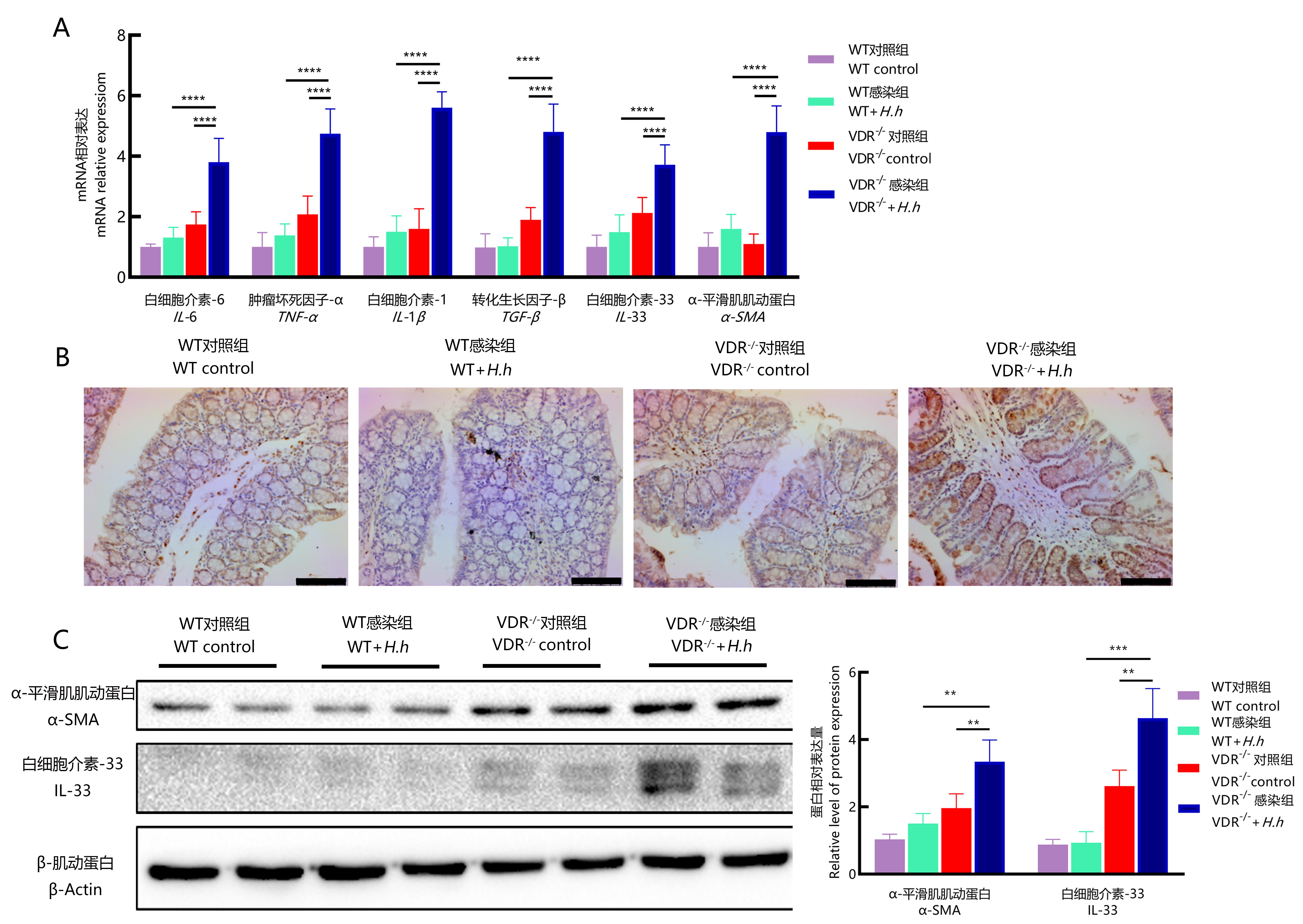
Figure 5 Detection of mRNA expression levels in colon tissue of mice infected with H. hepaticus after 16 weeks (A), IL-33 immunohistochemical staining (B), and Western blotting (C)Note: RT-PCR showed that, compared to the WT+H.h group, the transcription levels of pro-inflammatory and pro-fibrotic factors were significantly elevated in the VDR-/-+H.h group (****P<0.000 1,n=5). Immunohistochemistry and Western blotting also revealed an upregulation of fibrosis-related proteins such as IL-33 and α-SMA (**P<0.01, ***P<0.001). In Figure B, the magnification is ×200, and the scale bar is 100 μm.
| 1 | KHAN I, ULLAH N, ZHA L J, et al. Alteration of gut microbiota in inflammatory bowel disease (IBD): cause or consequence? IBD treatment targeting the gut microbiome[J]. Pathogens, 2019, 8(3):126. DOI: 10.3390/pathogens8030126 . |
| 2 | GEARRY R B. IBD and environment: are there differences between east and west[J]. Dig Dis, 2016, 34(1-2):84-89. DOI: 10.1159/000442933 . |
| 3 | OTT S J, MUSFELDT M, WENDEROTH D F, et al. Reduction in diversity of the colonic mucosa associated bacterial microflora in patients with active inflammatory bowel disease[J]. Gut, 2004, 53(5):685-693. DOI: 10.1136/gut.2003.025403 . |
| 4 | BETTENWORTH D, RIEDER F. Pathogenesis of intestinal fibrosis in inflammatory bowel disease and perspectives for therapeutic implication[J]. Dig Dis, 2017, 35(1-2):25-31. DOI: 10.1159/000449079 . |
| 5 | FOX J G, GE Z, WHARY M T, et al. Helicobacter hepaticus infection in mice: models for understanding lower bowel inflammation and cancer[J]. Mucosal Immunol, 2011, 4(1):22-30. DOI: 10.1038/mi.2010.61 . |
| 6 | NAGALINGAM N A, ROBINSON C J, BERGIN I L, et al. The effects of intestinal microbial community structure on disease manifestation in IL-10-/- mice infected with Helicobacter hepaticus[J]. Microbiome, 2013, 1(1):15. DOI: 10.1186/2049-2618-1-15 . |
| 7 | LU Y Y, CHEN Y E, LI Y H, et al. Monotropein inhibits colitis associated cancer through VDR/JAK1/STAT1 regulation of macrophage polarization[J]. Int Immunopharmacol, 2023, 124(Pt A):110838. DOI: 10.1016/j.intimp.2023.110838 . |
| 8 | ASSA A, VONG L, PINNELL L J, et al. Vitamin D deficiency promotes epithelial barrier dysfunction and intestinal inflammation[J]. J Infect Dis, 2014, 210(8):1296-1305. DOI: 10.1093/infdis/jiu235 . |
| 9 | CHETCUTI ZAMMIT S, ELLUL P, GIRARDIN G, et al. Vitamin D deficiency in a European inflammatory bowel disease inception cohort: an epi-IBD study[J]. Eur J Gastroenterol Hepatol, 2018, 30(11):1297-1303. DOI: 10.1097/MEG. 0000000000001238 . |
| 10 | 钱淼, 冯洁, 张泉, 等. 螺杆菌属LAMP快速检测方法的研究[J]. 扬州大学学报(农业与生命科学版), 2020,41(4):77-81. DOI:10.16872/j.cnki.1671-4652.2020.04.013 . |
| QIAN M, FENG J, ZHANG Q, et al. The study of rapid detection of Helicobacter species using loop-mediated isothermal amplification[J]. J Yangzhou Univ Agric Life Sci Ed, 2020,41(4):77-81. DOI:10.16872/j.cnki.1671-4652.2020.04.013 . | |
| 11 | WU Y, RAN L, YANG Y, et al. Deferasirox alleviates DSS-induced ulcerative colitis in mice by inhibiting ferroptosis and improving intestinal microbiota[J]. Life Sci, 2023, 314:121312. DOI: 10.1016/j.lfs.2022.121312 . |
| 12 | 孙靖谕, 谢灵志, 冯洁, 等. 啮齿类螺杆菌SYBR Green Ⅱ荧光定量PCR检测方法的建立[J]. 实验动物与比较医学, 2018, 38(2):91-97. DOI: 10.3969/j.issn.1674-5817.2018.02.003 . |
| SUN J Y, XIE L Z, FENG J, et al. Establishment of SYBR green Ⅱ based real-time PCR method for detection of Helicobacter rodentium[J]. Lab Anim Comp Med, 2018, 38(2):91-97. DOI: 10.3969/j.issn.1674-5817.2018.02.003 . | |
| 13 | WANG R, WANG D J, WANG H W, et al. Therapeutic targeting of Nrf2 signaling by maggot extracts ameliorates inflammation-associated intestinal fibrosis in chronic DSS-induced colitis[J]. Front Immunol, 2021, 12:670159. DOI: 10.3389/fimmu.2021.670159 . |
| 14 | ANDOH A, NISHIDA A. Pro- and anti-inflammatory roles of interleukin (IL)-33, IL-36, and IL-38 in inflammatory bowel disease[J]. J Gastroenterol, 2023, 58(2):69-78. DOI: 10.1007/s00535-022-01936-x . |
| 15 | XAVIER R J, PODOLSKY D K. Unravelling the pathogenesis of inflammatory bowel disease[J]. Nature, 2007, 448(7152):427-434. DOI: 10.1038/nature06005 . |
| 16 | CAHILL R J, FOLTZ C J, FOX J G, et al. Inflammatory bowel disease: an immunity-mediated condition triggered by bacterial infection with Helicobacter hepaticus[J]. Infect Immun, 1997, 65(8):3126-3131. DOI: 10.1128/iai.65.8.3126-3131.1997 . |
| 17 | BURICH A, HERSHBERG R, WAGGIE K, et al. Helicobacter-induced inflammatory bowel disease in IL-10- and T cell-deficient mice[J]. Am J Physiol Gastrointest Liver Physiol, 2001, 281(3): G764-G778. DOI: 10.1152/ajpgi.2001.281.3.G764 . |
| 18 | BATTISTINI C, BALLAN R, HERKENHOFF M E, et al. Vitamin D modulates intestinal microbiota in inflammatory bowel diseases[J]. Int J Mol Sci, 2020, 22(1):362. DOI: 10.3390/ijms22010362 . |
| 19 | DOMAZETOVIC V, IANTOMASI T, BONANOMI A G, et al. Vitamin D regulates claudin-2 and claudin-4 expression in active ulcerative colitis by p-Stat-6 and Smad-7 signaling[J]. Int J Colorectal Dis, 2020, 35(7):1231-1242. DOI: 10.1007/s00384-020-03576-0 . |
| 20 | ISMAILOVA A, WHITE J H. Vitamin D, infections and immunity[J]. Rev Endocr Metab Disord, 2022, 23(2):265-277. DOI: 10.1007/s11154-021-09679-5 . |
| 21 | SMILLIE C S, BITON M, ORDOVAS-MONTANES J, et al. Intra- and inter-cellular rewiring of the human colon during ulcerative colitis[J]. Cell, 2019, 178(3):714-730.e22. DOI: 10.1016/j.cell.2019.06.029 . |
| 22 | KOTSIOU O S, GOURGOULIANIS K I, ZAROGIANNIS S G. IL-33/ST2 axis in organ fibrosis[J]. Front Immunol, 2018, 9:2432. DOI: 10.3389/fimmu.2018.02432 . |
| [1] | LIU Rongle, CHENG Hao, SHANG Fusheng, CHANG Shufu, XU Ping. Study on Cardiac Aging Phenotypes of SHJH hr Mice [J]. Laboratory Animal and Comparative Medicine, 2025, 45(1): 13-20. |
| [2] | ZHANG Nan, LI Huaiyin, LIAN Xiaodi, WEI Juanpeng, GAO Ming. Effects of Different Durations of Light Exposure on Body Weight and Learning and Memory Abilities of NIH Mice [J]. Laboratory Animal and Comparative Medicine, 2025, 45(1): 73-78. |
| [3] | ZHAO Xiaona, WANG Peng, YE Maoqing, QU Xinkai. Establishment of a New Hyperglycemic Obesity Cardiac Dysfunction Mouse Model with Triacsin C [J]. Laboratory Animal and Comparative Medicine, 2024, 44(6): 605-612. |
| [4] | TAN He, YANG Xiaohui, ZHANG Daxiu, WANG Guicheng. Optimal Adaptation Period for Metabolic Cage Experiments in Mice at Different Developmental Stages [J]. Laboratory Animal and Comparative Medicine, 2024, 44(5): 502-510. |
| [5] | MENG Yu, LIANG Dongli, ZHENG Linlin, ZHOU Yuanyuan, WANG Zhaoxia. Optimization and Evaluation of Conditions for Orthotopic Nude Mouse Models of Human Liver Tumor Cells [J]. Laboratory Animal and Comparative Medicine, 2024, 44(5): 511-522. |
| [6] | Jing QIN, Yong ZHAO, Caiqin ZHANG, Bing BAI, Changhong SHI. Construction and Evaluation of Theranostic Near-infrared Fluorescent Probe for Targeting Inflammatory Brain Edema [J]. Laboratory Animal and Comparative Medicine, 2024, 44(3): 243-250. |
| [7] | Yisu ZHANG, Xinru LIU, Ruojie WU, Rui LIU, Hong OUYANG, Xiaohong LI. Establishment and Evaluation of Mouse Model of Pregnancy Pain-depression Comorbidity Induced by Chronic Unpredictable Stress, Complete Freund's Adjuvant and Formalin [J]. Laboratory Animal and Comparative Medicine, 2024, 44(3): 259-269. |
| [8] | Dong WU, Rui SHI, Peishan LUO, Ling'en LI, Xijing SHENG, Mengyang WANG, Lu NI, Sujuan WANG, Huixin YANG, Jing ZHAO. Effects of Different Pellet Feed Hardness on Growth and Reproduction, Feed Utilization Rate, and Environmental Dust in Laboratory Mice [J]. Laboratory Animal and Comparative Medicine, 2024, 44(3): 313-320. |
| [9] | Yun LIU, Tingting FENG, Wei TONG, Zhi GUO, Xia LI, Qi KONG, Zhiguang XIANG. Glycyrrhizic Acid Showed Therapeutic Effects on Severe Pulmonary Damages in Mice Induced by Pneumonia Virus of Mice Infection [J]. Laboratory Animal and Comparative Medicine, 2024, 44(3): 251-258. |
| [10] | Jinhua HU, Jingjie HAN, Min JIN, Bin HU, Yuefen LOU. Effects of Puerarin on Bone Density in Rats and Mice: A Meta-analysis [J]. Laboratory Animal and Comparative Medicine, 2024, 44(2): 149-161. |
| [11] | Min LIANG, Yang GUO, Jinjin WANG, Mengyan ZHU, Jun CHI, Yanjuan CHEN, Chengji WANG, Zhilan YU, Ruling SHEN. Construction of Dmd Gene Mutant Mice and Phenotype Verification in Muscle and Immune Systems [J]. Laboratory Animal and Comparative Medicine, 2024, 44(1): 42-51. |
| [12] | Jianhua ZHENG, Yunzhi FA, Qiaoyan DONG, Yefeng QIU, Jingqing CHEN. Construction and Evaluation of a Mouse Model with Intestinal Injury by Acute Hypoxic Stress in Plateau [J]. Laboratory Animal and Comparative Medicine, 2024, 44(1): 31-41. |
| [13] | Qianqian TANG, Xiuli ZHANG, Zai CHANG. Statistical Analysis of the Leakage Situation in the Automated Watering System for Mice in Tsinghua University Laboratory Animal Resources Center [J]. Laboratory Animal and Comparative Medicine, 2024, 44(1): 85-91. |
| [14] | Xin LIU, Shaobo SHI, Cui ZHANG, Bo YANG, Chuan QU. Construction and Evaluation of End-to-side Anastomosis Model of Autologous Arteriovenous Fistula in Mice [J]. Laboratory Animal and Comparative Medicine, 2023, 43(6): 595-603. |
| [15] | Dan WANG, Xiaolu ZHANG, Yan WANG, Bo FU, Wendong WANG, Jing LIU, Suyin ZHANG, Yihe WU, Deguo WU, Xiaoyan DU, Dawei ZHAN, Xiulin ZHANG, Changlong LI. Study on the Antibody Production Efficiency in Modified Big-BALB/c Mice [J]. Laboratory Animal and Comparative Medicine, 2023, 43(6): 612-618. |
| Viewed | ||||||
|
Full text |
|
|||||
|
Abstract |
|
|||||