
Laboratory Animal and Comparative Medicine ›› 2025, Vol. 45 ›› Issue (3): 279-289.DOI: 10.12300/j.issn.1674-5817.2024.128
• Animal Models of Human Diseases • Previous Articles Next Articles
PAN Yicong( ), JIANG Wenhong()(
 ), HU Ming, QIN Xiao
Received:2024-08-28
Revised:2024-12-18
Online:2025-06-25
Published:2025-06-25
Contact:
JIANG Wenhong
CLC Number:
PAN Yicong,JIANG Wenhong,HU Ming,et al. Optimization of Surgical Procedure and Efficacy Evaluation of Aortic Calcification Model in Rats with Chronic Kidney Disease[J]. Laboratory Animal and Comparative Medicine, 2025, 45(3): 279-289. DOI: 10.12300/j.issn.1674-5817.2024.128.
Add to citation manager EndNote|Ris|BibTeX
URL: https://www.slarc.org.cn/dwyx/EN/10.12300/j.issn.1674-5817.2024.128
Figure1 Flowchart of the experimental process for modeling and evaluating aortic calcification in SD rats with chronic kidney disease
目标基因名称 Target gene name | NCBI基因序列号 NCBI reference sequence | 引物序列信息 Primer sequence information | 扩增片段大小/bp Amplification fragment size/bp |
|---|---|---|---|
| Sm22 (Tagln) | NC-086026.1 | F:5’-CAGATGGAACAGGTGGCTCAA-3’ | 161 |
| R:5’-GCCCAAAGCCATTACAGTCCTC-3’ | |||
| OPN (Spp1) | NC-086032.1 | F:5’-GCCGAGGTGATAGCTTGGCTTA-3’ | 145 |
| R:5’-TTGATAGCCTCATCGGACTCCTG-3’ | |||
| Runx2 | NC-086027.1 | F:5’-GGATGCCTTAGTGCCCAAATG-3’ | 120 |
| R:5’-CACCCTGTGAGGTGGCTGAA-3’ | |||
| β-actin | NC-086030.1 | F:5’-CACCCGCGAGTACAACCTTC-3’ | 207 |
| R:5’-CCCATACCCACCATCACACC-3’ |
Table 1 Primers for real-time fluorescent quantitative PCR
目标基因名称 Target gene name | NCBI基因序列号 NCBI reference sequence | 引物序列信息 Primer sequence information | 扩增片段大小/bp Amplification fragment size/bp |
|---|---|---|---|
| Sm22 (Tagln) | NC-086026.1 | F:5’-CAGATGGAACAGGTGGCTCAA-3’ | 161 |
| R:5’-GCCCAAAGCCATTACAGTCCTC-3’ | |||
| OPN (Spp1) | NC-086032.1 | F:5’-GCCGAGGTGATAGCTTGGCTTA-3’ | 145 |
| R:5’-TTGATAGCCTCATCGGACTCCTG-3’ | |||
| Runx2 | NC-086027.1 | F:5’-GGATGCCTTAGTGCCCAAATG-3’ | 120 |
| R:5’-CACCCTGTGAGGTGGCTGAA-3’ | |||
| β-actin | NC-086030.1 | F:5’-CACCCGCGAGTACAACCTTC-3’ | 207 |
| R:5’-CCCATACCCACCATCACACC-3’ |
Figure 2 Kaplan-Meier survival curve analysis of rats in chronic renal insufficiency model groups constructed using different surgical methods
血生化指标 Biochemistry index | 实验组(n=9) Experimental group | 对照组(n=12) Control group | t值 t value | P值 P value |
|---|---|---|---|---|
血清钙 c/(mmol·L-1) Serum calcium ions | 1.876±0.036 | 1.929±0.042 | 3.431 | 0.004 |
血清磷c/(mmol·L-1) Serum phosphate ions | 2.059±0.333 | 1.580±0.271 | 3.046 | 0.014 |
血清肌酐c/(μmol·L-1) Serum creatinine | 119.960±35.640 | 21.164±8.076 | 8.984 | <0.000 1 |
血清尿素氮c/(mmol·L-1) Serum urea nitrogen | 57.991±8.745 | 22.609±8.058 | 6.895 | <0.000 1 |
Table 2 Comparison of biochemical indicators between the experimental group and the control group
血生化指标 Biochemistry index | 实验组(n=9) Experimental group | 对照组(n=12) Control group | t值 t value | P值 P value |
|---|---|---|---|---|
血清钙 c/(mmol·L-1) Serum calcium ions | 1.876±0.036 | 1.929±0.042 | 3.431 | 0.004 |
血清磷c/(mmol·L-1) Serum phosphate ions | 2.059±0.333 | 1.580±0.271 | 3.046 | 0.014 |
血清肌酐c/(μmol·L-1) Serum creatinine | 119.960±35.640 | 21.164±8.076 | 8.984 | <0.000 1 |
血清尿素氮c/(mmol·L-1) Serum urea nitrogen | 57.991±8.745 | 22.609±8.058 | 6.895 | <0.000 1 |
Figure 3 HE staining observation of kidneys from rats in the experimental and control groups
Figure 4 Observation of von Kossa and Alizarin red S staining in the aorta of rats from the experimental and control groups
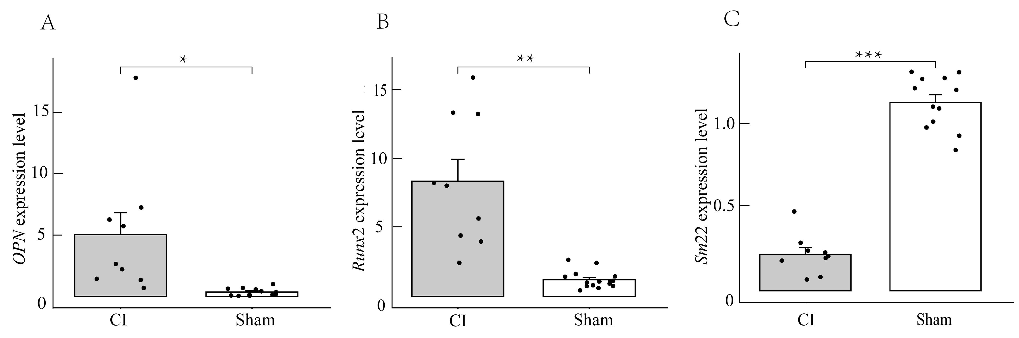
Figure 5 Real-time fluorescence quantitative PCR detection of OPN, Runx2, and Sm22 gene expression in aortic tissue of rats from the experimental and control groups
| [1] | HUANG A R, GUO G Y, YU Y Q, et al. The roles of collagen in chronic kidney disease and vascular calcification[J]. J Mol Med, 2021, 99(1):75-92. DOI:10.1007/s00109-020-02014-6 . |
| [2] | 李贵森. 2019年«中国慢性肾脏病矿物质和骨异常诊治指南»解读[J]. 诊断学理论与实践, 2020, 19(3):229-231. DOI: 10.16150/j.1671-2870.2020.03.005 . |
| LI G S. Interpretation of Chinese Guideline for Diagnosis and Treatment of Chronic Kidney Disease-Mineral and Bone Disorder (2019 version)[J]. J Diagn Concepts Pract, 2020, 19(3):229-231. DOI: 10.16150/j.1671-2870.2020.03.005 . | |
| [3] | YE Y Z, CHEN A, LI L, et al. Repression of the antiporter SLC7A11/glutathione/glutathione peroxidase 4 axis drives ferroptosis of vascular smooth muscle cells to facilitate vascular calcification[J]. Kidney Int. 2022 102(6):1259-1275. DOI:10.1016/j.kint.2022.07.034 . |
| [4] | 柳潇雨. Shox2促进血管钙化的作用及机制研究[D]. 广州: 南方医科大学, 2023. DOI:10.27003/d.cnki.gojyu.2023.000180 . |
| LIU X Y. The role and mechanism of Shox2 in promoting vascular calcification[D]. Guangzhou: Southern Medical University, 2023. DOI:10.27003/d.cnki.gojyu.2023.000180 . | |
| [5] | 阙冬冬. 五羟色胺对慢性肾脏病大鼠血管钙化的作用及其机制研究[D]. 广州: 南方医科大学, 2023. DOI:10.27003/d.cnki.gojyu.2023.000170 . |
| QUE D D. Study on the effect and mechanism of serotonin on vascular calcification in rats with chronic kidney disease [D]. Guangzhou: Southern Medical University, 2023. DOI:10.27003/d.cnki.gojyu.2023.000170 . | |
| [6] | 冯丽芸. 二氢杨梅素抑制慢性肾脏病大鼠血管钙化及其机制研究[D]. 广州: 南方医科大学, 2022. DOI:10.27003/d.cnki.gojyu.2022.000227 . |
| FENG L Y. Study on the inhibition of vascular calcification and its mechanism by dihydromyricetin in rats with chronic kidney disease [D]. Guangzhou: Southern Medical University, 2022. DOI:10.27003/d.cnki.gojyu.2022.000227 . | |
| [7] | SHOBEIRI N, ADAMS M A, HOLDEN R M. Vascular calcification in animal models of CKD: a review[J]. Am J Nephrol, 2010, 31(6):471-481. DOI:10.1159/000299794 . |
| [8] | LIU Y J, GUO Y, BAO S M, et al. Bone marrow mesenchymal stem cell-derived exosomal microRNA-381-3p alleviates vascular calcification in chronic kidney disease by targeting NFAT5[J]. Cell Death Dis, 2022, 13(3):278. DOI:10.1038/s41419-022-04703-1 . |
| [9] | 苏培培. 不同剂量骨化三醇对慢性肾衰竭大鼠主动脉钙化的影响[D]. 唐山: 华北理工大学, 2021. DOI:10.27108/d.cnki.ghelu.2021.000272 . |
| SU P P. Effects of different doses of calcitriol on aortic calcification in rats with chronic renal failure [D]. Tangshan: North China University of Science and Technology, 2021. DOI:10.27108/d.cnki.ghelu.2021.000272 . | |
| [10] | WU X J, SHEN S J, WU J Y, et al. ENPP1 ameliorates vascular calcification via inhibiting the osteogenic transformation of VSMCs and generating PPi[J]. Open Med, 2023, 18(1):20230861. DOI:10.1515/med-2023-0861 . |
| [11] | CHEN C Z, LI Y D, LU H L, et al. Curcumin attenuates vascular calcification via the exosomal miR-92b-3p/KLF4 axis[J]. Exp Biol Med, 2022, 247(16):1420-1432. DOI:10.1177/1535370222 1095456 . |
| [12] | LIU X Y, CHEN A, LIANG Q C, et al. Spermidine inhibits vascular calcification in chronic kidney disease through modulation of SIRT1 signaling pathway[J]. Aging Cell, 2021, 20(6): e13377. DOI:10.1111/acel.13377 . |
| [13] | ZHANG X L, LI Y N, YANG P Z, et al. Trimethylamine-N-oxide promotes vascular calcification through activation of NLRP3 (nucleotide-binding domain, leucine-rich-containing family, pyrin domain-containing-3) inflammasome and NF-κB (nuclear factor κB) signals[J]. Arterioscler Thromb Vasc Biol, 2020, 40(3):751-765. DOI:10.1161/ATVBAHA.119.313414 . |
| [14] | ROWE P S, MCCARTHY E M, YU A L, et al. Correction of vascular calcification and hyperphosphatemia in CKD rats treated with ASARM peptide[J]. Kidney360, 2022, 3(10):1683-1698. DOI:10.34067/KID.0002782022 . |
| [15] | MA W Q, SUN X J, ZHU Y, et al. PDK4 promotes vascular calcification by interfering with autophagic activity and metabolic reprogramming[J]. Cell Death Dis, 2020, 11(11):991. DOI:10.1038/s41419-020-03162-w . |
| [16] | SATO H, GOTO M, NISHIMURA G, et al. Upacicalcet, a positive allosteric modulator of the calcium-sensing receptor, prevents vascular calcification and bone disorder in a rat adenine-induced secondary hyperparathyroidism model[J]. Bone, 2023, 167:116613. DOI:10.1016/j.bone.2022.116613 . |
| [17] | LI Z H, WU J, ZHANG X L, et al. CDC42 promotes vascular calcification in chronic kidney disease[J]. J Pathol, 2019, 249(4):461-471. DOI:10.1002/path.5334 . |
| [18] | CHANG J R, GUO J, WANG Y, et al. Intermedin1-53 attenuates vascular calcification in rats with chronic kidney disease by upregulation of α-Klotho[J]. Kidney Int, 2016, 89(3):586-600. DOI:10.1016/j.kint.2015.12.029 . |
| [19] | LEE S J, LEE I K, JEON J H. Vascular calcification-new insights into its mechanism[J]. Int J Mol Sci, 2020, 21(8):2685. DOI:10.3390/ijms21082685 . |
| [20] | 张佳莉, 张岩. 急性肾损伤动物模型构建方法与研究现状[J]. 中国实验动物学报, 2022, 30(7):955-965. DOI: 10.3969/j.issn.1005-4847.2022.07.011 . |
| ZHANG J L, ZHANG Y. Research approaches and status of animal models for acute kidney injury[J]. Acta Lab Anim Sci Sin, 2022, 30(7):955-965. DOI: 10.3969/j.issn.1005-4847.2022.07.011 . | |
| [21] | 陈娟, 易香伶, 罗佳, 等. 急性肾损伤动物模型及体外模型研究进展[J]. 华西医学, 2023, 38(5):777-783. DOI:10.7507/1002-0179.202210185 . |
| CHEN J, YI X L, LUO J, et al. Advances in animal models and in vitro models of acute kidney injury[J]. West China Med J, 2023, 38(5):777-783. DOI:10.7507/1002-0179.202210185 . | |
| [22] | HERRMANN J, BABIC M, TÖLLE M, et al. Research models for studying vascular calcification[J]. Int J Mol Sci, 2020, 21(6):2204. DOI:10.3390/ijms21062204 . |
| [23] | BAO Y W, YUAN Y, CHEN J H, et al. Kidney disease models: tools to identify mechanisms and potential therapeutic targets[J]. Zool Res, 2018, 39(2):72-86. DOI:10.24272/j.issn.2095-8137.2017.055 . |
| [24] | FU Y, TANG C Y, CAI J, et al. Rodent models of AKI-CKD transition[J]. Am J Physiol Renal Physiol, 2018, 315(4):F1098-F1106. DOI:10.1152/ajprenal.00199.2018 . |
| [25] | HERRMANN J, GUMMI M R, XIA M D, et al. Vascular calcification in rodent models-keeping track with an extented method assortment[J]. Biology, 2021, 10(6):459. DOI:10.3390/biology10060459 . |
| [1] | LI Hui. Advances in Animal Models for Biolinguistic Research [J]. Laboratory Animal and Comparative Medicine, 2026, 46(2): 297-305. |
| [2] | LIAO Wangyue, LEI Shuang, LI Xuan, GUO Min, ZHOU Ruoran. Literature Analysis and Validity Assessment for Animal Models of Attention Deficit and Hyperactive Disorder [J]. Laboratory Animal and Comparative Medicine, 2026, 46(1): 66-80. |
| [3] | CHEN Ziyi, SUN Hongyan, KANG Pinfang, WU Wenjuan. Research Advances in Construction Methods and Novel Technologies for Animal Models of Pulmonary Hypertension [J]. Laboratory Animal and Comparative Medicine, 2026, 46(1): 81-93. |
| [4] | GAO Chaoqi, ZHU Zhibo, SUN Xiandong. Application Progress and Classification Analysis of Rat Vascular Remodeling Models [J]. Laboratory Animal and Comparative Medicine, 2025, 45(5): 542-550. |
| [5] | LIU Yang, CHENG Laiyang, GUO Zhongkun. Progress on Animal Models of Perimenopausal Syndrome Based on Traditional Chinese Medicine Disease-Syndrome Combination [J]. Laboratory Animal and Comparative Medicine, 2025, 45(5): 586-595. |
| [6] | LIU Yayi, JIA Yunfeng, ZUO Yiming, ZHANG Junping, LÜ Shichao. Progress and Evaluation of Animal Model of Heart Qi-Yin Deficiency Syndrome [J]. Laboratory Animal and Comparative Medicine, 2025, 45(4): 411-421. |
| [7] | QIN Chao, LI Shuangxing, ZHAO Tingting, JIANG Chenchen, ZHAO Jing, YANG Yanwei, LIN Zhi, WANG Sanlong, WEN Hairuo. Study on the 90-day Feeding Experimental Background Data of SD Rats for Drug Safety Evaluation [J]. Laboratory Animal and Comparative Medicine, 2025, 45(4): 439-448. |
| [8] | LIAN Hui, JIANG Yanling, LIU Jia, ZHANG Yuli, XIE Wei, XUE Xiaoou, LI Jian. Construction and Evaluation of a Rat Model of Abnormal Uterine Bleeding [J]. Laboratory Animal and Comparative Medicine, 2025, 45(2): 130-146. |
| [9] | YANG Jiahao, DING Chunlei, QIAN Fenghua, SUN Qi, JIANG Xusheng, CHEN Wen, SHEN Mengwen. Research Progress on Animal Models of Sepsis-Related Organ Injury [J]. Laboratory Animal and Comparative Medicine, 2024, 44(6): 636-644. |
| [10] | HUANG Dongyan, WU Jianhui. Establishment Methods and Application Evaluation of Animal Models in Reproductive Toxicology Research [J]. Laboratory Animal and Comparative Medicine, 2024, 44(5): 550-559. |
| [11] | ZHENG Yiqing, DENG Yasheng, FAN Yanping, LIANG Tianwei, HUANG Hui, LIU Yonghui, NI Zhaobing, LIN Jiang. Application Analysis of Animal Models for Pelvic Inflammatory Disease Based on Data Mining [J]. Laboratory Animal and Comparative Medicine, 2024, 44(4): 405-418. |
| [12] | WU Yue, LI Lu, ZHANG Yang, WANG Jue, FENG Tingting, LI Yitong, WANG Kai, KONG Qi. Integrative Analysis of Omics Data in Animal Models of Coronavirus Infection [J]. Laboratory Animal and Comparative Medicine, 2024, 44(4): 357-373. |
| [13] | Committee of Experts on Medical Animal Experiments, Chinese Research Hospital Association. Guidelines for the Selection of Animal Models and Preclinical Drug Trials for Spontaneous Intracerebral Hemorrhage (2024 Edition) [J]. Laboratory Animal and Comparative Medicine, 2024, 44(1): 3-30. |
| [14] | Shuwu XIE, Ruling SHEN, Jinxing LIN, Chun FAN. Progress in Establishment and Application of Laboratory Animal Models Related to Development of Male Infertility Drugs [J]. Laboratory Animal and Comparative Medicine, 2023, 43(5): 504-511. |
| [15] | Rui ZHANG, Meiyu LÜ, Jianjun ZHANG, Jinlian LIU, Yan CHEN, Zhiqiang HUANG, Yao LIU, Lanhua ZHOU. Research Progress on Establishing and Evaluation of Acne Animal Models [J]. Laboratory Animal and Comparative Medicine, 2023, 43(4): 398-405. |
| Viewed | ||||||
|
Full text |
|
|||||
|
Abstract |
|
|||||