
Laboratory Animal and Comparative Medicine ›› 2023, Vol. 43 ›› Issue (5): 574-578.DOI: 10.12300/j.issn.1674-5817.2023.095
• Research Reports • Previous Articles Next Articles
Chengji WANG1, Jue WANG1, Haijie WANG1, Weisheng LU1, Yan SHI2, Zhengye GU1, Mingqiu WAN1, Ruling SHEN1( )(
)( )
)
Received:2023-06-30
Revised:2023-10-02
Online:2023-10-25
Published:2023-10-25
Contact:
Ruling SHEN
CLC Number:
Chengji WANG,Jue WANG,Haijie WANG,et al. Application of Optimized Latex Perfusion Technique in the Establishment of Craniofacial Venous Model in Mice[J]. Laboratory Animal and Comparative Medicine, 2023, 43(5): 574-578. DOI: 10.12300/j.issn.1674-5817.2023.095.
Add to citation manager EndNote|Ris|BibTeX
URL: https://www.slarc.org.cn/dwyx/EN/10.12300/j.issn.1674-5817.2023.095
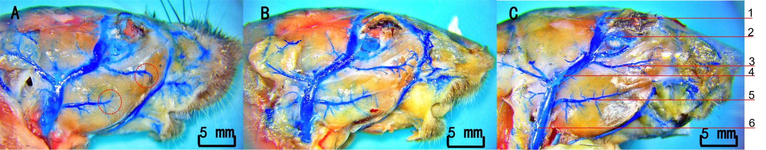
Figure 1 Results of latex perfusion in the head and facial veins of miceNote:A, 60% latex saline group; B, 60% latex heparin group; C, 30% latex heparin group. 1, Superior palpebral vein; 2, Orbital venous sinus; 3, Facial transverse vein; 4, Superficial temporal vein; 5, Masseteric vein; 6, External jugular vein.
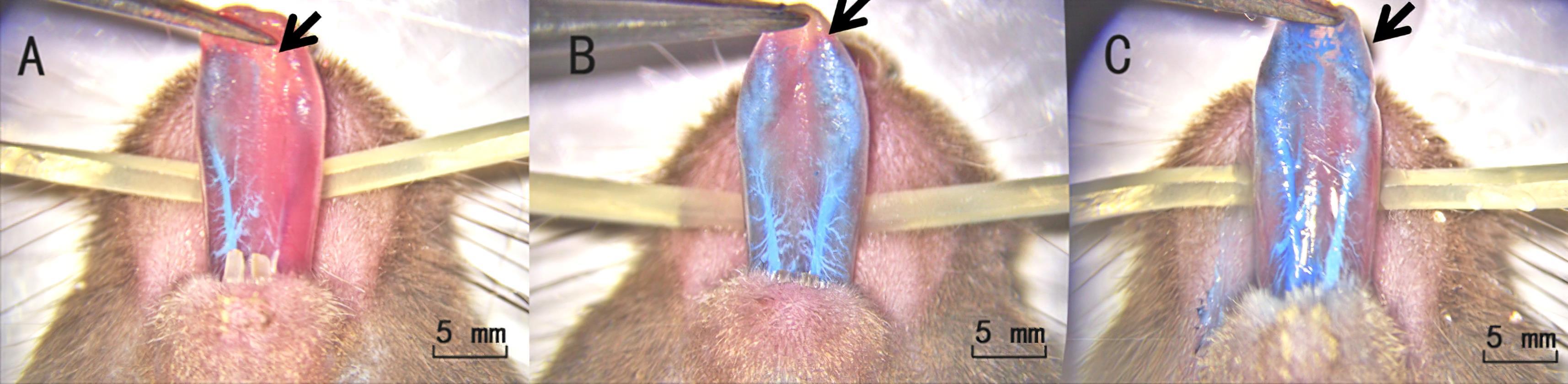
Figure 2 Comparison of lingual vein perfusion effects among different groups of miceNote: A, 60% latex saline group; B, 60% latex heparin group; C, 30% latex heparin group.
| 1 | 余琛琳, 赵乐, 汤球, 等. 一项小鼠采血新技术: 颌下静脉丛采血法[J]. 中国比较医学杂志, 2008, 18(12):53-54, 95. DOI: 10.3969/j.issn.1671-7856.2008.12.014 . |
| YU C L, ZHAO L, TANG Q, et al. A new method of blood collection from the submandibular vein plexus in mice[J]. Chin J Comp Med, 2008, 18(12):53-54, 95. DOI: 10.3969/j.issn.1671-7856.2008.12.014 . | |
| 2 | 车兆义, 邹悦, 宋清斌. 大鼠实验中几种常用的采血方法探讨[J]. 局解手术学杂志, 2008, 17(2): 84-85. DOI: 10.3969/j.issn.1672-5042.2008.02.005 . |
| CHE Z Y, ZOU Y, SONG Q B. Several experiments in rat blood collection methods commonly used[J]. J Reg Anat Oper Surg, 2008, 17(2): 84-85. DOI: 10.3969/j.issn.1672-5042.2008.02.005 . | |
| 3 | 杨健莉, 刘佳, 郑志红. 常用实验大小鼠采血方法及其对实验动物福利的影响[J]. 中国比较医学杂志, 2019, 29(1): 90-94. DOI: 10.3969/j.issn.1671-7856.2019.01.016 . |
| YANG J L, LIU J, ZHENG Z H. Comparison and analysis of blood sampling methods from rats and mice[J]. Chin J Comp Med, 2019, 29(1): 90-94. DOI: 10.3969/j.issn.1671-7856.2019.01.016 . | |
| 4 | 邹天乐, 龚瑜, 林爱娣, 等. 硫酸钡血管造影术的优化[J]. 中国临床解剖学杂志, 2010, 28(1):104-106. DOI: 10.13418/j.issn.1001-165x.2010.01.017 . |
| ZOU T L, GONG Y, LIN A D, et al. Modified Barium Sulfate-latex injection technique for angiography[J]. Chin J Clin Anat, 2010, 28(1):104-106. DOI: 10.13418/j.issn.1001-165x.2010.01.017 . | |
| 5 | 钱凤涛, 王成稷, 王恺, 等. 优化硫酸钡造影剂对小鼠血管造影效果观察[J]. 实验动物与比较医学, 2019, 39(4): 319-322. DOI: 10.3969/j.issn.1674-5817.2019.04.012 . |
| QIAN F T, WANG C J, WANG K, et al. Study on angiography in mice with Barium sulfate[J]. Lab Anim Comp Med, 2019, 39(4): 319-322. DOI: 10.3969/j.issn.1674-5817.2019.04.012 . | |
| 6 | 杨曦, 徐永清, 何晓清, 等. 数字化技术制备大鼠跨区穿支皮瓣微小血管模型的实验研究[J]. 中国修复重建外科杂志, 2017, 31(12):1485-1489. |
| YANG X, XU Y Q, HE X Q, et al. Establishment of micro-vessels model of cross-boundary perforator flap in rat via digital technology[J]. Chin J Reparative Reconstr Surg, 2017, 31(12):1485-1489. | |
| 7 | 张习高, 陈超, 王建武, 等. 采用颈内静脉逆行插管行乳胶灌注翼丛标本制作法[J]. 解剖学杂志, 2003, 26(4): 359. DOI: 10.3969/j.issn.1001-1633.2003.04.035 . |
| ZHANG X G, CHEN C, WANG J W, et al. Preparation of pterygium plexus by retrograde intubation of internal jugular vein[J]. Chin J Anat, 2003, 26(4): 359. DOI: 10.3969/j.issn.1001-1633.2003.04.035 . | |
| 8 | 甘承, 田佳, 刘立强, 等. 人体头面部解剖标本的防腐固定技术及血管内乳胶灌注技术研究[J]. 中国美容医学, 2015, 24(12):44-49. DOI: 10.15909/j.cnki.cn61-1347/r.000515 . |
| GAN C, TIAN J, LIU L Q, et al. Research of anticorrosion and latex perfusion technology in human craniofacial specimens[J]. Chin J Aesthetic Med, 2015, 24(12):44-49. DOI: 10.15909/j.cnki.cn61-1347/r.000515 . | |
| 9 | 罗东辉, 罗坤. 乳胶灌注和硅胶灌注在头颅血管教学标本制作中的对比研究[J]. 新疆医学, 2014, 44(9): 171-173. |
| LUO D H, LUO K. Comparative study of latex perfusion and silica gel perfusion in making teaching specimens of cranial blood vessels[J]. Xinjiang Med J, 2014, 44(9): 171-173. | |
| 10 | 周文逊, 王刚. 乳胶灌注在心的血管标本制作中的应用[J]. 中国医药导报, 2010, 7(36): 134. DOI: 10.3969/j.issn.1673-7210.2010.36.071 . |
| ZHOU W X, WANG G. Application of latex perfusion in blood vessel specimen making of heart[J]. China Med Her, 2010, 7(36): 134. DOI: 10.3969/j.issn.1673-7210.2010.36.071 . | |
| 11 | 成亮, 陈铿, 柴益民, 等. 手指末节指掌侧浅静脉的显微解剖及临床应用[J]. 中华显微外科杂志, 2011, 34(2):131-133, F0003. |
| CHENG L, CHEN K, CHAI Y M, et al. Microanatomy study and clinical application of superficial palmar digital veins in fingertip replantation[J]. Chin J Microsurg, 2011, 34(2):131-133, F0003. | |
| 12 | ONISHI A, ANGE K ST, DORDICK J S, et al. Heparin and anticoagulation[J]. Front Biosci (Landmark Ed), 2016, 21(7):1372-1392. DOI: 10.2741/4462 . |
| 13 | 漆光平, 杜亚政, 潘爱华, 等. 乳胶血管灌注的技术探讨[J]. 湖南医科大学学报, 2000, 25(4):411-412. |
| QI G P, DU Y Z, PAN A H, et al. Filling blood vessels with latex and its application[J]. Bull Hunan Med Univ, 2000, 25(4):411-412. |
| [1] | JIAO Qingzhen, WU Guihua, TANG Wen, FAN Fan, FENG Kai, YANG Chunxiang, QIAO Jian, DENG Sufang. Dynamic Monitoring and Analysis of Ammonia Concentration in Laboratory Animal Facilities Under Suspension of Heating Ventilation and Air Conditioning System [J]. Laboratory Animal and Comparative Medicine, 2025, 45(4): 490-495. |
| [2] | LIU Wentao, LUO Yanhong, LONG Yongxia, LUO Qihui, CHEN Zhengli, LIU Lida. Common Environmental Problems and Testing Experiences in Laboratory Animal Facilities in Sichuan Province [J]. Laboratory Animal and Comparative Medicine, 2025, 45(4): 483-489. |
| [3] | ZHAO Xin, WANG Chenxi, SHI Wenqing, LOU Yuefen. Advances in the Application of Zebrafish in the Research of Inflammatory Bowel Disease Mechanisms and Drug Development [J]. Laboratory Animal and Comparative Medicine, 2025, 45(4): 422-431. |
| [4] | GONG Leilei, WANG Xiaoxia, FENG Xuewei, LI Xinlei, ZHAO Han, ZHANG Xueyan, FENG Xin. A Mouse Model and Mechanism Study of Premature Ovarian Insufficiency Induced by Different Concentrations of Cyclophosphamide [J]. Laboratory Animal and Comparative Medicine, 2025, 45(4): 403-410. |
| [5] | LIN Zhenhua, CHU Xiangyu, WEI Zhenxi, DONG Chuanjun, ZHAO Zenglin, SUN Xiaoxia, LI Qingyu, ZHANG Qi. Evaluation of the Safety and Efficacy of Bone Cement in Experimental Pigs Using Vertebroplasty [J]. Laboratory Animal and Comparative Medicine, 2025, 45(4): 466-472. |
| [6] | JIANG Juan, SONG Ning, LIAN Wenbo, SHAO Congcong, GU Wenwen, SHI Yan. Comparison of Histopathological and Molecular Pathological Phenotypes in Mouse Models of Intrauterine Adhesions Induced by Two Concentrations of Ethanol Perfusion [J]. Laboratory Animal and Comparative Medicine, 2025, 45(4): 393-402. |
| [7] | LIU Yueqin, XUE Weiguo, WANG Shuyou, SHEN Yaohua, JIA Shuyong, WANG Guangjun, SONG Xiaojing. Observation of Digestive Tract Tissue Morphology in Mice Using Probe-Based Confocal Laser Endomicroscopy [J]. Laboratory Animal and Comparative Medicine, 2025, 45(4): 457-465. |
| [8] | ZHENG Qingyong, YANG Donghua, MA Zhichao, ZHOU Ziyu, LU Yang, WANG Jingyu, XING Lina, KANG Yingying, DU Li, ZHAO Chunxiang, DI Baoshan, TIAN Jinhui. Recommendations for Standardized Reporting of Systematic Reviews and Meta-Analysis of Animal Experiments [J]. Laboratory Animal and Comparative Medicine, 2025, 45(4): 496-507. |
| [9] | WANG Tingjun, LUO Hao, CHEN Qi. Discussion on AI-Based Digital Upgrade and Application Practice of Laboratory Animal Centers [J]. Laboratory Animal and Comparative Medicine, 2025, 45(4): 473-482. |
| [10] | WANG Jiaoxiang, ZHANG Lu, CHEN Shuhan, JIAO Deling, ZHAO Heng, WEI Taiyun, GUO Jianxiong, XU Kaixiang, WEI Hongjiang. Construction and Functional Validation of GTKO/hCD55 Gene-Edited Xenotransplant Donor Pigs [J]. Laboratory Animal and Comparative Medicine, 2025, 45(4): 379-392. |
| [11] | QIN Chao, LI Shuangxing, ZHAO Tingting, JIANG Chenchen, ZHAO Jing, YANG Yanwei, LIN Zhi, WANG Sanlong, WEN Hairuo. Study on the 90-day Feeding Experimental Background Data of SD Rats for Drug Safety Evaluation [J]. Laboratory Animal and Comparative Medicine, 2025, 45(4): 439-448. |
| [12] | LIU Kun, LAN Qing, YI Bing, XIE Xiaojie. Key Challenges and Mitigation Strategies for Animal Pregnancy in Non-clinical Reproductive Toxicity Testing of Drugs [J]. Laboratory Animal and Comparative Medicine, 2025, 45(4): 449-456. |
| [13] | . [J]. Laboratory Animal and Comparative Medicine, 2025, 45(4): 508-514. |
| [14] | CHEN Ziyi, SUN Hongyan, KANG Pinfang, WU Wenjuan. Research Advances in Animal Experimental Models of Pulmonary Hypertension [J]. Laboratory Animal and Comparative Medicine, 2025, (): 1-12. |
| [15] | XU Yingtao, WANG Mengmeng, LIN Ping, CHI Haitao, WANG Yi, BAI Ying. Exosomes Improve Ischemic Stroke by Regulation of Ferroptosis Through the NRF2/SLC7A11/GPX4 Pathway in Mice [J]. Laboratory Animal and Comparative Medicine, 2025, (): 1-11. |
| Viewed | ||||||
|
Full text |
|
|||||
|
Abstract |
|
|||||