

Laboratory Animal and Comparative Medicine
• XXXX XXXX •
XU Haochen( ), LE Qingqing, HE Xiwen, CAI Bing, YANG Longhe(
 )()
)()
Online:2026-09-07
Correspondence to:
YANG Longhe
CLC Number:
XU Haochen,LE Qingqing,HE Xiwen,et al. Evaluation of the Efficacy of an Eye-Protective Compound Formulation on the Retina of Mice with Blue Light-Induced Damage[J]. Laboratory Animal and Comparative Medicine. DOI: 10.12300/j.issn.1674-5817.2026.010.
Add to citation manager EndNote|Ris|BibTeX
URL: https://www.slarc.org.cn/dwyx/EN/10.12300/j.issn.1674-5817.2026.010
The sequences of primers used in RT-qPCR
| 基因名称 Gene | 引物序列 Sequences (5' - 3') |
|---|---|
| IL-1β | F-GAAATGCCACCTTTTGACAGTG R-TGGATGCTCTCATCAGGACAG |
| IL-6 | F-AATTAAGCCTCCGACTTGTGAAG R-CTTCCATCCAGTTGCCTTCTTG |
| TNF-α | F-GTAGCAAACCACCAAGTGGAGG R-CAGCCTTGTCCCTTGAAGAGAA |
| GAPDH | F-GGTGAAGGTCGGTGTGAACG R-CTCGCTCCTGGAAGATGGTG |
Table 1
The sequences of primers used in RT-qPCR
| 基因名称 Gene | 引物序列 Sequences (5' - 3') |
|---|---|
| IL-1β | F-GAAATGCCACCTTTTGACAGTG R-TGGATGCTCTCATCAGGACAG |
| IL-6 | F-AATTAAGCCTCCGACTTGTGAAG R-CTTCCATCCAGTTGCCTTCTTG |
| TNF-α | F-GTAGCAAACCACCAAGTGGAGG R-CAGCCTTGTCCCTTGAAGAGAA |
| GAPDH | F-GGTGAAGGTCGGTGTGAACG R-CTCGCTCCTGGAAGATGGTG |
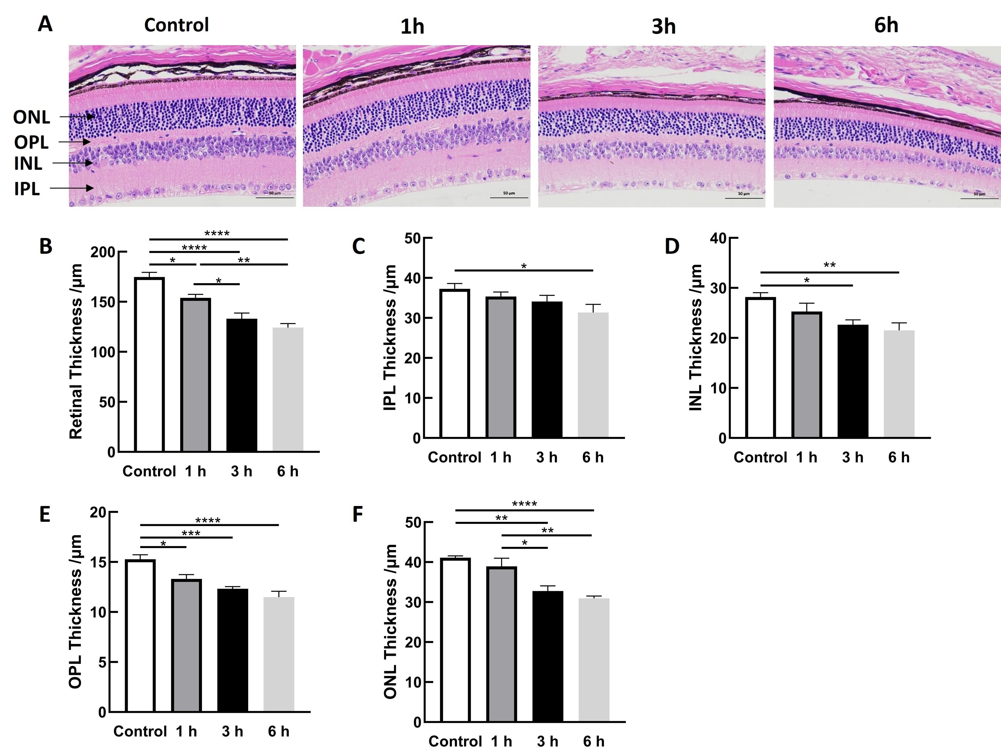
Figure 1
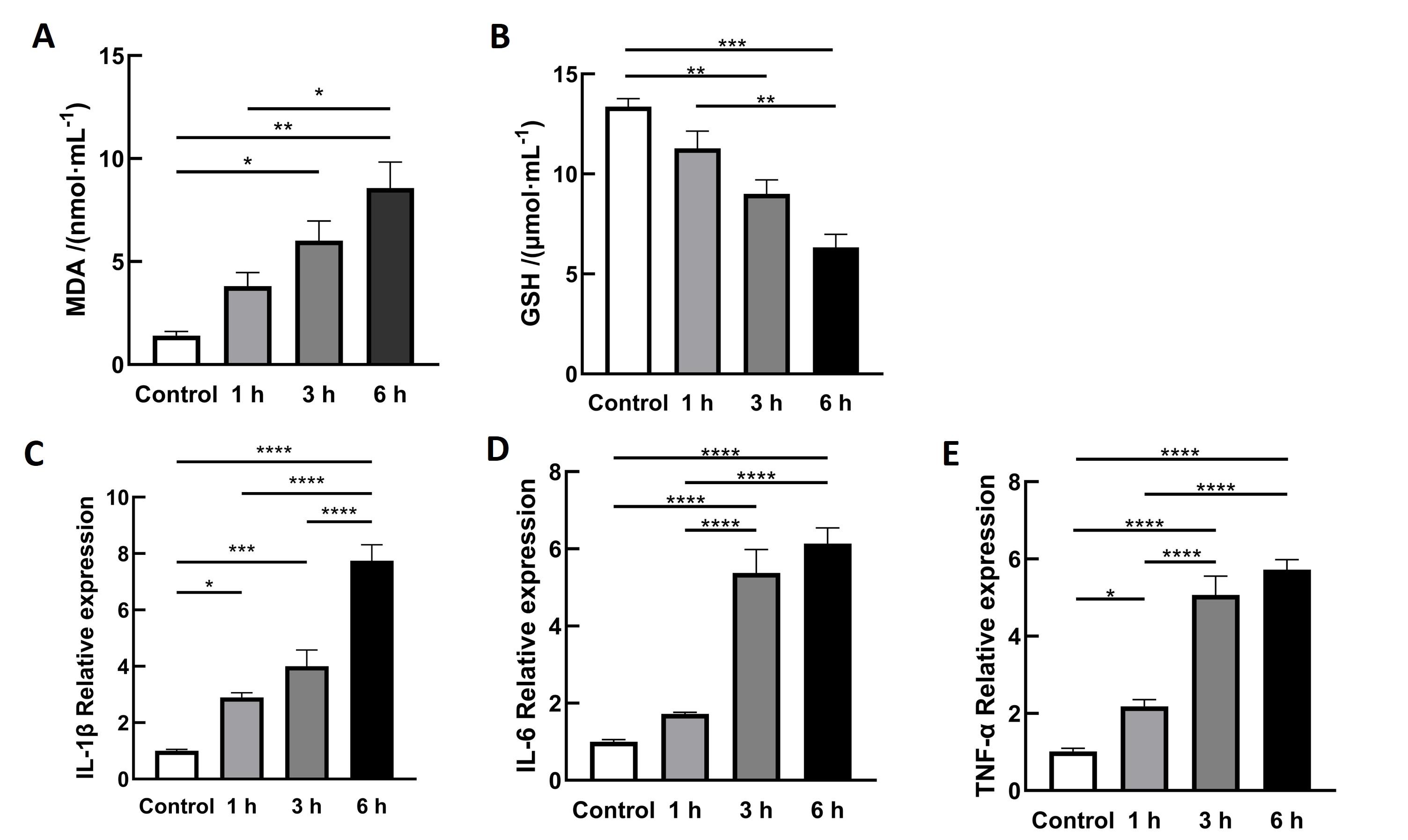
Figure 2
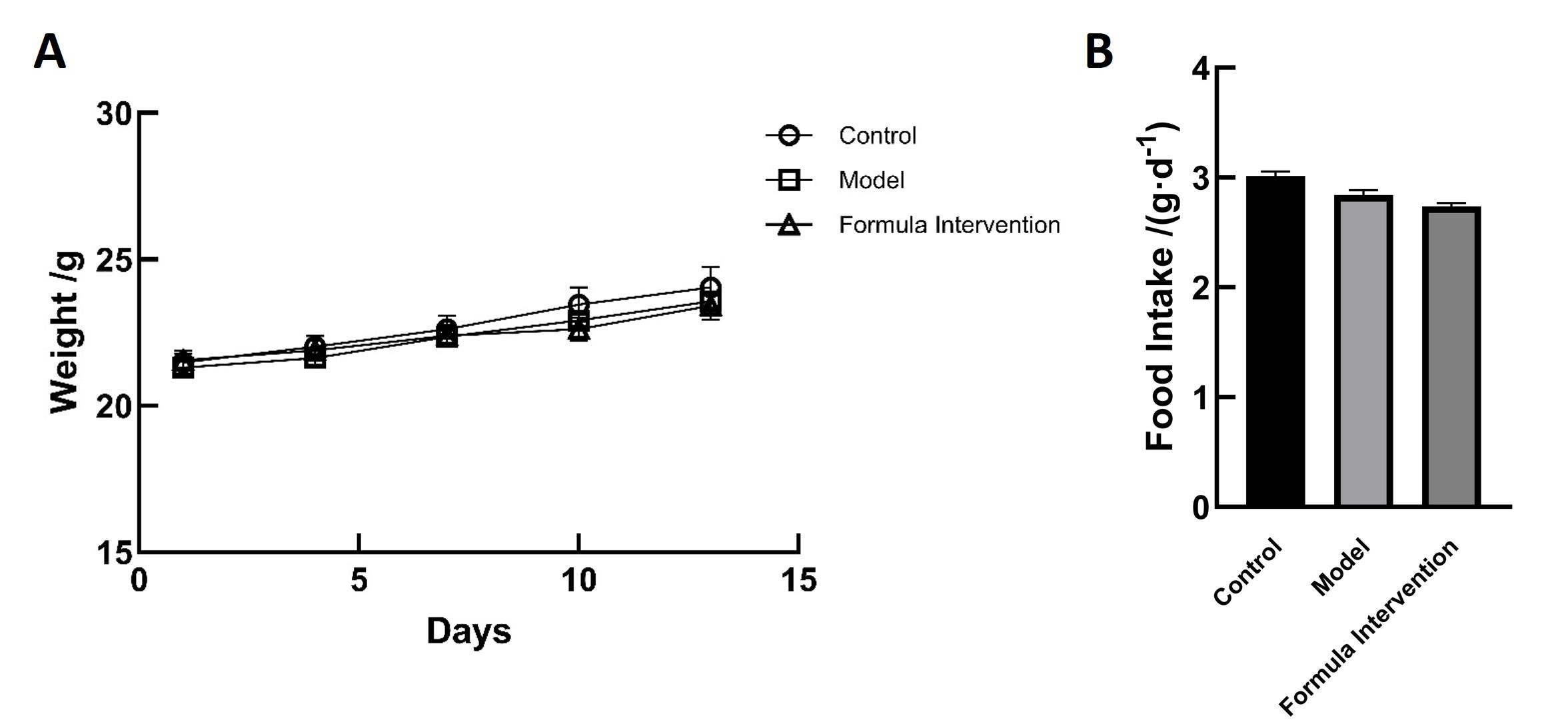
Figure 3
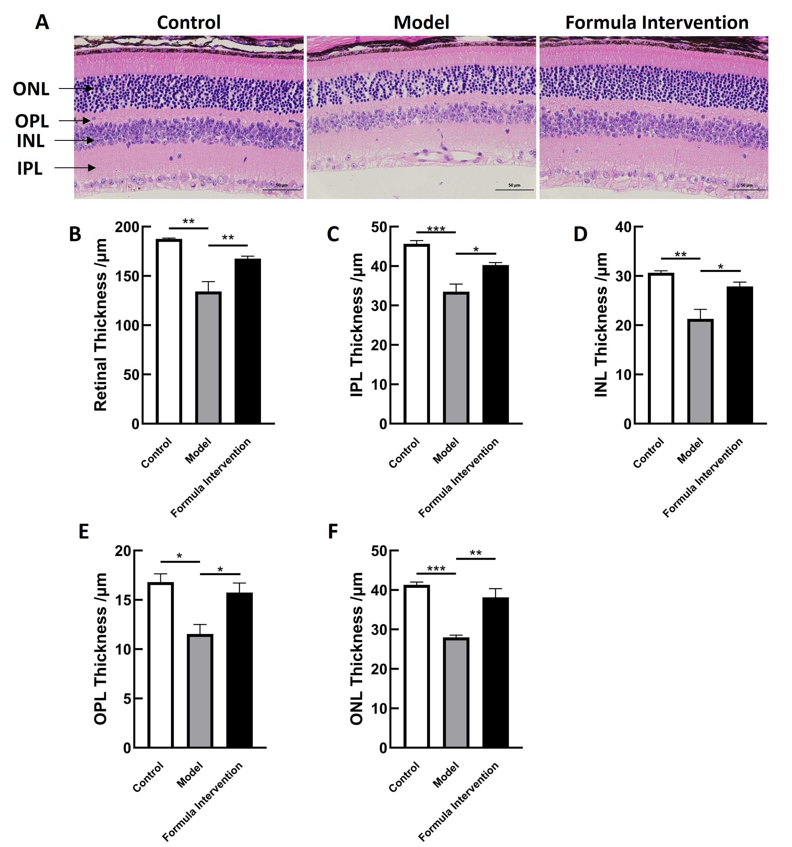
Figure 4
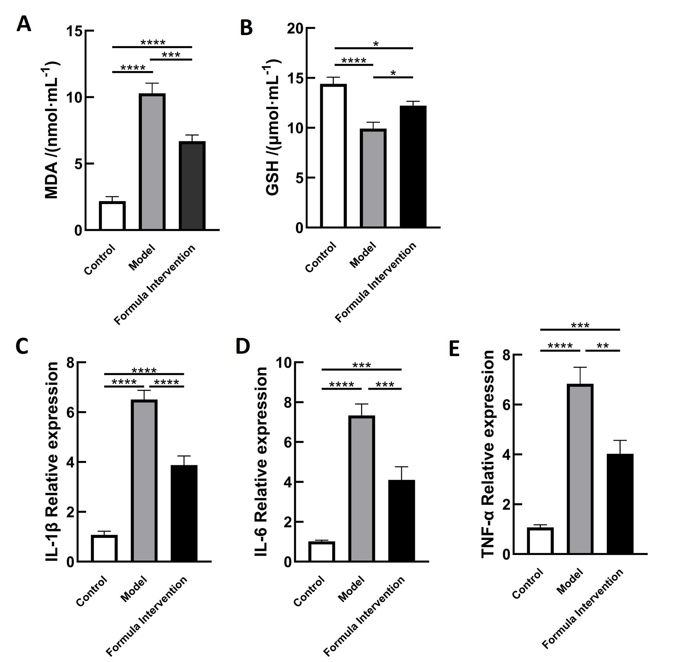
Figure 5
| [1] |
|
| [2] |
|
| [3] |
|
| [4] |
|
| [5] |
|
| [6] |
|
| [7] |
|
| [8] |
|
| [9] |
|
| [10] |
|
| [11] |
|
| [12] |
|
| [13] |
|
| [14] |
庞雨菲. 黑果枸杞多酚对丙烯酰胺诱导大鼠纹状体氧化应激损伤的保护作用及机制[D]. 银川: 宁夏医科大学, 2023.
|
|
|
|
| [15] |
|
| [16] |
娄解南, 谭书明, 谢国芳, 等. 刺梨多酚对丙烯酰胺致肝脏氧化损伤的保护作用[J]. 食品安全质量检测学报, 2023, 14(17): 122-128.
|
|
|
|
| [17] |
张丹, 葛颖颖, 杨琼琼, 等. 刺梨果抗氧化活性成分与体外抗氧化活性研究[C]//中国食品科学技术学会第十五届年会论文摘要集. 2018: 283-284.
|
|
|
|
| [18] |
|
| [19] |
|
| [20] |
|
| [21] |
|
| [22] |
|
| [23] |
马修彬, 陈松. 蓝光对视网膜色素上皮细胞的损伤机制[J]. 国际眼科纵览, 2007, 31(6): 415-419. DOI: 10.3760/cma.j.issn.1673-5803.2007.06.015 .
|
|
|
|
| [24] |
|
| [25] |
|
| [26] |
|
| [27] |
|
| [28] |
|
| [29] |
|
| [30] |
|
| [31] |
|
| [32] |
|
| [33] |
|
| [34] |
|
| [35] |
|
| [36] |
|
| [37] |
朱恒杏, 邵孟茹, 洪天畅, 等. 黑枸杞花青素、EGCG对中波紫外线诱导HaCaT细胞氧化损伤的协同保护作用[J]. 现代食品科技, 2024, 40(8): 106-115. DOI: 10.13982/j.mfst.1673-9078.2024.8.0918 .
|
|
|
|
| [38] |
邓楷, 欧阳健, 胡娜, 等. 黑果枸杞花青素结构差异对其稳定性及细胞抗氧化活性的影响[J]. 天然产物研究与开发, 2022, 34(2): 213-219. DOI: 10.16333/j.1001-6880.2022.2.005 .
|
|
|
|
| [39] |
李婧, 苏蓉, 刘振华, 等. 黑果枸杞果实中抗炎活性成分的筛选及作用机制研究[J]. 中国野生植物资源, 2023, 42(5): 9-15, 38. DOI: 10.3969/j.issn.1006-9690.2023.05.002 .
|
|
|
|
| [40] |
薛才华, 武强, 王梦杰, 等. 黑枸杞花青素对巨噬细胞RAW264.7炎性与自噬交互作用的影响[J]. 中国畜牧杂志, 2021, 57(12): 244-249.
|
|
|
|
| [41] |
|
| [42] |
|
| [43] |
|
| [44] |
|
| [45] |
|
| [1] | QIANG Sujing, LI Dongting, XU Jinhua, CAI Li, WEI Siyu, ZOU Jialei, DING Yuqiang. Identification and Analysis of the Klebsiella pneumoniae Species Complex in Laboratory Rats and Mice [J]. Laboratory Animal and Comparative Medicine, 2026, 46(4): 564-572. |
| [2] | DU Hongzhi, FAN Jin, WEI Xiaolu, WANG Lianmei, LIU Yan, CHEN Weiya, CHEN Tengfei, GAO Yunhang, SONG Ling, ZHANG Guangping, HOU Hongping. Multimodal Assessment of a Hypoxic Pulmonary Hypertension Mouse Model [J]. Laboratory Animal and Comparative Medicine, 2026, 46(4): 476-486. |
| [3] | WANG Juan, XU Jiahui, TIAN Yunyuan, ZHANG Mengmeng, LI Min, WANG Siwang, LI Yao. Comparison and Behavioral Observation of Two Female Mice Models of Ulcerative Colitis [J]. Laboratory Animal and Comparative Medicine, 2026, 46(3): 332-343. |
| [4] | LI Jiafei, ZHANG Zhenhao, WANG Shuo, TIAN Ge, WEN Shuang, YAN Yuxue, CUI Ran, YE Zhen, CUI Yongchun. Effects of Autonomic Neuromodulators on Atrial Electrical Remodeling and Histopathological Changes in a Rat Model of Atrial Fibrillation [J]. Laboratory Animal and Comparative Medicine, 2026, 46(3): 321-331. |
| [5] | LI Longxue, WAN Chongfan, ZHANG Qi, LEI Ruting, WANG Xiaoyue, CHENG Leyan, LAI Qi, LIU Ronghua, LIU Xuan, XU Tielong. Molecular Mechanisms of Qingfei Paidu Decoction in the Prevention and Treatment of Acute Lung Injury in Mice Based on miRNA Sequencing [J]. Laboratory Animal and Comparative Medicine, 2026, 46(3): 311-320. |
| [6] | AI Xiufeng, ZHANG Lizong, FANG Mingsun, LÜ Dongying, CHEN Chu, CAI Zhaowei, WANG Dejun. Analysis of Differences in the Intestinal Flora of Rats and Mice after Drinking Chlorinated Water Based on 16S rRNA Sequencing [J]. Laboratory Animal and Comparative Medicine, 2026, 46(3): 437-445. |
| [7] | LIU Sai, FU Bin, LI Sidi, CHEN Zhida, ZHANG Yue, GUO Zhongkun, WANG Yongan, WANG Kezhou. Adra2a Regulates LPS-Induced Inflammation in Hepatocytes of Lbp-/- Mice via the MAPK Signaling Pathway [J]. Laboratory Animal and Comparative Medicine, 2026, 46(2): 212-221. |
| [8] | ZHAO He, ZHANG Tao, LI Li, XIAO Yuzhou, AN Xuefang, ZHANG Fan. Enhancement Effect of Porcine Inhibin Polyclonal Antibody on Superovulation in C57BL/6J Mice [J]. Laboratory Animal and Comparative Medicine, 2026, 46(2): 271-278. |
| [9] | TANG Xiaohang, GU Yingmin, LÜ Yangyang, HUANG Mingshu, TIAN Xuesong. Evaluation of the Histological Staining Performance of Rat Eyeball Sections Prepared Using a Self-Developed Fixative [J]. Laboratory Animal and Comparative Medicine, 2026, 46(2): 261-270. |
| [10] | CUI Changwan, LU Yiping, YU Miao, WANG Shuang, WU Si, SUN Zhengrong. Inhibitory Effect of Solute Carrier Family 7 Member 5 Inhibitor JPH203 on Renal Fibrosis Induced by Unilateral Ureteral Obstruction in Mice [J]. Laboratory Animal and Comparative Medicine, 2026, 46(2): 205-211. |
| [11] | WU Xianwen, LIU Lili, CHEN Ye, XU Guoheng. Optimization of Cage-Changing Intervals and Wood Shavings Usage for Mice During the Growth Phase in Breeding Systems [J]. Laboratory Animal and Comparative Medicine, 2026, 46(2): 251-260. |
| [12] | RONG Wenshuang, NIU Yuanfei, LIU Meiting, YANG Mengyuan, CUI Shuang, MA Lina, FU Yao, WANG Lianmei, CAO Junling. Influence of Antigen Type on the Establishment of an Induced Sjögren Syndrome Mouse Model [J]. Laboratory Animal and Comparative Medicine, 2026, 46(2): 178-190. |
| [13] | XU Yingtao, WANG Mengmeng, LIN Ping, CHI Haitao, WANG Yi, BAI Ying. Exosomes Treat Ischemic Stroke by Regulation of Ferroptosis Through the NRF2/SLC7A11/GPX4 Pathway in Mice [J]. Laboratory Animal and Comparative Medicine, 2026, 46(1): 20-31. |
| [14] | YANG Lin, JIN Meng, WU Hanqing, LI Shun, ZHOU Xiaohui. Establishment and Preliminary Analysis of an AG6 Mouse Encephalopathy Model Induced by Vaccinia Virus Tiantan Strain Infection [J]. Laboratory Animal and Comparative Medicine, 2026, 46(1): 3-10. |
| [15] | LIU Chang, XIANG Xuesong, HE Huihuang, CHEN Xiaoqing, QIU Wenhong. Establishment and Evaluation of an Oxidative Stress Model of Atopic Dermatitis Induced by 2,4-dinitrofluorobenzene [J]. Laboratory Animal and Comparative Medicine, 2026, 46(1): 46-54. |
| Viewed | ||||||
|
Full text |
|
|||||
|
Abstract |
|
|||||
