
Laboratory Animal and Comparative Medicine ›› 2026, Vol. 46 ›› Issue (2): 261-270.DOI: 10.12300/j.issn.1674-5817.2025.083
• Animal Experimental Techniques and Methods • Previous Articles Next Articles
TANG Xiaohang1( ), GU Yingmin1, LÜ Yangyang1, HUANG Mingshu1()(
 ), TIAN Xuesong2()()
Received:2025-02-27
Revised:2025-10-18
Online:2026-04-25
Published:2026-04-18
Contact:
HUANG Mingshu, TIAN Xuesong
CLC Number:
TANG Xiaohang,GU Yingmin,Lü Yangyang,et al. Evaluation of the Histological Staining Performance of Rat Eyeball Sections Prepared Using a Self-Developed Fixative[J]. Laboratory Animal and Comparative Medicine, 2026, 46(2): 261-270. DOI: 10.12300/j.issn.1674-5817.2025.083.
Add to citation manager EndNote|Ris|BibTeX
URL: https://www.slarc.org.cn/dwyx/EN/10.12300/j.issn.1674-5817.2025.083
分值(程度) Score(grade) | 评分标准 Scoring criteria |
|---|---|
| 0(正常) | 各层组织结构完整,无裂痕、皱褶或脱片现象;各层细胞染色清晰、排列整齐、形态完整 |
| 1(轻微) | 裂痕、褶皱范围在10%以下,各层结构无分离,断裂;细胞染色清楚、形态完整、无变形 |
| 2(轻度) | 裂痕、褶皱范围在10%~30%,各层结构排列整齐,无分离;细胞染色清楚、组织完整、无变形 |
| 3(中度) | 裂痕、褶皱范围在30%~50%,各层结构分离、断裂;细胞界限不明显,颜色对比度降低,组织明显变形、破碎 |
| 4(重度) | 裂痕、褶皱在50%以上,大范围脱片、破碎明显;细胞界限不明显,颜色对比度降低,组织变形严重、收缩明显、结构不完整 |
Table 1 Quantitative scoring criteria for eyeball HE staining quality
分值(程度) Score(grade) | 评分标准 Scoring criteria |
|---|---|
| 0(正常) | 各层组织结构完整,无裂痕、皱褶或脱片现象;各层细胞染色清晰、排列整齐、形态完整 |
| 1(轻微) | 裂痕、褶皱范围在10%以下,各层结构无分离,断裂;细胞染色清楚、形态完整、无变形 |
| 2(轻度) | 裂痕、褶皱范围在10%~30%,各层结构排列整齐,无分离;细胞染色清楚、组织完整、无变形 |
| 3(中度) | 裂痕、褶皱范围在30%~50%,各层结构分离、断裂;细胞界限不明显,颜色对比度降低,组织明显变形、破碎 |
| 4(重度) | 裂痕、褶皱在50%以上,大范围脱片、破碎明显;细胞界限不明显,颜色对比度降低,组织变形严重、收缩明显、结构不完整 |
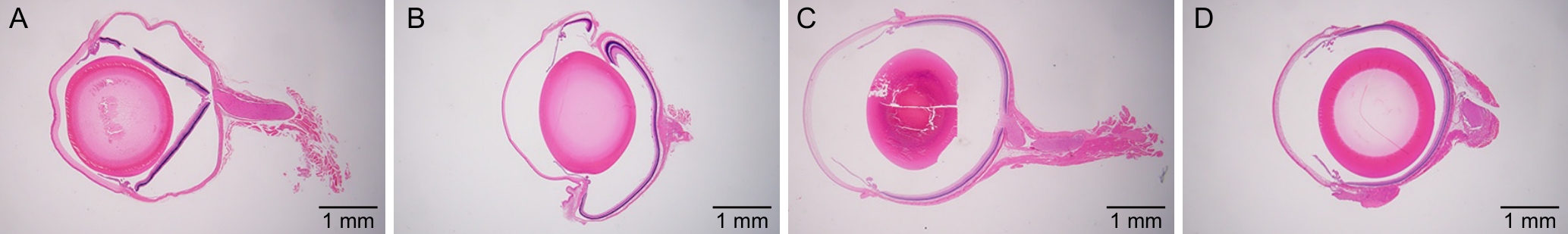
Figure 1 Overall morphology and tissue structure of rat eyeballs in different fixative groups (×12.5)
固定液种类 Fixative types | 眼球圆润度 Sphericality of eyeball | 角膜完整度 Corneal integrity | 晶状体完整度 Lens integrity | 视网膜完整度 Retinal integrity |
|---|---|---|---|---|
| 10%甲醛组(10% Formaldehyde group) | 皱缩、凹陷 | 可见裂隙 | 基本完整 | 完全分离、断裂 |
| 戊二醛-甲醛组(Glutaraldehyde-formaldehyde group) | 皱缩、凹陷 | 完整 | 完整 | 部分分离、断裂 |
| 改良Davidson固定液组(Modified Davidson’s fixative group) | 圆润、无变形 | 明显裂隙 | 破碎 | 完整 |
| 自研固定液组(Self-developed fixative group) | 圆润、无变形 | 有裂隙 | 完整 | 完整 |
Table 2 Comparison of overall morphology and tissue structure of rat eyeballs in different fixative groups
固定液种类 Fixative types | 眼球圆润度 Sphericality of eyeball | 角膜完整度 Corneal integrity | 晶状体完整度 Lens integrity | 视网膜完整度 Retinal integrity |
|---|---|---|---|---|
| 10%甲醛组(10% Formaldehyde group) | 皱缩、凹陷 | 可见裂隙 | 基本完整 | 完全分离、断裂 |
| 戊二醛-甲醛组(Glutaraldehyde-formaldehyde group) | 皱缩、凹陷 | 完整 | 完整 | 部分分离、断裂 |
| 改良Davidson固定液组(Modified Davidson’s fixative group) | 圆润、无变形 | 明显裂隙 | 破碎 | 完整 |
| 自研固定液组(Self-developed fixative group) | 圆润、无变形 | 有裂隙 | 完整 | 完整 |
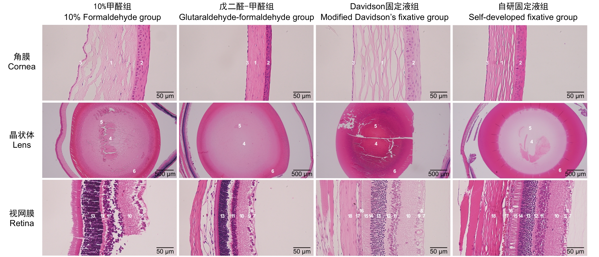
Figure 2 Microscopic observation of rat eyeballs structures in different fixative groups
观察指标 Observation indicators | 10%甲醛组 10% Formaldehyde group | 戊二醛-甲醛组 Glutaraldehyde-formaldehyde group | 改良Davidson固定液组 Modified Davidson’s fixative group | 自研固定液组 Self-developed fixative group | |
|---|---|---|---|---|---|
角膜 Cornea | 结构完整度 | 基质层明显裂痕 | 紧密 | 基质层明显裂痕 | 基质层轻微裂痕 |
| 各层结构 | 褶皱不规则、完整性差,上皮层与基质层分离 | 整齐、紧密无断裂 | 褶皱不规则、完整性差,上皮层与基质层分离明显 | 完整性尚可,基质层结构不紧密 | |
| 细胞紧密程度 | 裂隙较多 | 整齐无裂隙 | 裂隙较多 | 裂隙轻微 | |
| 染色情况 | 淡染 | 鲜艳 | 淡染 | 鲜艳 | |
晶状体 Lens | 结构完整度 | 赤道和皮质有裂痕 | 完整 | 大面积破碎、脱片 | 外围轻微褶皱 |
| 细胞紧密程度 | 有裂隙 | 整齐无裂隙 | 有裂隙 | 整齐无裂隙 | |
| 染色情况 | 鲜艳 | 鲜艳 | 深染 | 鲜艳 | |
视网膜 Retina | 完整度 | 断裂,部分结构脱落 | 较少断裂 | 完整无断裂 | 完整无断裂 |
| 各层结构 | 与脉络膜/巩膜严重分离 | 外网层分离肿胀,神经纤维层分离肿胀 | 各层完整无分离 | 各层完整无分离 | |
| 细胞紧密程度 | 大面积裂隙 | 部分有裂隙 | 整齐无裂隙 | 整齐无裂隙 | |
| 染色情况 | 鲜艳 | 鲜艳 | 淡染 | 鲜艳 | |
Table 3 Comparison of the histological structure of rat eyeballs in different fixative groups
观察指标 Observation indicators | 10%甲醛组 10% Formaldehyde group | 戊二醛-甲醛组 Glutaraldehyde-formaldehyde group | 改良Davidson固定液组 Modified Davidson’s fixative group | 自研固定液组 Self-developed fixative group | |
|---|---|---|---|---|---|
角膜 Cornea | 结构完整度 | 基质层明显裂痕 | 紧密 | 基质层明显裂痕 | 基质层轻微裂痕 |
| 各层结构 | 褶皱不规则、完整性差,上皮层与基质层分离 | 整齐、紧密无断裂 | 褶皱不规则、完整性差,上皮层与基质层分离明显 | 完整性尚可,基质层结构不紧密 | |
| 细胞紧密程度 | 裂隙较多 | 整齐无裂隙 | 裂隙较多 | 裂隙轻微 | |
| 染色情况 | 淡染 | 鲜艳 | 淡染 | 鲜艳 | |
晶状体 Lens | 结构完整度 | 赤道和皮质有裂痕 | 完整 | 大面积破碎、脱片 | 外围轻微褶皱 |
| 细胞紧密程度 | 有裂隙 | 整齐无裂隙 | 有裂隙 | 整齐无裂隙 | |
| 染色情况 | 鲜艳 | 鲜艳 | 深染 | 鲜艳 | |
视网膜 Retina | 完整度 | 断裂,部分结构脱落 | 较少断裂 | 完整无断裂 | 完整无断裂 |
| 各层结构 | 与脉络膜/巩膜严重分离 | 外网层分离肿胀,神经纤维层分离肿胀 | 各层完整无分离 | 各层完整无分离 | |
| 细胞紧密程度 | 大面积裂隙 | 部分有裂隙 | 整齐无裂隙 | 整齐无裂隙 | |
| 染色情况 | 鲜艳 | 鲜艳 | 淡染 | 鲜艳 | |
观察指标 Observation indicators | 分值/程度 Score/grade | 角膜 Cornea | 晶状体 Lens | 视网膜 Retina |
|---|---|---|---|---|
10%甲醛组 10% Formaldehyde group | 0(正常) | 0 | 0 | 0 |
| 1(轻微) | 0 | 1 | 0 | |
| 2(轻度) | 4 | 5 | 0 | |
| 3(中度) | 4 | 4 | 1 | |
| 4(重度) | 2 | 0 | 9 | |
戊二醛-甲醛组 Glutaraldehyde-formaldehyde group | 0(正常) | 6 | 2 | 0 |
| 1(轻微) | 4 | 5 | 0 | |
| 2(轻度) | 0 | 3 | 4 | |
| 3(中度) | 0 | 0 | 5 | |
| 4(重度) | 0 | 0 | 1 | |
改良Davidson固定液组 Modified Davidson’s fixative group | 0(正常) | 0 | 0 | 8 |
| 1(轻微) | 0 | 0 | 2 | |
| 2(轻度) | 0 | 0 | 0 | |
| 3(中度) | 2 | 0 | 0 | |
| 4(重度) | 8 | 10 | 0 | |
自研固定液组 Self-developed fixative group | 0(正常) | 0 | 2 | 9 |
| 1(轻微) | 0 | 6 | 1 | |
| 2(轻度) | 0 | 2 | 0 | |
| 3(中度) | 3 | 0 | 0 | |
| 4(重度) | 7 | 0 | 0 |
Table 4 Histological scoring results of rat eyeballs in different fixative groups
观察指标 Observation indicators | 分值/程度 Score/grade | 角膜 Cornea | 晶状体 Lens | 视网膜 Retina |
|---|---|---|---|---|
10%甲醛组 10% Formaldehyde group | 0(正常) | 0 | 0 | 0 |
| 1(轻微) | 0 | 1 | 0 | |
| 2(轻度) | 4 | 5 | 0 | |
| 3(中度) | 4 | 4 | 1 | |
| 4(重度) | 2 | 0 | 9 | |
戊二醛-甲醛组 Glutaraldehyde-formaldehyde group | 0(正常) | 6 | 2 | 0 |
| 1(轻微) | 4 | 5 | 0 | |
| 2(轻度) | 0 | 3 | 4 | |
| 3(中度) | 0 | 0 | 5 | |
| 4(重度) | 0 | 0 | 1 | |
改良Davidson固定液组 Modified Davidson’s fixative group | 0(正常) | 0 | 0 | 8 |
| 1(轻微) | 0 | 0 | 2 | |
| 2(轻度) | 0 | 0 | 0 | |
| 3(中度) | 2 | 0 | 0 | |
| 4(重度) | 8 | 10 | 0 | |
自研固定液组 Self-developed fixative group | 0(正常) | 0 | 2 | 9 |
| 1(轻微) | 0 | 6 | 1 | |
| 2(轻度) | 0 | 2 | 0 | |
| 3(中度) | 3 | 0 | 0 | |
| 4(重度) | 7 | 0 | 0 |
组别 Group | 角膜 Cornea | 晶状体 Lens | 视网膜 Retina |
|---|---|---|---|
| 改良Davidson固定液组(Modified Davidson’s fixative group) | 3.80±0.42 | 4.00±0.00 | 0.20±0.42 |
| 10%甲醛组(10% Formaldehyde group) | 2.80±0.79** | 2.30±0.67** | 3.90±0.32** |
| 戊二醛-甲醛组(Glutaraldehyde-formaldehyde group) | 0.40±0.52** | 1.10±0.74** | 2.70±0.67** |
| 自研固定液组(Self-developed fixative group) | 3.70±0.48 | 1.00±0.67** | 0.10±0.32 |
Table 5 Evaluation of sectioning quality of different parts of rat eyeballs in different fixative groups
组别 Group | 角膜 Cornea | 晶状体 Lens | 视网膜 Retina |
|---|---|---|---|
| 改良Davidson固定液组(Modified Davidson’s fixative group) | 3.80±0.42 | 4.00±0.00 | 0.20±0.42 |
| 10%甲醛组(10% Formaldehyde group) | 2.80±0.79** | 2.30±0.67** | 3.90±0.32** |
| 戊二醛-甲醛组(Glutaraldehyde-formaldehyde group) | 0.40±0.52** | 1.10±0.74** | 2.70±0.67** |
| 自研固定液组(Self-developed fixative group) | 3.70±0.48 | 1.00±0.67** | 0.10±0.32 |
| [1] | 成前, 李月, 王伟进, 等. 中国老年人口健康状况及其家庭照料需求预测[J]. 人口学刊, 2024, 46(5):73-89. DOI: 10.16405/j.cnki.1004-129X.2024.05.005 . |
| CHENG Q, LI Y, WANG W J, et al. The prediction of health status and family care needs of the elderly people in China[J]. Popul J, 2024, 46(5):73-89. DOI: 10.16405/j.cnki.1004-129X.2024.05.005 . | |
| [2] | 王余萍, 袁源智. 青光眼视神经损害机制[J]. 中国临床医学, 2016, 23(5):667-671. DOI: 10.12025/j.issn.1008-6358.2016.2016 0290 . |
| WANG Y P, YUAN Y Z. Mechanisms of glaucomatous optic neuropathy[J]. Chin J Clin Med, 2016, 23(5):667-671. DOI: 10.12025/j.issn.1008-6358.2016.20160290 . | |
| [3] | 蔡永民. 白内障发病机制及治疗进展研究[J]. 医学理论与实践, 2020, 33(15):2450-2452. DOI: 10.19381/j.issn.1001-7585.2020.15.012 . |
| CAI Y M. Research on pathogenesis and therapeutic advances of cataract[J]. J Med Theory Pract, 2020, 33(15):2450-2452. DOI: 10.19381/j.issn.1001-7585.2020.15.012 . | |
| [4] | 葛军, 朱思泉. 中医药治疗老年性黄斑变性的研究进展[J]. 中医临床研究, 2023, 15(21):74-78. DOI: 10.3969/j.issn.1674-7860.2023.21.014 . |
| GE J, ZHU S Q. A review on the TCM treatment of age-related macular degeneration[J]. Clin J Chin Med, 2023, 15(21):74-78. DOI: 10.3969/j.issn.1674-7860.2023.21.014 . | |
| [5] | 裴希. 螺内酯与缬沙坦对氧诱导小鼠视网膜病变新生血管抑制作用及机制的初步探讨[D]. 广州: 南方医科大学, 2014. |
| PEI X. A Preliminary Study on the Inhibitory effects and mechanisms of spironolactone and valsartan on oxygen-induced retinopathy neovascularization in mice[D]. Guangzhou: Southern Medical University, 2014. | |
| [6] | 孙河龙, 李耀洋, 李丹, 等. 组织固定液与固定方法选择的探讨[J]. 甘肃医药, 2019, 38(12):1061-1064, 1069. DOI: 10.15975/j.cnki.gsyy.2019.12.002 . |
| SUN H L, LI Y Y, LI D, et al. Selection of tissue fixation fluid and fixation method[J]. Gansu Med J, 2019, 38(12):1061-1064, 1069. DOI: 10.15975/j.cnki.gsyy.2019.12.002 . | |
| [7] | SCHAFER K A, EIGHMY J, FIKES J D, et al. Use of severity grades to characterize histopathologic changes[J]. Toxicol Pathol, 2018, 46(3): 256-265. DOI: 10.1177/0192623318761348 . |
| [8] | 李晶晶, 朱鸿, 施彩虹. 三种方法对大鼠视网膜固定效果的比较研究[J]. 上海交通大学学报(医学版), 2011, 31(8):1105-1107. DOI: 10.3969/j.issn.1674-8115.2011.08.013 . |
| LI J J, ZHU H, SHI C H. Outcomes of rat retina fixation with three different methods[J]. J Shanghai Jiaotong Univ Med Sci, 2011, 31(8):1105-1107. DOI: 10.3969/j.issn.1674-8115.2011.08.013 . | |
| [9] | 王松涛, 肖虹蕾, 王敏, 等. 四种不同固定液固定小鼠视网膜效果的比较研究[J]. 眼科新进展, 2016, 36(8):709-712. DOI: 10.13389/j.cnki.rao.2016.0188 . |
| WANG S T, XIAO H L, WANG M, et al. Comparative study of mouse retinal fixation outcomes with four different fixation solutions[J]. Recent Adv Ophthalmol, 2016, 36(8):709-712. DOI: 10.13389/j.cnki.rao.2016.0188 . | |
| [10] | 张遐, 鞠躬, 董光皎. 甲醛和戊二醛应用于神经系统免疫组织化学组织固定的特性分析[J]. 神经解剖学杂志, 1990, 6(2):249-256. |
| ZHANG X, JU G, DONG G J. Analysis of the properties of formaldehyde and glutaraldehyde for tissue fixation in neural system immunohistochemistry[J]. Chin J Neuroanat, 1990, 6(2):249-256. | |
| [11] | 宋惠欣, 蒋文君, 毕宏生. 三种不同固定液对豚鼠眼球的固定效果比较[J]. 国际眼科杂志, 2018, 18(6):1010-1013. DOI: 10.3980/j.issn.1672-5123.2018.6.07 . |
| SONG H X, JIANG W J, BI H S. A comparative study on the effect of fixation for guinea pigs eyeballs among three different fixation solution[J]. Int Eye Sci, 2018, 18(6):1010-1013. DOI: 10.3980/j.issn.1672-5123.2018.6.07 . | |
| [12] | 张文忻, 李永平, 林健贤, 等. 冰醋酸固定液对视网膜组织固定效果的探讨[J]. 眼科学报, 2006, 22(2):112-114. DOI: 10.3969/j.issn.1000-4432.2006.02.012 . |
| ZHANG W X, LI Y P, LIN J X, et al. The investigation of FFA fixative solution for retina[J]. Eye Sci, 2006, 22(2):112-114. DOI: 10.3969/j.issn.1000-4432.2006.02.012 . | |
| [13] | 赵宝忠, 刘俊华, 高俊琴, 等. 乙醇类固定剂对组织细胞处理性能的实验评价[J]. 临床与实验病理学杂志, 2012, 28(2):223-225. DOI: 10.3969/j.issn.1001-7399.2012.02.033 . |
| ZHAO B Z, LIU J H, GAO J Q, et al. Experimental evaluation of ethanol-based fixatives for the processing of tissues and cells[J]. Chin J Clin Exp Pathol, 2012, 28(2):223-225. DOI: 10.3969/j.issn.1001-7399.2012.02.033 . | |
| [14] | TOKUDA K, BARON B, KURAMITSU Y, et al. Optimization of fixative solution for retinal morphology: a comparison with Davidson's fixative and other fixation solutions[J]. Jpn J Ophthalmol, 2018, 62(4):481-490. DOI: 10.1007/s10384-018-0592-7 . |
| [15] | WANG H, YANG L L, JI Y L,et al. Different fixative methods influence histological morphology and TUNEL staining in mouse testes[J].Reprod Toxicol, 2016, 60:53-61.DOI: 10.1016/j.reprotox.2016.01.006 . |
| [16] | BAK S Y, LEE S W, CHOI C H, et al. Assessment of the influence of acetic acid residue on typeⅠcollagen during isolation and characterization[J]. Materials, 2018, 11(12):2518. DOI: 10.3390/ma11122518 . |
| [17] | 郭志鲲. 蛋白质变性作用研究动向[J]. 生物化学与生物物理进展, 1974, 1(4):16-22, 49. DOI: CNKI:SUN:SHSW.0.1974-04-006 . |
| GUO Z K. Advances in the study of protein denaturation[J]. Prog Biochem Biophys, 1974, 1(4):16-22, 49. DOI: CNKI:SUN:SHSW.0.1974-04-006 . | |
| [18] | POSOKHOV Y O, KYRYCHENKO A. Effect of acetone accumulation on structure and dynamics of lipid membranes studied by molecular dynamics simulations[J]. Comput Biol Chem, 2013, 46:23-31. DOI: 10.1016/j.compbiolchem.2013. 04.005 . |
| [19] | AL-GHOUL K J, COSTELLO M J. Fiber cell morphology and cytoplasmic texture in cataractous and normal human lens nuclei[J]. Curr Eye Res, 1996, 15(5):533-542. DOI: 10.3109/0271 3689609000764 . |
| [20] | 常晓杰, 徐颖超, 刘畅. 不同实验方法检测常用有机溶剂对细菌活性的影响及其安全使用限量[J]. 微生物学通报, 2016, 43(7):1635-1645. DOI: 10.13344/j.microbiol.china.150595 . |
| CHANG X J, XU Y C, LIU C. Effects of common solvent concentrations on bacterial activities[J]. Microbiol China, 2016, 43(7):1635-1645. DOI: 10.13344/j.microbiol.china.150595 . | |
| [21] | 黄熙泰, 于自然, 李翠凤. 现代生物化学[M]. 3版. 北京: 化学工业出版社, 2012. |
| HUANG X T, YU Z R, LI C F. Modern Biochemistry[M]. 3rd. Beijing: Chemical Industry Press, 2012. | |
| [22] | ZAZERI G, POVINELLI A P R, PAVAN N M, et al. Solvent-induced lag phase during the formation of lysozyme amyloid fibrils triggered by sodium dodecyl sulfate: biophysical experimental and in silico study of solvent effects[J]. Molecules, 2023, 28(19):6891. DOI: 10.3390/molecules 2819 6891 . |
| [23] | SUN N, SHIBATA B, HESS J F, et al. An alternative means of retaining ocular structure and improving immunoreactivity for light microscopy studies[J]. Mol Vis, 2015, 21:428-442. |
| [24] | 秦川. 实验动物比较组织学彩色图谱[M]. 北京: 科学出版社, 2017: 234-241, 247. |
| Qin C. Color atlas of comparative histology of laboratory animals[M]. Beijing: Science Press, 2017: 234-241, 247. |
| [1] | SONG Jing, YANG Zongtong, LI Xiaojing, LI Zifa, SU Fengyun, XU Dongchuan, SUI Zaiyun. Effects of Xiebai San on the Morphological Structures of Lung and Intestinal Tissues and Expression Levels of PI3K and Akt in Rats with Allergic Asthma [J]. Laboratory Animal and Comparative Medicine, 2026, 46(2): 191-204. |
| [2] | JIANG Haitao, YUAN Hantao, HUANG Wenting, YANG Rongrong, CHEN Xiaochun, YU Baoqing, LI Sibo. Regulation of Rat Intervertebral Disc Annulus Fibrosus Cell Proliferation and Apoptosis by Yaoshu Zhuyu Fang via miR-17-5P/MDM2/p53 Pathway [J]. Laboratory Animal and Comparative Medicine, 2026, 46(1): 55-65. |
| [3] | GAO Chaoqi, ZHU Zhibo, SUN Xiandong. Application Progress and Classification Analysis of Rat Vascular Remodeling Models [J]. Laboratory Animal and Comparative Medicine, 2025, 45(5): 542-550. |
| [4] | LUO Yifan, ZHANG Zhenwei, MEI Lu, SHI Yeping, XING Yitong, ZHANG Zeqi, LI Chuxin, HAN Chunxia, YANG Pingshun, CHEN Qiusheng. Telocytes-Mediated Effects and Mechanisms of Anointing and Massage Therapy Using Oligopeptide-Herbal Medicine Composite Against Obesity in Rats [J]. Laboratory Animal and Comparative Medicine, 2025, 45(5): 551-560. |
| [5] | QIN Chao, LI Shuangxing, ZHAO Tingting, JIANG Chenchen, ZHAO Jing, YANG Yanwei, LIN Zhi, WANG Sanlong, WEN Hairuo. Study on the 90-day Feeding Experimental Background Data of SD Rats for Drug Safety Evaluation [J]. Laboratory Animal and Comparative Medicine, 2025, 45(4): 439-448. |
| [6] | LIU Liyu, JI Bo, LIU Xiaoxuan, FANG Yang, ZHANG Ling, GUO Tingting, QUAN Ye, LI Hewen, LIU Yitian. Exploration of Rat Fetal Lung Tissue Fixation Methods [J]. Laboratory Animal and Comparative Medicine, 2025, 45(4): 432-438. |
| [7] | PAN Yicong, JIANG Wenhong, HU Ming, QIN Xiao. Optimization of Surgical Procedure and Efficacy Evaluation of Aortic Calcification Model in Rats with Chronic Kidney Disease [J]. Laboratory Animal and Comparative Medicine, 2025, 45(3): 279-289. |
| [8] | LIU Zhiwei, YANG Ran, LIAN Hao, ZHANG Yu, JIN Lilun. Cartilage Protection and Anti-Inflammatory Effects of Fraxetin on Monosodium Iodoacetate-Induced Rat Model of Osteoarthritis [J]. Laboratory Animal and Comparative Medicine, 2025, 45(3): 259-268. |
| [9] | JIANG Meng, HAO Shulan, TONG Liguo, ZHONG Qiming, GAO Zhenfei, WANG Yonghui, WANG Xixing, JI Haijie. Dynamic Evaluation of Vinorelbine-Induced Phlebitis of Dorsalis Pedis Vein in a Rat Model [J]. Laboratory Animal and Comparative Medicine, 2025, 45(3): 251-258. |
| [10] | LIAN Hui, JIANG Yanling, LIU Jia, ZHANG Yuli, XIE Wei, XUE Xiaoou, LI Jian. Construction and Evaluation of a Rat Model of Abnormal Uterine Bleeding [J]. Laboratory Animal and Comparative Medicine, 2025, 45(2): 130-146. |
| [11] | WU Haifeng, ZHOU Xiaojiang, LI Chenjiang, LI Huaiyin, GAO Ming. Comparison of the Fixation Effects of Six Composite Fixatives on Retinal Tissue of Golden Hamsters [J]. Laboratory Animal and Comparative Medicine, 2024, 44(6): 675-681. |
| [12] | YIN Yulian, MA Lina, TU Siyuan, CHEN Ling, YE Meina, CHEN Hongfeng. Establishment and Evaluation of a Rat Model of Non-Puerperal Mastitis [J]. Laboratory Animal and Comparative Medicine, 2024, 44(6): 587-596. |
| [13] | YANG Jin, YU Shiya, LIN Nan, FANG Yongchao, ZHAO Hu, QIU Jinwei, LIN Hongming, CHEN Huiyan, WANG Yu, WU Weihang. Effect of Modified Duodenal Exclusion Surgery on Glucose Metabolism in Rats with Type 2 Diabetes Mellitus [J]. Laboratory Animal and Comparative Medicine, 2024, 44(5): 523-530. |
| [14] | QI Longju, CHEN Shiyuan, LIAO Zehua, SHI Yuanhu, SUN Yuyu, WANG Qinghua. Transcriptomic Analysis of Menstrual Blood-Derived Stem Cells Transplantation Combined with Exercise Training in Promoting Spinal Cord Injury Recovery in Rats [J]. Laboratory Animal and Comparative Medicine, 2024, 44(5): 531-542. |
| [15] | ZHANG Naiqun, YUAN Piaopiao, CAO Linrong, YING Na, YANG Taotao. Application of PNR Detection in the Diagnosis and Drug-efficacy Evaluation of Diabetic Kidney Disease in Rats [J]. Laboratory Animal and Comparative Medicine, 2024, 44(5): 543-549. |
| Viewed | ||||||
|
Full text |
|
|||||
|
Abstract |
|
|||||