
Laboratory Animal and Comparative Medicine ›› 2026, Vol. 46 ›› Issue (2): 191-204.DOI: 10.12300/j.issn.1674-5817.2025.084
• Animal Models of Human Diseases • Previous Articles Next Articles
SONG Jing1,2,3, YANG Zongtong2( )(
)( ), LI Xiaojing2, LI Zifa1, SU Fengyun3, XU Dongchuan2, SUI Zaiyun2()()
Received:2025-06-10
Revised:2025-08-21
Online:2026-04-25
Published:2026-04-18
Correspondence to:
YANG Zongtong, SUI Zaiyun
CLC Number:
SONG Jing,YANG Zongtong,LI Xiaojing,et al. Effects of Xiebai San on the Morphological Structures of Lung and Intestinal Tissues and Expression Levels of PI3K and Akt in Rats with Allergic Asthma[J]. Laboratory Animal and Comparative Medicine, 2026, 46(2): 191-204. DOI: 10.12300/j.issn.1674-5817.2025.084.
Add to citation manager EndNote|Ris|BibTeX
URL: https://www.slarc.org.cn/dwyx/EN/10.12300/j.issn.1674-5817.2025.084
基因名称 Gene name | 引物序列(5'→3') Primer sequence (5'→3') | 产物大小/bp Product size/bp |
|---|---|---|
| PI3K | F: GCGTGACATGTAGGCTCTCG | 349 |
| R: GGGCAGTGCTGGTGGAT | ||
| Akt | F: CCGCCTGATCAAGTTCTCCT | 118 |
| R: TTCAGATGATCCATGCGGGG | ||
| β-actin | F: CGCAGCCACTGTCGAGTC | 96 |
| R: GTCATCCATGGCGAACTGGT |
Table 1 Primer design and synthesis
基因名称 Gene name | 引物序列(5'→3') Primer sequence (5'→3') | 产物大小/bp Product size/bp |
|---|---|---|
| PI3K | F: GCGTGACATGTAGGCTCTCG | 349 |
| R: GGGCAGTGCTGGTGGAT | ||
| Akt | F: CCGCCTGATCAAGTTCTCCT | 118 |
| R: TTCAGATGATCCATGCGGGG | ||
| β-actin | F: CGCAGCCACTGTCGAGTC | 96 |
| R: GTCATCCATGGCGAACTGGT |
程序 Program | 温度/℃ Temperature/℃ | 时间 Time |
|---|---|---|
预变性 Initial denaturation | 95 | 3 min |
扩增 Amplification | 95 | 20 s |
45个循环 45 cycles | 61 | 15 s |
熔解曲线 Melting curve | 95 | 5 s |
| 65 | 1 min | |
| 97 | 10 s |
Table 2 PCR cycling program
程序 Program | 温度/℃ Temperature/℃ | 时间 Time |
|---|---|---|
预变性 Initial denaturation | 95 | 3 min |
扩增 Amplification | 95 | 20 s |
45个循环 45 cycles | 61 | 15 s |
熔解曲线 Melting curve | 95 | 5 s |
| 65 | 1 min | |
| 97 | 10 s |
组别 Group | 肺气肿积分 Pulmonary emphysema score | 肺充/出血积分 Pulmonary congestion/bleeding score | 肺间质增厚积分 Pulmonary interstitial thickening score | 炎细胞浸润积分 Inflammatory cell infiltration score | 总积分 Total score |
|---|---|---|---|---|---|
空白组 Control group | 0.00±0.00 | 0.67±0.58 | 0.67±0.58 | 0.67±0.58 | 2.00±1.00 |
模型组 Model group | 2.56±0.53** | 1.56±0.53* | 1.89±0.60* | 1.89±0.78* | 7.89±1.05** |
阳性药组 Positive control group | 1.86±0.69## | 1.43±0.53 | 2.14±0.38 | 1.57±0.53 | 7.00±1.15 |
泻白散组 Xiebai San group | 1.46±0.88## | 1.54±0.52 | 1.46±0.52 | 1.46±0.52 | 5.92±1.26# |
Table 3 Comparison of McGuigan pathological scores in lung tissues of rats after Xiebai San administration
组别 Group | 肺气肿积分 Pulmonary emphysema score | 肺充/出血积分 Pulmonary congestion/bleeding score | 肺间质增厚积分 Pulmonary interstitial thickening score | 炎细胞浸润积分 Inflammatory cell infiltration score | 总积分 Total score |
|---|---|---|---|---|---|
空白组 Control group | 0.00±0.00 | 0.67±0.58 | 0.67±0.58 | 0.67±0.58 | 2.00±1.00 |
模型组 Model group | 2.56±0.53** | 1.56±0.53* | 1.89±0.60* | 1.89±0.78* | 7.89±1.05** |
阳性药组 Positive control group | 1.86±0.69## | 1.43±0.53 | 2.14±0.38 | 1.57±0.53 | 7.00±1.15 |
泻白散组 Xiebai San group | 1.46±0.88## | 1.54±0.52 | 1.46±0.52 | 1.46±0.52 | 5.92±1.26# |
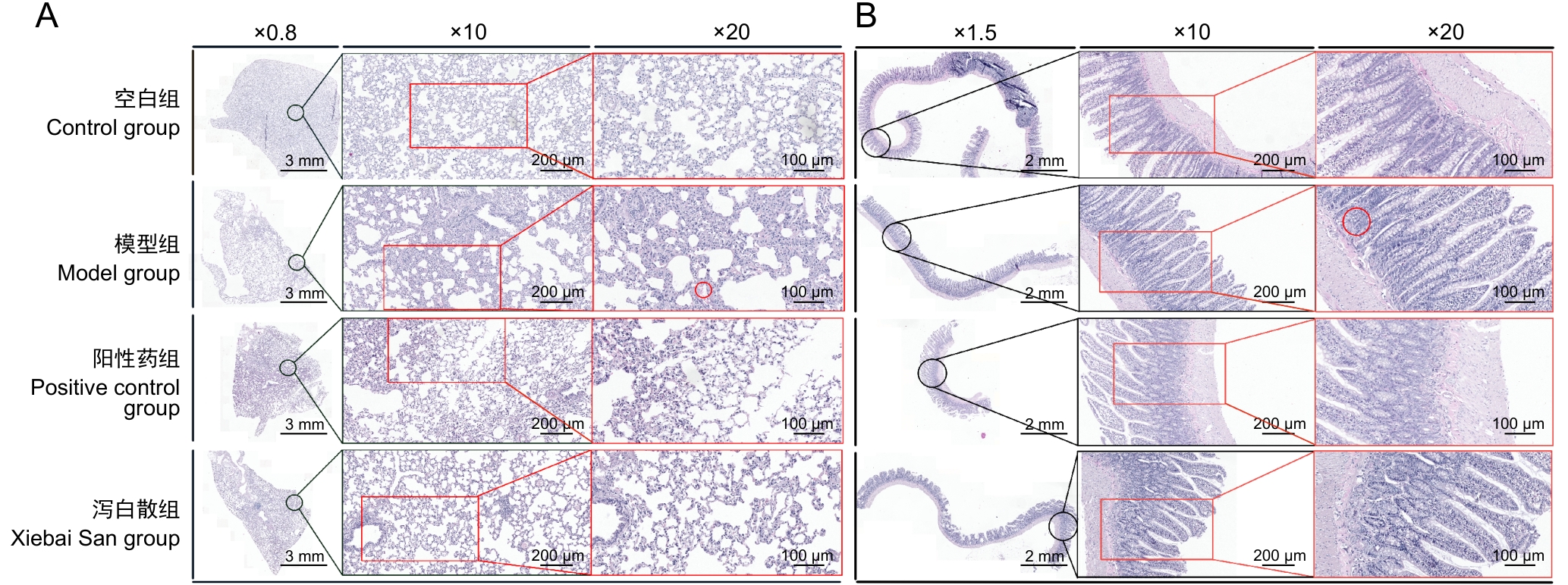
Figure 1 Effects of Xiebai San on histopathological changes in lung and intestinal tissues of asthmatic rats
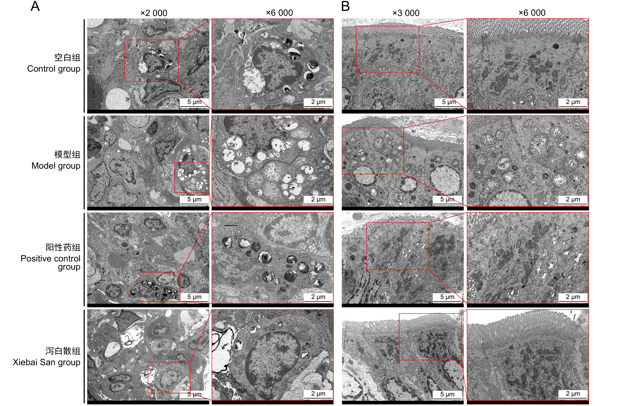
Figure 2 Effects of Xiebai San administration on ultrastructural pathology of lung and intestinal tissues in asthmatic rats
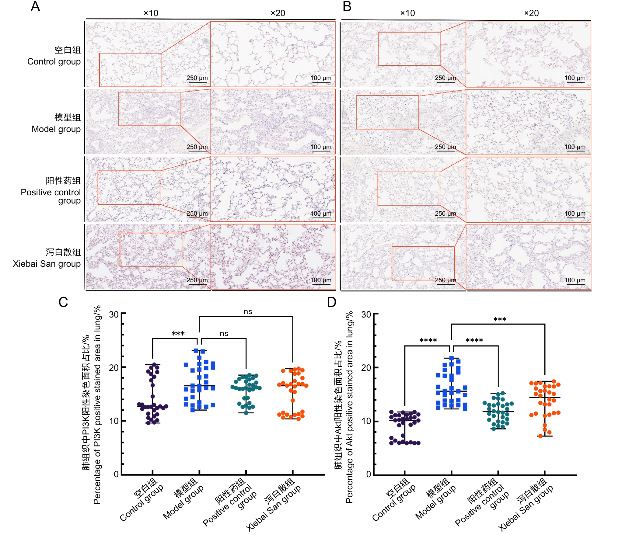
Figure 3 Effects of Xiebai San administration on PI3K and Akt protein distribution in lung tissues of asthmatic rats
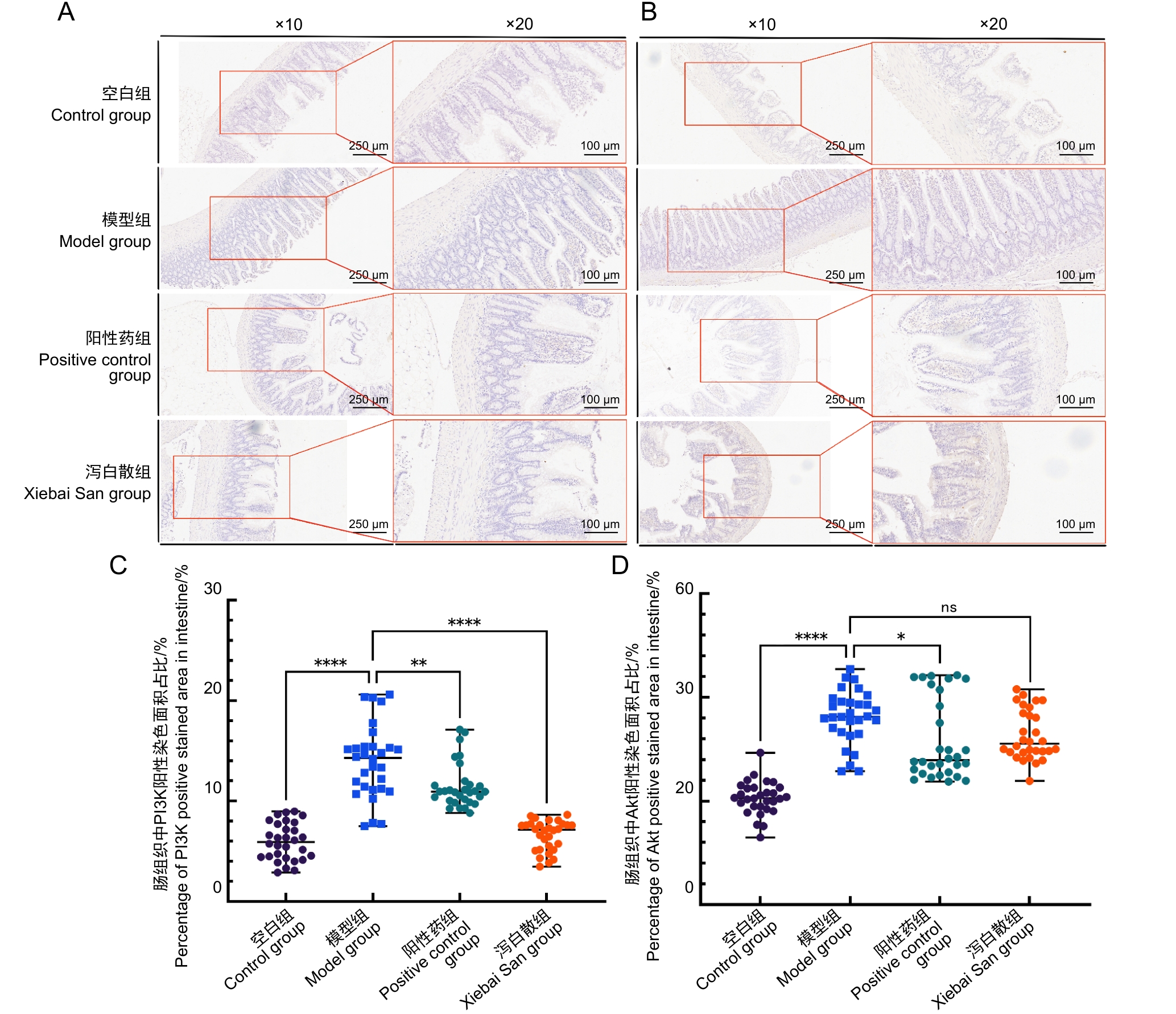
Figure 4 Effects of Xiebai San administration on PI3K and Akt protein distribution in intestinal tissues of asthmatic rats
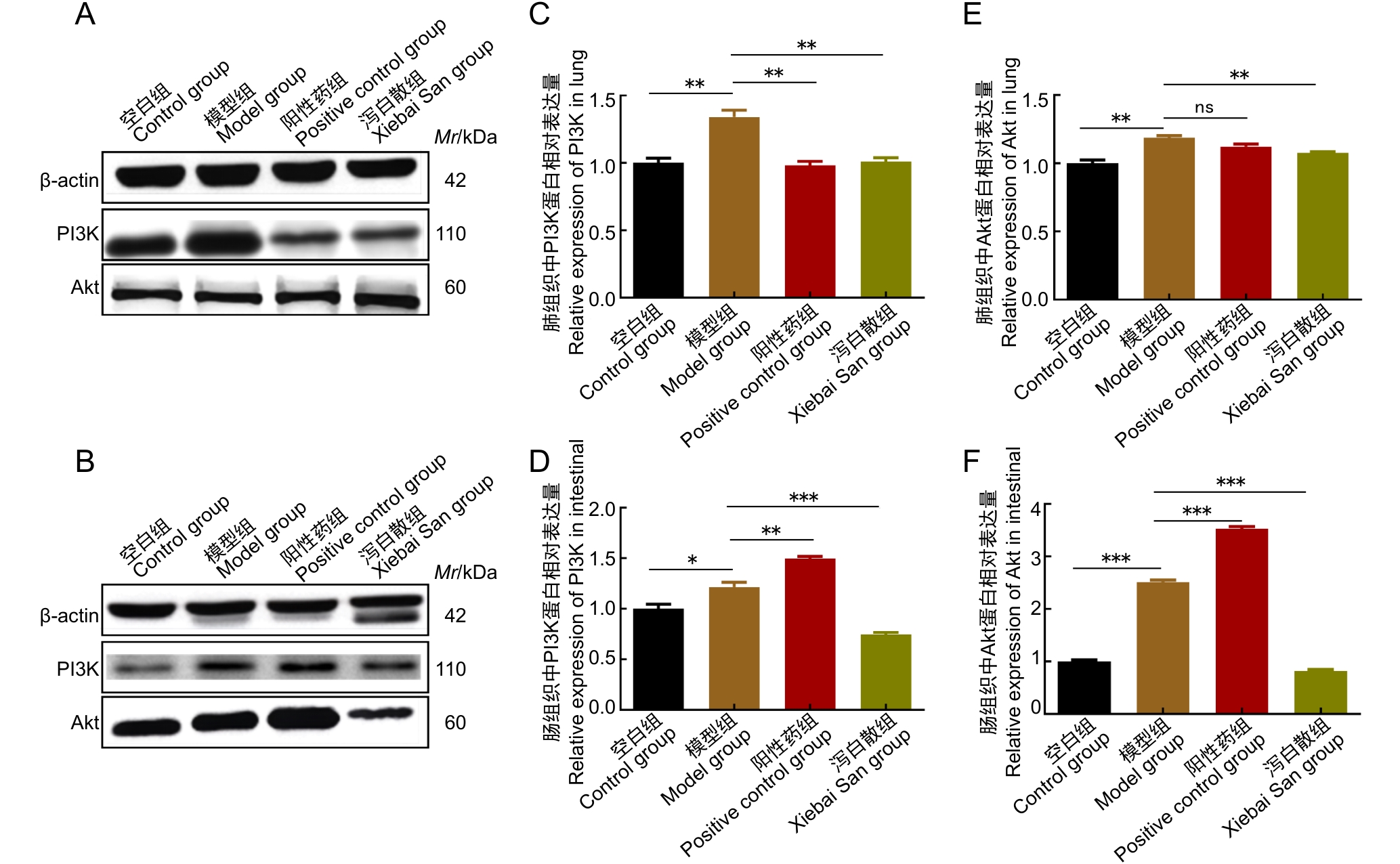
Figure 5 Effects of Xiebai San administration on PI3K and Akt protein expression in lung and intestinal tissues of asthmatic rats
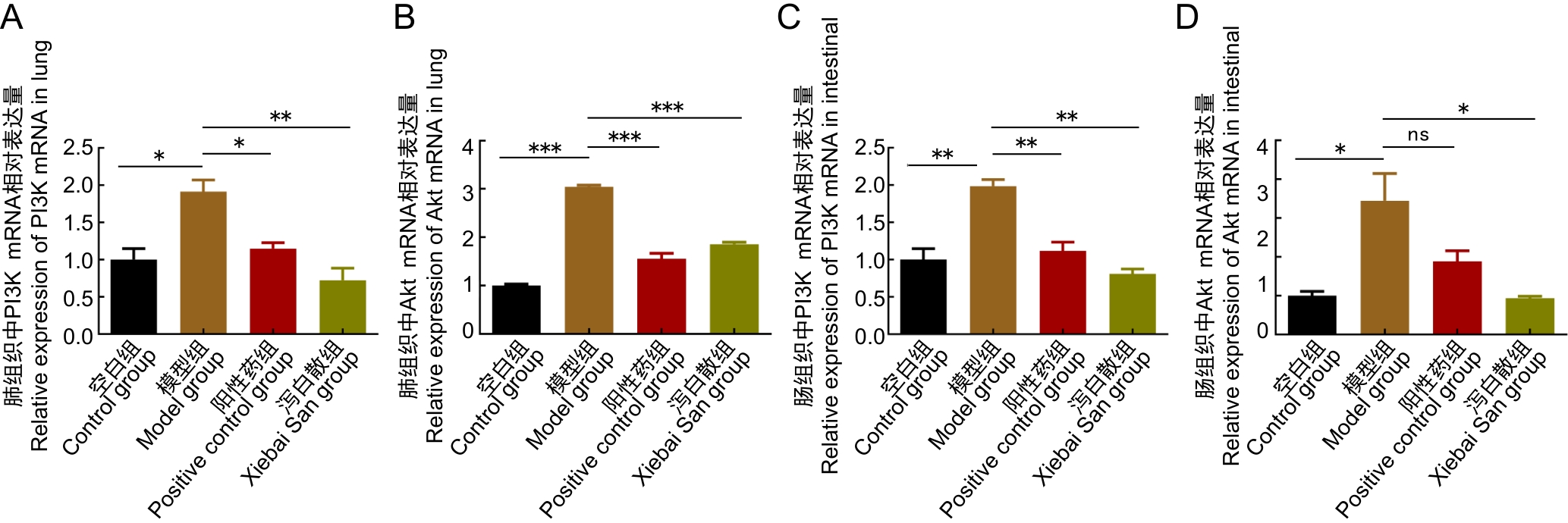
Figure 6 Effects of Xiebai San administration on PI3K and Akt gene expression in lung and intestinal tissues of asthmatic rats
| [1] | 任雪交, 黄巍, 邱菲菲, 等. 支气管哮喘动物模型研究的文献分析与评价[J]. 实验动物与比较医学, 2022, 42(1): 74-80. DOI:10.12300/j.issn.1674-5817.2021.044 . |
| REN X J, HUANG W, QIU F F, et al. Progress in animal models for bronchial asthma[J]. Lab Anim Comp Med, 2022, 42(1): 74-80. DOI:10.12300/j.issn.1674-5817.2021.044 . | |
| [2] | 罗世雄, 张赛, 陈慧. 常见哮喘动物模型的建立方法与评价研究进展[J]. 实验动物与比较医学, 2025, 45(2): 167-175. DOI:10.12300/j.issn.1674-5817.2024.120 . |
| LUO S X, ZHANG S, CHEN H. Research progress in establishment and evaluation of common asthma animal models[J]. Lab Anim Comp Med, 2025, 45(2): 167-175. DOI:10.12300/j.issn.1674-5817.2024.120 . | |
| [3] | 兰露莎, 赵兵兵, 杨红宇, 等. 氧化苦参碱对小鼠哮喘模型保护作用的初步研究[J]. 实验动物与比较医学, 2017, 37(4): 320-323. DOI:10.3969/j.issn.1674-5817.2017.04.013 . |
| LAN L S, ZHAO B B, YANG H Y, et al. The protective effect of oxymatrine on asthma mice[J]. Lab Anim Comp Med, 2017, 37(4): 320-323. DOI:10.3969/j.issn.1674-5817.2017.04.013 . | |
| [4] | AN J, LIU Y Q, WANG Y Q, et al. The role of intestinal mucosal barrier in autoimmune disease: a potential target[J]. Front Immunol, 2022, 13:871713. DOI:10.3389/fimmu.2022.871713 . |
| [5] | CERVANTES-GARCÍA D, JIMÉNEZ M, RIVAS-SANTIAGO C E, et al. Lactococcus lactis NZ9000 prevents asthmatic airway inflammation and remodelling in rats through the improvement of intestinal barrier function and systemic TGF-β production[J]. Int Arch Allergy Immunol, 2021, 182(4):277-291. DOI:10.1159/000511146 . |
| [6] | 洪天一. 蝎黄解痉治哮颗粒影响NF-κB和HIF-1α信号传导通路治疗哮喘的调控机制[D]. 长春: 长春中医药大学, 2020. DOI: 10.26980/d.cnki.gcczc.2020.000459 . |
| HONG T Y. Regulatory mechanism of Xiehuang Jiezhuan Zhixiao granules on the NF-κB and HIF-1α signaling pathways in the treatment of asthma[D]. Changchun: Changchun University of Chinese Medicine, 2020. DOI: 10.26980/d.cnki.gcczc.2020.000459 . | |
| [7] | 马兰兰, 陈玲, 王琴, 等. 肺通气功能正常的儿童支气管哮喘控制情况及急性发作随访的研究[J]. 中国当代儿科杂志, 2024, 26(5): 476-480. DOI:10.7499/j.issn.1008-8830.2311149 . |
| MA L L, CHEN L, WANG Q, et al. Control status and follow-up of acute attacks in children with bronchial asthma with normal pulmonary ventilation function[J]. Chin J Contemp Pediatr, 2024, 26(5): 476-480. DOI:10.7499/j.issn.1008-8830.2311149 . | |
| [8] | 丁云录, 郑明昱, 南敏伦, 等. 基于ERK信号通路探讨鹿茸大补汤颗粒对哮喘缓解期豚鼠的调控及作用机制[J]. 中国老年学杂志, 2023, 43(21):5309-5313. DOI:10.3969/j.issn.1005-9202.2023.21.054 . |
| DING Y L, ZHENG M Y, NAN M L, et al. Based on ERK signal pathway, this paper discusses the regulation and mechanism of Lulong Dabutang Granule on guinea pigs with asthma in remission stage[J]. Chin J Gerontol, 2023, 43(21):5309-5313. DOI:10.3969/j.issn.1005-9202.2023.21.054 . | |
| [9] | 李玉丽, 易腾达, 谭志强, 等. 经典名方泻白散的源流及古今应用考究[J]. 中国实验方剂学杂志, 2021, 27(4):168-174. DOI:10.13422/j.cnki.syfjx.20202328 . |
| LI Y L, YI T D, TAN Z Q, et al. Literature research on origin and application of classical prescription xiebaisan[J]. Chin J Exp Tradit Med Formulae, 2021, 27(4):168-174. DOI:10.13422/j.cnki.syfjx.20202328 . | |
| [10] | 刘凌志, 梁敏兰, 郭栋伟. 黄芩泻白散对支气管哮喘小鼠的治疗作用及Wnt/β-catenin信号轴的影响[J]. 湖南中医杂志, 2025, 41(6):140-146. DOI:10.16808/j.cnki.issn1003-7705.2025.06.028 . |
| LIU L Z, LIANG M L, GUO D W. Therapeutic effect of Huangqin Xiebai powder on rats with bronchial asthma and its effect on the Wnt/β-catenin signaling axis[J]. Hunan J Tradit Chin Med, 2025, 41(6):140-146. DOI:10.16808/j.cnki.issn1003-7705.2025.06.028 . | |
| [11] | 张天柱, 张景龙, 樊湘泽, 等. 泻白散对小鼠过敏性哮喘气道炎症的作用及机制[J]. 中国实验方剂学杂志, 2014, 20(20):173-177. DOI:10.13422/j.cnki.syfjx.2014200173 . |
| ZHANG T Z, ZHANG J L, FAN X Z, et al. Effects and its mechanism of Xiebai San on allergic airway inflammation in asthma mouse[J]. Chin J Exp Tradit Med Formulae, 2014, 20(20):173-177. DOI:10.13422/j.cnki.syfjx.2014200173 . | |
| [12] | JUTEL M, MOSNAIM G S, BERNSTEIN J A, et al. The One Health approach for allergic diseases and asthma[J]. Allergy, 2023, 78(7):1777-1793. DOI:10.1111/all.15755 . |
| [13] | JOHNSON C C, OWNBY D R. The infant gut bacterial microbiota and risk of pediatric asthma and allergic diseases[J]. Transl Res, 2017, 179:60-70. DOI:10.1016/j.trsl.2016.06.010 . |
| [14] | 杨阳, 张桂信, 杨琦, 等. 从肠-肺轴相关微环境角度诠释中医"肺与大肠相表里"的新内涵[J]. 中国中西医结合外科杂志, 2025, 31(02): 295-299. DOI:10.3969/j.issn.1007-6948.2025.02.027 . |
| YANG Y, ZHANG G X, YANG Q, et al. Interpretation of the new connotation of "lung and large intestine are exterior and interior" in traditional Chinese medicine from the perspective of microenvironment related to intestine-lung axis[J]. Chin J Surg Integr Tradit West Med, 2025, 31(2): 295-299. DOI:10.3969/j.issn.1007-6948.2025.02.027 . | |
| [15] | 刘寻, 周婷婷, 朱紫冰, 等. 槟榔碱通过上调PI3K/Akt/mTOR信号通路促进口腔黏膜下纤维化的体内外实验研究[J]. 中国临床药理学与治疗学, 2025, 30(07): 865-875. DOI:10.12092/j.issn.1009-2501.2025.07.001 . |
| LIU X, ZHOU T T, ZHU Z B, et al. Arecoline promotes oral submucous fibrosis by upregulating PI3K/Akt/mTOR signaling pathway in vivo and in vitro [J]. Chin J Clin Pharmacol Ther, 2025, 30(07): 865-875. DOI:10.12092/j.issn.1009-2501.2025.07.001 . | |
| [16] | YAO T, WU Y H, FU L Y, et al. Christensenellaceae minuta modulates epithelial healing via PI3K-AKT pathway and macrophage differentiation in the colitis[J]. Microbiol Res, 2024, 289:127927. DOI:10.1016/j.micres.2024.127927 . |
| [17] | ACOSTA-MARTINEZ M, CABAIL M Z. The PI3K/Akt pathway in meta-inflammation[J]. Int J Mol Sci, 2022, 23(23):15330. DOI:10.3390/ijms232315330 . |
| [18] | 徐东川. 基于网络药理学方法研究泻白散治疗哮喘的作用机制[D]. 济南: 山东中医药大学, 2022. DOI:10.27282/d.cnki.gsdzu.2022.000841 . |
| XU D C. Investigating the mechanism of Xie Bai San in the treatment of asthma using network pharmacology methods[D]. Jinan: Shandong University of Traditional Chinese Medicine, 2022. DOI:10.27282/d.cnki.gsdzu.2022.000841 . | |
| [19] | MCGUIGAN R M, MULLENIX P, NORLUND L L, et al. Acute lung injury using oleic acid in the laboratory rat: establishment of a working model and evidence against free radicals in the acute phase[J]. Curr Surg, 2003, 60(4):412-417. DOI:10.1016/S0149-7944(02)00775-4 . |
| [20] | 覃芳芳, 丘琴, 韦红杏, 等. 泻白散的研究进展及质量标志物(Q-Marker)预测分析[J]. 中华中医药学刊, 2025, 43(11): 155-161. DOI:10.13193/j.issn.1673-7717.2025.11.031 . |
| QIN F F, QIU Q, WEI H X, et al. Research progress of Xiebai Powder and prediction analysis of quality markers (Q-markers)[J]. Chin Arch Tradit Chin Med, 2025, 43(11): 155-161. DOI:10.13193/j.issn.1673-7717.2025.11.031 . | |
| [21] | LAMPIASI N. Macrophage polarization: learning to manage it 2.0[J]. Int J Mol Sci, 2023, 24(24):17409. DOI: 10.3390/ijms242417409 . |
| [22] | 张会敏, 杨宗统, 苏本正, 等. 高效液相色谱指纹图谱评价泻白散及其3种单味药内在质量[J]. 化学分析计量, 2021, 30(9): 48-53. DOI:10.3969/j.issn.1008 –6145.2021.09.011. |
| ZHANG H M, YANG Z T, SU B Z, et al. Evaluation of internal quality of Xiebai powder and its three single herbs by HPLC fingerprint[J]. Chem Anal Meterage, 2021, 30(9): 48-53. DOI:10.3969/j.issn.1008 –6145.2021.09.011. | |
| [23] | 徐东川, 刘瑾, 李晓晶, 等. 泻白散大鼠体内入血成分研究[J]. 中国药房, 2022, 33(1):38-45. DOI:10.6039/j.issn.1001-0408.2022.01.07 . |
| XU D C, LIU J, LI X J, et al. Study on absorbed components of Xiebai powder in rat blood[J]. China Pharm, 2022, 33(1):38-45. DOI:10.6039/j.issn.1001-0408.2022.01.07 . | |
| [24] | 杨宗统, 徐东川, 刘瑾, 等. 基于非靶向代谢组学和肠道菌群探究经典名方泻白散对过敏性哮喘大鼠的保护作用[J]. 中国实验动物学报, 2024, 32(2): 177-189. DOI:10.3969/j.issn.1005-4847.2024.02.005 . |
| YANG Z T, XU D C, LIU J, et al. Protective effect of Xiebaisan on allergic asthma in rats based on non-targeted metabolomics and intestinal bacterial flora[J]. Acta Lab Anim Sci Sin, 2024, 32(2): 177-189. DOI:10.3969/j.issn.1005-4847.2024.02.005 . | |
| [25] | WANG J, HE M, YANG M, et al. Gut microbiota as a key regulator of intestinal mucosal immunity[J]. Life Sci, 2024, 345:122612. DOI: 10.1016/j.lfs.2024.122612 . |
| [26] | ZHENG S H, XUE T Y, WANG B, et al. Chinese medicine in the treatment of ulcerative colitis: the mechanisms of signaling pathway regulations[J]. Am J Chin Med, 2022, 50(7): 1781-1798. DOI: 10.1142/S0192415X22500756 . |
| [27] | YAO T, WU Y H, FU L Y, et al. Christensenellaceae minuta modulates epithelial healing via PI3K-AKT pathway and macrophage differentiation in the colitis[J]. Microbiol Res, 2024, 289:127927. DOI: 10.1016/j.micres.2024.127927 . |
| [28] | ACOSTA-MARTINEZ M, CABAIL M Z. The PI3K/Akt pathway in meta-inflammation[J]. Int J Mol Sci, 2022, 23(23): 15330. DOI: 10.3390/ijms232315330 . |
| [29] | ZHANG M, SONG X, LIU S B, et al. Magnolin inhibits intestinal epithelial cell apoptosis alleviating Crohn's disease-like colitis by suppressing the PI3K/AKT signaling pathway[J]. Int Immunopharmacol, 2024, 134: 112181. DOI: 10.1016/j.intimp. 2024.112181 |
| [30] | 王百乔, 林小茹, 韩敏, 等. 二甲双胍对阿尔茨海默症模型大鼠认知功能障碍及PI3K/Akt通路的影响[J]. 实验动物与比较医学, 2021, 41(4): 313-320. DOI:10.12300/j.issn.1674-5817.2020.185 . |
| WANG B Q, LIN X R, HAN M, et al. Effects of metformin on cognitive dysfunction and PI3K/Akt pathway in Alzheimer's disease rats[J]. Lab Anim Comp Med, 2021, 41(4): 313-320. DOI:10.12300/j.issn.1674-5817.2020.185 . |
| [1] | TANG Jianping, ZHAO Liya, ZHAO Ying. Screening and Analysis of Microsatellite Genetic Markers in Commonly Used Inbred Rat Strains [J]. Laboratory Animal and Comparative Medicine, 2026, 46(3): 388-396. |
| [2] | LI Jiafei, ZHANG Zhenhao, WANG Shuo, TIAN Ge, WEN Shuang, YAN Yuxue, CUI Ran, YE Zhen, CUI Yongchun. Effects of Autonomic Neuromodulators on Atrial Electrical Remodeling and Histopathological Changes in a Rat Model of Atrial Fibrillation [J]. Laboratory Animal and Comparative Medicine, 2026, 46(3): 321-331. |
| [3] | AI Xiufeng, ZHANG Lizong, FANG Mingsun, LÜ Dongying, CHEN Chu, CAI Zhaowei, WANG Dejun. Analysis of Differences in the Intestinal Flora of Rats and Mice after Drinking Chlorinated Water Based on 16S rRNA Sequencing [J]. Laboratory Animal and Comparative Medicine, 2026, 46(3): 437-445. |
| [4] | TANG Xiaohang, GU Yingmin, LÜ Yangyang, HUANG Mingshu, TIAN Xuesong. Evaluation of the Histological Staining Performance of Rat Eyeball Sections Prepared Using a Self-Developed Fixative [J]. Laboratory Animal and Comparative Medicine, 2026, 46(2): 261-270. |
| [5] | JIANG Haitao, YUAN Hantao, HUANG Wenting, YANG Rongrong, CHEN Xiaochun, YU Baoqing, LI Sibo. Regulation of Rat Intervertebral Disc Annulus Fibrosus Cell Proliferation and Apoptosis by Yaoshu Zhuyu Fang via miR-17-5P/MDM2/p53 Pathway [J]. Laboratory Animal and Comparative Medicine, 2026, 46(1): 55-65. |
| [6] | LUO Yifan, ZHANG Zhenwei, MEI Lu, SHI Yeping, XING Yitong, ZHANG Zeqi, LI Chuxin, HAN Chunxia, YANG Pingshun, CHEN Qiusheng. Telocytes-Mediated Effects and Mechanisms of Anointing and Massage Therapy Using Oligopeptide-Herbal Medicine Composite Against Obesity in Rats [J]. Laboratory Animal and Comparative Medicine, 2025, 45(5): 551-560. |
| [7] | GAO Chaoqi, ZHU Zhibo, SUN Xiandong. Application Progress and Classification Analysis of Rat Vascular Remodeling Models [J]. Laboratory Animal and Comparative Medicine, 2025, 45(5): 542-550. |
| [8] | LIU Liyu, JI Bo, LIU Xiaoxuan, FANG Yang, ZHANG Ling, GUO Tingting, QUAN Ye, LI Hewen, LIU Yitian. Exploration of Rat Fetal Lung Tissue Fixation Methods [J]. Laboratory Animal and Comparative Medicine, 2025, 45(4): 432-438. |
| [9] | QIN Chao, LI Shuangxing, ZHAO Tingting, JIANG Chenchen, ZHAO Jing, YANG Yanwei, LIN Zhi, WANG Sanlong, WEN Hairuo. Study on the 90-day Feeding Experimental Background Data of SD Rats for Drug Safety Evaluation [J]. Laboratory Animal and Comparative Medicine, 2025, 45(4): 439-448. |
| [10] | LIU Zhiwei, YANG Ran, LIAN Hao, ZHANG Yu, JIN Lilun. Cartilage Protection and Anti-Inflammatory Effects of Fraxetin on Monosodium Iodoacetate-Induced Rat Model of Osteoarthritis [J]. Laboratory Animal and Comparative Medicine, 2025, 45(3): 259-268. |
| [11] | JIANG Meng, HAO Shulan, TONG Liguo, ZHONG Qiming, GAO Zhenfei, WANG Yonghui, WANG Xixing, JI Haijie. Dynamic Evaluation of Vinorelbine-Induced Phlebitis of Dorsalis Pedis Vein in a Rat Model [J]. Laboratory Animal and Comparative Medicine, 2025, 45(3): 251-258. |
| [12] | PAN Yicong, JIANG Wenhong, HU Ming, QIN Xiao. Optimization of Surgical Procedure and Efficacy Evaluation of Aortic Calcification Model in Rats with Chronic Kidney Disease [J]. Laboratory Animal and Comparative Medicine, 2025, 45(3): 279-289. |
| [13] | LIAN Hui, JIANG Yanling, LIU Jia, ZHANG Yuli, XIE Wei, XUE Xiaoou, LI Jian. Construction and Evaluation of a Rat Model of Abnormal Uterine Bleeding [J]. Laboratory Animal and Comparative Medicine, 2025, 45(2): 130-146. |
| [14] | LUO Shixiong, ZHANG Sai, CHEN Hui. Research Progress in Establishment and Evaluation of Common Asthma Animal Models [J]. Laboratory Animal and Comparative Medicine, 2025, 45(2): 167-175. |
| [15] | YIN Yulian, MA Lina, TU Siyuan, CHEN Ling, YE Meina, CHEN Hongfeng. Establishment and Evaluation of a Rat Model of Non-Puerperal Mastitis [J]. Laboratory Animal and Comparative Medicine, 2024, 44(6): 587-596. |
| Viewed | ||||||
|
Full text |
|
|||||
|
Abstract |
|
|||||