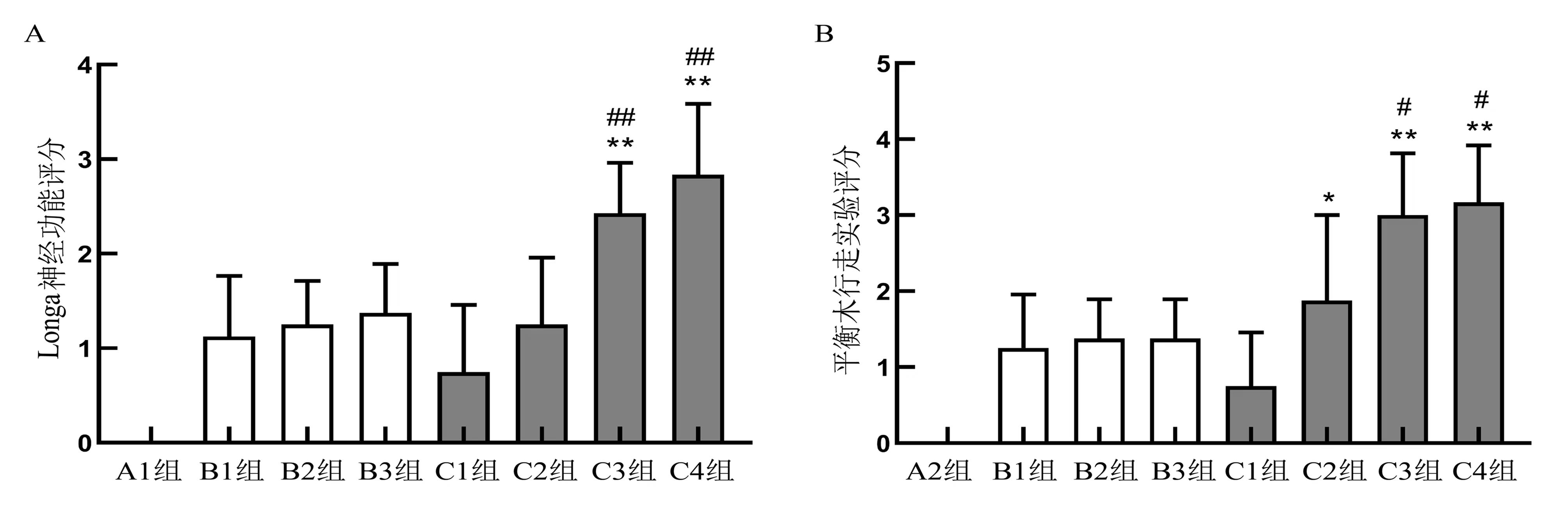
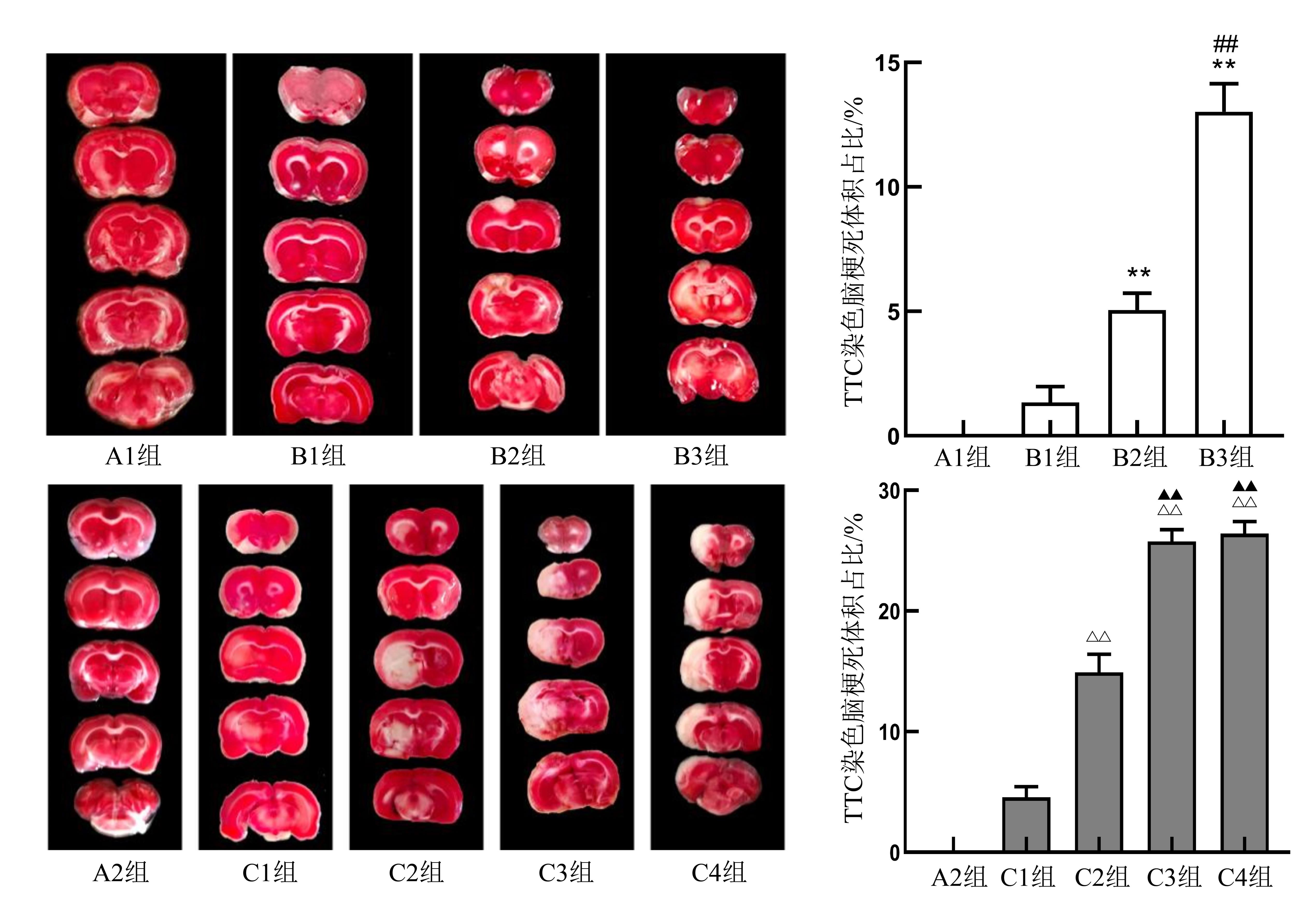
| 1 |
HERPICH F, RINCON F. Management of acute ischemic stroke[J]. Crit Care Med, 2020, 48(11):1654-1663. DOI:10.1097/CCM.0000000000004597 .
|
| 2 |
SOMMER C J. Ischemic stroke: experimental models and reality[J]. Acta Neuropathol, 2017, 133(2):245-261. DOI:10.1007/s00401-017-1667-0 .
|
| 3 |
SINGH D, WASAN H, REETA K H. Preclinical stroke research and translational failure: a bird's eye view on preventable variables[J]. Cell Mol Neurobiol, 2021:2021 Mar 30. DOI:10.1007/s10571-021-01083-6 .
|
| 4 |
YAN T, CHOPP M, CHEN J L. Experimental animal models and inflammatory cellular changes in cerebral ischemic and hemorrhagic stroke[J]. Neurosci Bull, 2015, 31(6):717-734. DOI:10.1007/s12264-015-1567-z .
|
| 5 |
DURUKAN A, TATLISUMAK T. Acute ischemic stroke: overview of major experimental rodent models, pathophysiology, and therapy of focal cerebral ischemia[J]. Pharmacol Biochem Behav, 2007, 87(1):179-197. DOI:10.1016/j.pbb.2007.04.015 .
|
| 6 |
MCCABE C, ARROJA M M, REID E, et al. Animal models of ischaemic stroke and characterisation of the ischaemic penumbra[J]. Neuropharmacology, 2018, 134(Pt B):169-177. DOI:10.1016/j.neuropharm.2017.09.022 .
|
| 7 |
YANG Y, KIMURA-OHBA S, THOMPSON J, et al. Rodent models of vascular cognitive impairment[J]. Transl Stroke Res, 2016, 7(5):407-414. DOI:10.1007/s12975-016-0486-2 .
|
| 8 |
FLURI F, SCHUHMANN M K, KLEINSCHNITZ C. Animal models of ischemic stroke and their application in clinical research[J]. Drug Des Devel Ther, 2015, 9:3445-3454. DOI:10.2147/DDDT.S56071 .
|
| 9 |
MACRAE I M. Preclinical stroke research: advantages and disadvantages of the most common rodent models of focal ischaemia[J]. Br J Pharmacol, 2011, 164(4):1062-1078. DOI:10.1111/j.1476-5381.2011.01398.x .
|
| 10 |
SHAHI M, ABEDELAHI A, MOHAMMADNEJAD D, et al. Exact location of sensorimotor cortex injury after photochemical modulation; evidence of stroke based on stereological and morphometric studies in mice[J]. Lasers Med Sci, 2021, 36(1):91-98. DOI:10.1007/s10103-020-03017-y .
|
| 11 |
KHATEEB K, YAO Z J, KHARAZIA V N, et al. A practical method for creating targeted focal ischemic stroke in the cortex of nonhuman Primates[J]. Annu Int Conf IEEE Eng Med Biol Soc, 2019, 2019:3515-3518. DOI:10.1109/EMBC.2 019.8857741 .
|
| 12 |
LIU N W, KE C C, ZHAO Y H, et al. Evolutional characterization of photochemically induced stroke in rats: a multimodality imaging and molecular biological study[J]. Transl Stroke Res, 2017, 8(3):244-256. DOI:10.1007/s12975-016-0512-4 .
|
| 13 |
丁惠洁, 刘珂舟, 杨珊珊, 等. 大鼠脑皮层血管栓塞和再生过程的实时动态记录方法[J]. 生理学报, 2019, 71(4):581-587. DOI:10.13294/j.aps.2019.0015 .
|
| 14 |
YAO Z J, YAZDAN-SHAHMORAD A. A quantitative model for estimating the scale of photochemically induced ischemic stroke[J]. Annu Int Conf IEEE Eng Med Biol Soc, 2018, 2018:2744-2747. DOI:10.1109/EMBC.2018.8512880 .
|
| 15 |
MA D C, ZHANG N N, ZHANG Y N, et al. Salvianolic Acids for Injection alleviates cerebral ischemia/reperfusion injury by switching M1/M2 phenotypes and inhibiting NLRP3 inflammasome/pyroptosis axis in microglia in vivo and in vitro [J]. J Ethnopharmacol, 2021, 270:113776. DOI:10.1016/j.jep. 2021.113776 .
|
| 16 |
WANG F J, LI R Y, TU P F, et al. Total glycosides of Cistanche deserticola promote neurological function recovery by inducing neurovascular regeneration via nrf-2/keap-1 pathway in MCAO/R rats[J]. Front Pharmacol, 2020, 11:236. DOI:10.3389/fphar.2020.00236 .
|
| 17 |
XIAO X T, LUO C, YUAN Y, et al. Systematic evaluation during early-phase ischemia predicts outcomes in middle cerebral artery occlusion mice[J]. Neuroreport, 2021, 32(1):29-37. DOI:10.1097/WNR.0000000000001553 .
|
| 18 |
TANG Y N, ZHANG G F, CHEN H L, et al. Selective brain hypothermia-induced neuroprotection against focal cerebral ischemia/reperfusion injury is associated with Fis1 inhibition[J]. Neural Regen Res, 2020, 15(5):903-911. DOI:10.4103/1673-5374.268973 .
|
| 19 |
GAO B Y, SUN C C, XIA G H, et al. Paired associated magnetic stimulation promotes neural repair in the rat middle cerebral artery occlusion model of stroke[J]. Neural Regen Res, 2020, 15(11):2047-2056. DOI:10.4103/1673-5374.282266 .
|
| 20 |
CHEN K H, CHAI H T, CHEN C H, et al. Synergic effect of combined cyclosporin and melatonin protects the brain against acute ischemic reperfusion injury[J]. Biomed Pharmacother, 2021, 136:111266. DOI:10.1016/j.biopha. 2021.111266 .
|
| 21 |
卢宝全, 尚小明, 杨峰, 等. 应用手持激光器建立光化学法脑梗死动物模型[J]. 中国比较医学杂志, 2012, 22(9):63-67, 80, 后插4. DOI:10.3969/j.issn.1671-7856.2012.009.014 .
|
| 22 |
VITAL S A, GAVINS F N E. Surgical approach for middle cerebral artery occlusion and reperfusion induced stroke in mice[J]. JoVE, 2016(116):54302. DOI:10.3791/54302 .
|
| 23 |
TAKANO K, TATLISUMAK T, BERGMANN A G, et al. Reproducibility and reliability of middle cerebral artery occlusion using a silicone-coated suture (Koizumi) in rats[J]. J Neurol Sci, 1997, 153(1):8-11. DOI:10.1016/s0022-510x(97)00184-6 .
|
| 24 |
WATSON B D, DIETRICH W D, BUSTO R, et al. Induction of reproducible brain infarction by photochemically initiated thrombosis[J]. Ann Neurol, 1985, 17(5):497-504. DOI:10.1002/ana.410170513 .
|
| 25 |
马浚宁, 高俊玮, 侯博儒, 等. 光化学栓塞法建立缺血性脑卒中动物模型[J]. 中国组织工程研究, 2015, 19(49):7951-7957. DOI:10.3969/j.issn.2095-4344.2015.49.014 .
|

 ), Hongshi ZHANG2, Yufeng WANG3, Meng JIA4, Deyu CONG1,3(