
Laboratory Animal and Comparative Medicine ›› 2026, Vol. 46 ›› Issue (3): 321-331.DOI: 10.12300/j.issn.1674-5817.2025.125
• Animal Models of Human Diseases • Previous Articles Next Articles
LI Jiafei1( ), ZHANG Zhenhao1(), WANG Shuo1, TIAN Ge1, WEN Shuang1, YAN Yuxue1, CUI Ran1, YE Zhen2()(
 ), CUI Yongchun1()()
Received:2025-07-25
Revised:2025-12-05
Online:2026-06-25
Published:2026-06-19
Contact:
YE Zhen, CUI Yongchun
CLC Number:
LI Jiafei,ZHANG Zhenhao,WANG Shuo,et al. Effects of Autonomic Neuromodulators on Atrial Electrical Remodeling and Histopathological Changes in a Rat Model of Atrial Fibrillation[J]. Laboratory Animal and Comparative Medicine, 2026, 46(3): 321-331. DOI: 10.12300/j.issn.1674-5817.2025.125.
Add to citation manager EndNote|Ris|BibTeX
URL: https://www.slarc.org.cn/dwyx/EN/10.12300/j.issn.1674-5817.2025.125
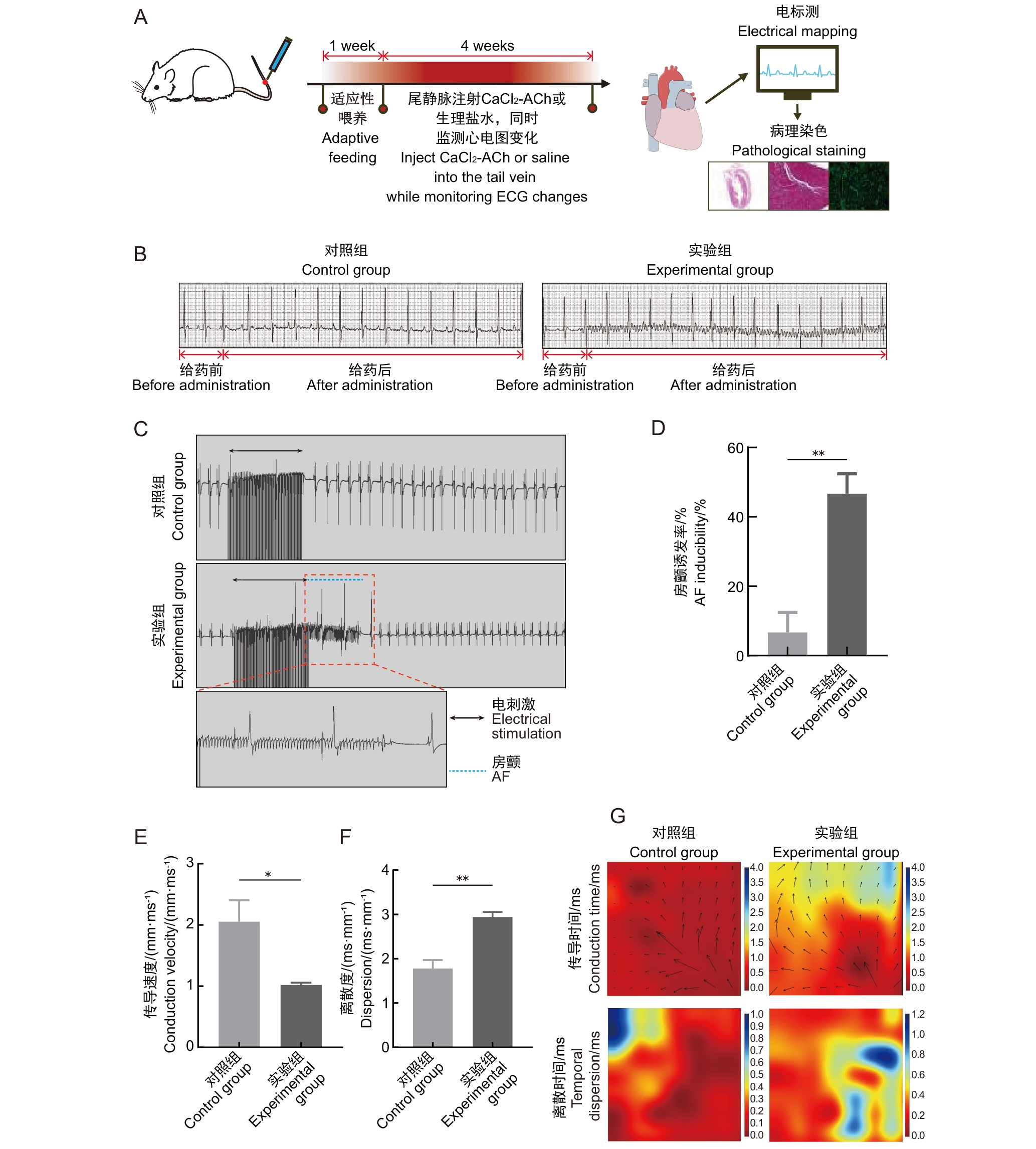
Figure 1 Autonomic neuromodulators induce changes in atrial electrophysiological characteristics in rats
参数 Parameter | 对照组 Control group (n=5) | 实验组 Experimental group (n=5) |
|---|---|---|
| LAD/mm | 4.04±0.19 | 4.89±0.08∗ |
| LVEF/% | 81.47±1.19 | 71.57±1.31∗∗ |
| FS/% | 52.40±1.78 | 42.14±1.15∗∗ |
| LVEDD/mm | 6.86±0.23 | 7.31±0.15 |
| LVESD/mm | 3.35±0.05 | 3.87±0.27 |
Table 1 Comparison of echocardiographic parameters among groups of rats
参数 Parameter | 对照组 Control group (n=5) | 实验组 Experimental group (n=5) |
|---|---|---|
| LAD/mm | 4.04±0.19 | 4.89±0.08∗ |
| LVEF/% | 81.47±1.19 | 71.57±1.31∗∗ |
| FS/% | 52.40±1.78 | 42.14±1.15∗∗ |
| LVEDD/mm | 6.86±0.23 | 7.31±0.15 |
| LVESD/mm | 3.35±0.05 | 3.87±0.27 |
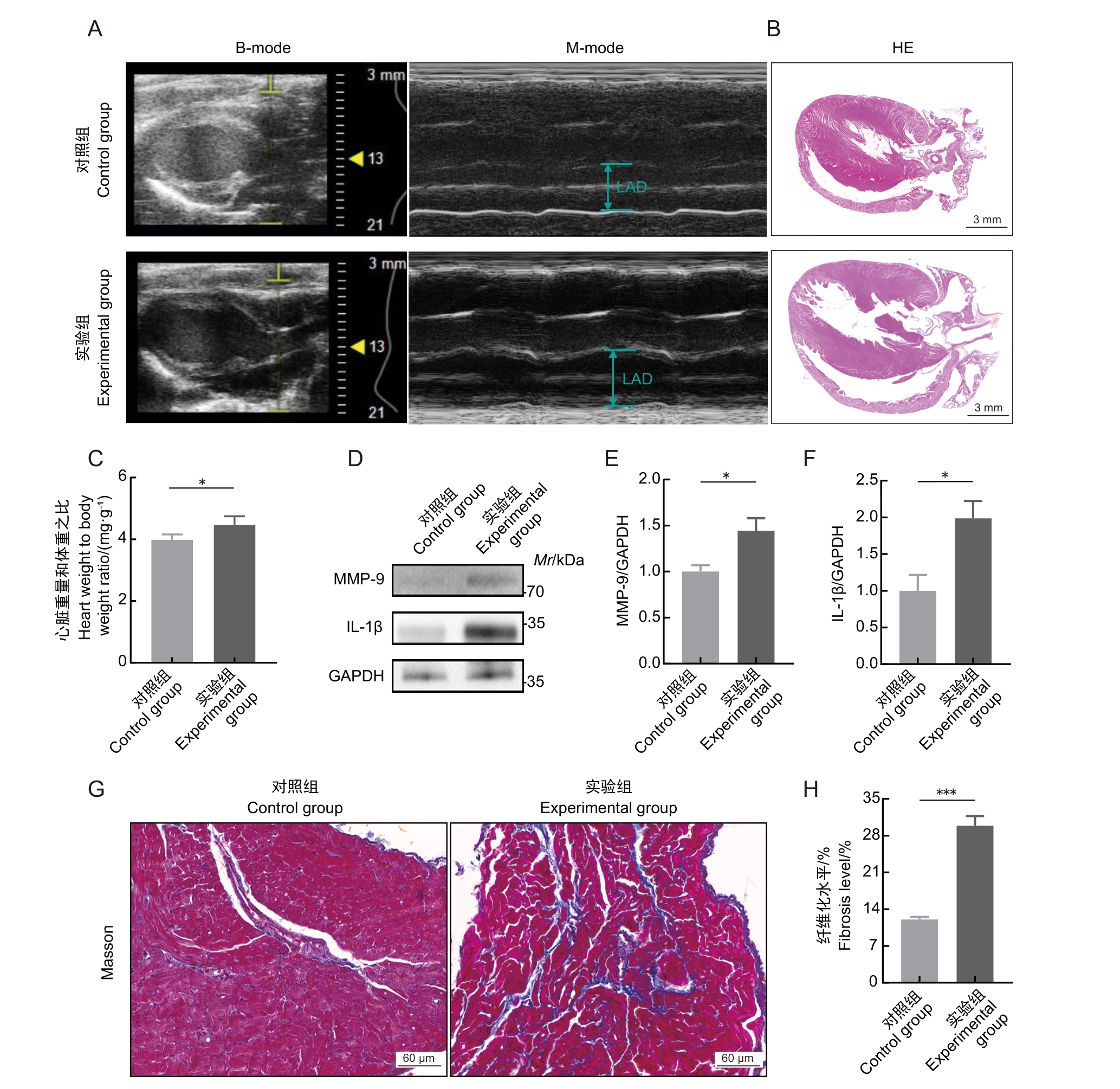
Figure 2 Autonomic neuromodulators induce structural changes in rat atrial myocardium
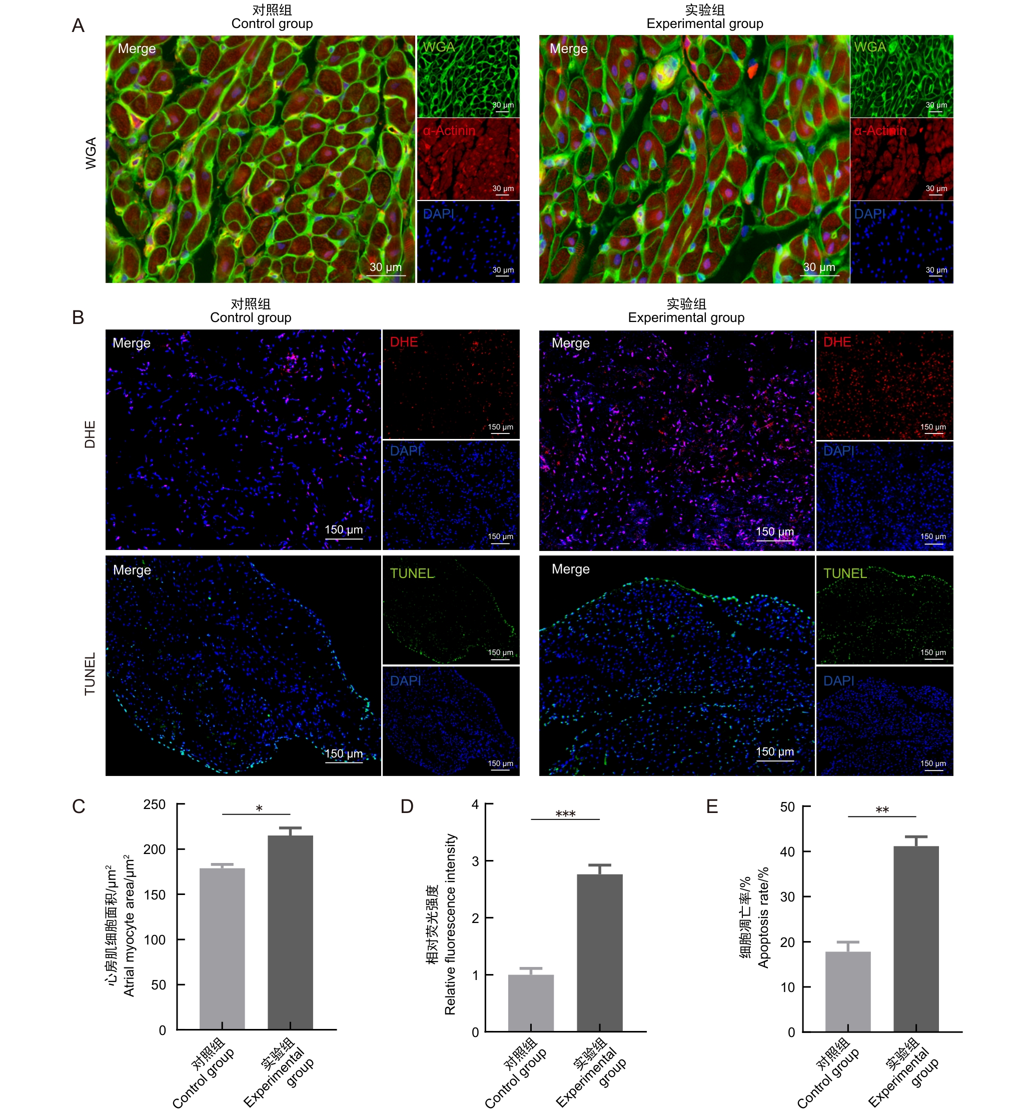
Figure 3 Autonomic neuromodulators induce morphological and oxidative stress changes in rat atrial cardiomyocytes
| [1] | SINGH J P, KANDALA J, CAMM A J. Non-pharmacological modulation of the autonomic tone to treat heart failure[J]. Eur Heart J, 2014, 35(2): 77-85. DOI: 10.1093/eurheartj/eht436 . |
| [2] | HERRING N, KALLA M, PATERSON D J. The autonomic nervous system and cardiac arrhythmias: current concepts and emerging therapies[J]. Nat Rev Cardiol, 2019, 16(12): 707-726. DOI: 10.1038/s41569-019-0221-2 . |
| [3] | 赵庆彦, 黄从新. 迷走神经对心房颤动影响的离子通道基础[J]. 中国心脏起搏与心电生理杂志, 2005, 19(2): 147-149. DOI: 10.13333/j.cnki.cjcpe.2005.02.026 . |
| ZHAO Q Y, HUANG C X. Ion channel basis of vagus nerve on atrial fibrillation[J]. Chin J Card Pacing Electrophysiol, 2005, 19(2): 147-149. DOI: 10.13333/j.cnki.cjcpe.2005.02.026 . | |
| [4] | FU Y P, JIANG T N, SUN H K, et al. Necroptosis is required for atrial fibrillation and involved in aerobic exercise-conferred cardioprotection[J]. J Cell Mol Med, 2021, 25(17): 8363-8375. DOI: 10.1111/jcmm.16796 . |
| [5] | KORNEJ J, BÖRSCHEL C S, BENJAMIN E J, et al. Epidemiology of atrial fibrillation in the 21st century: novel methods and new insights[J]. Circ Res, 2020, 127(1): 4-20. DOI: 10.1161/CIRCRESAHA.120.316340 . |
| [6] | 雷力, 戴磊, 张秋霞, 等. 基于国家基本公共卫生服务项目构建与评价新发心房颤动风险列线图[J]. 临床心血管病杂志, 2022, 38(3): 216-221. DOI: 10.13201/j.issn.1001-1439.2022.03.011 . |
| LEI L, DAI L, ZHANG Q X, et al. Development and evaluation of a nomogram for incident atrial fibrillation based on national basic public health service project[J]. J Clin Cardiol, 2022, 38(3): 216-221. DOI: 10.13201/j.issn.1001-1439.2022.03.011 . | |
| [7] | TANG J, ZHANG Q W, PENG S X, et al. Differences in global, regional, and national time trends in disability-adjusted life years for atrial fibrillation and flutter, 1990-2019: an age-period-cohort analysis from the 2019 global burden of disease study[J]. Front Cardiovasc Med, 2024, 11: 1401722. DOI: 10.3389/fcvm.2024.1401722 . |
| [8] | TAKAHASHI Y, YAMAGUCHI T, OTSUBO T, et al. Histological validation of atrial structural remodelling in patients with atrial fibrillation[J]. Eur Heart J, 2023, 44(35): 3339-3353. DOI: 10.1093/eurheartj/ehad396 . |
| [9] | ZHENG D L, WU Q R, ZENG P, et al. Advanced glycation end products induce senescence of atrial myocytes and increase susceptibility of atrial fibrillation in diabetic mice[J]. Aging Cell, 2022, 21(12): e13734. DOI: 10.1111/acel.13734 . |
| [10] | REBECCHI M, DE RUVO E, SGUEGLIA M, et al. Atrial fibrillation and sympatho-vagal imbalance: from the choice of the antiarrhythmic treatment to patients with syncope and ganglionated plexi ablation[J]. Eur Heart J , 25(Suppl C): C1-C6. DOI: 10.1093/eurheartjsupp/suad075 . |
| [11] | GONG Q, LE X, YU P C, et al. Therapeutic advances in atrial fibrillation based on animal models[J]. J Zhejiang Univ Sci B, 2024, 25(2): 135-152. DOI: 10.1631/jzus.B2300285 . |
| [12] | 黄燕, 黄从新. 钙离子通道与心房颤动关系的研究进展[J]. 医学综述, 2015, 21(13): 2353-2356. DOI: 10.3969/j.issn.1006-2084.2015.13.019 . |
| HUANG Y, HUANG C X. Research advance of the relationship between Ca2+ channels and the occurrence of atrial fibrillation[J]. Med Recap, 2015, 21(13): 2353-2356. DOI: 10.3969/j.issn.1006-2084.2015.13.019 . | |
| [13] | XIE D Y, XIONG K, DONG N G, et al. An endogenous cholinergic system controls electrical conduction in the heart[J]. Eur Heart J, 2025, 46(13): 1232-1246. DOI: 10.1093/eurheartj/ehae699 . |
| [14] | SHI S Q, MAO X X, LV J Y, et al. Qi-Po-Sheng-Mai granule ameliorates Ach-CaCl2-induced atrial fibrillation by regulating calcium homeostasis in cardiomyocytes[J]. Phytomedicine, 2023, 119: 155017. DOI: 10.1016/j.phymed. 2023.155017 . |
| [15] | 张婷, 陈帅, 刘星, 等. 氯化钙-乙酰胆碱混合液诱导小鼠心房颤动心房纤维化模型的构建[J]. 中国循证心血管医学杂志, 2023, 15(4): 440-442. DOI: 10.3969/j.issn.1674-4055.2023.04.11 . |
| ZHANG T, CHEN S, LIU X, et al. Model of atrial fibrosis induced by mixed CaCl2-Ach solution in mice with atrial fibrillation[J]. Chin J Evid Based Cardiovasc Med, 2023, 15(4): 440-442. DOI: 10.3969/j.issn.1674-4055.2023.04.11 . | |
| [16] | RODRIGO R. Prevention of postoperative atrial fibrillation: novel and safe strategy based on the modulation of the antioxidant system[J]. Front Physiol, 2012, 3: 93. DOI: 10.3389/fphys.2012.00093 . |
| [17] | QUAGLIARIELLO V, DE LAURENTIIS M, REA D, et al. The SGLT-2 inhibitor empagliflozin improves myocardial strain, reduces cardiac fibrosis and pro-inflammatory cytokines in non-diabetic mice treated with doxorubicin[J]. Cardiovasc Diabetol, 2021, 20(1): 150. DOI: 10.1186/s12933-021-01346-y . |
| [18] | MESUBI O O, ANDERSON M E. Atrial remodelling in atrial fibrillation: CaMKⅡ as a nodal proarrhythmic signal[J]. Cardiovasc Res, 2016, 109(4): 542-557. DOI: 10.1093/cvr/cvw002 . |
| [19] | YU L M, DONG X, HUANG T, et al. Inhibition of ferroptosis by icariin treatment attenuates excessive ethanol consumption-induced atrial remodeling and susceptibility to atrial fibrillation, role of SIRT1[J]. Apoptosis, 2023, 28(3-4): 607-626. DOI: 10.1007/s10495-023-01814-8 . |
| [20] | YUAN Y, MARTSCH P, CHEN X H, et al. Atrial cardiomyocyte-restricted cleavage of gasdermin D promotes atrial arrhythmogenesis[J]. Eur Heart J, 2025, 46(13): 1250-1262. DOI: 10.1093/eurheartj/ehaf024 . |
| [21] | SRIDHAR A, DESANTIAGO J, CHEN H N, et al. Modulation of NOX2 causes obesity-mediated atrial fibrillation[J]. J Clin Invest, 2024, 134(18): e175447. DOI: 10.1172/JCI175447 . |
| [22] | SCHÜTTLER D, BAPAT A, KÄÄB S, et al. Animal models of atrial fibrillation[J]. Circ Res, 2020, 127(1): 91-110. DOI: 10.1161/circresaha.120.316366 . |
| [23] | YANG L, XIE G H, WANG Y G, et al. Metabolic behaviors of aconitum alkaloids in different concentrations of Aconiti Lateralis Radix Praeparata and effects of aconitine in healthy human and long QT syndrome cardiomyocytes[J]. Molecules, 2022, 27(13): 4055. DOI: 10.3390/molecules27134055 . |
| [24] | 张冬, 张敏, 朱瑾彦, 等. 自发持续性心房颤动动物实验模型的探索研究[J]. 实验动物科学, 2023, 40(3): 61-70. DOI: 10.3969/j.issn.1006-6179.2023.03.010 . |
| ZHANG D, ZHANG M, ZHU J Y, et al. Experimental study on animal models of spontaneously persistent atrial fibrillation[J]. Lab Anim Sci, 2023, 40(3): 61-70. DOI: 10.3969/j.issn.1006-6179.2023.03.010 . | |
| [25] | 冯凯, 杨蕾熙, 成祎琳, 等. 乙酰胆碱合并氯化钙静脉注射致兔和大鼠房颤模型的比较[J]. 实验动物与比较医学, 2019, 39(5): 371-376. DOI: 10.3969/j.issn.1674-5817.2019.05.006 . |
| FENG K, YANG L X, CHENG Y L, et al. Comparison on rabbit and rat atrial fibrillation models induced by intravenous injection of acetylcholine-calcium chloride mixed solution[J]. Lab Anim Comp Med, 2019, 39(5): 371-376. DOI: 10.3969/j.issn.1674-5817.2019.05.006 . |
| [1] | TANG Jianping, ZHAO Liya, ZHAO Ying. Screening and Analysis of Microsatellite Genetic Markers in Commonly Used Inbred Rat Strains [J]. Laboratory Animal and Comparative Medicine, 2026, 46(3): 388-396. |
| [2] | AI Xiufeng, ZHANG Lizong, FANG Mingsun, LÜ Dongying, CHEN Chu, CAI Zhaowei, WANG Dejun. Analysis of Differences in the Intestinal Flora of Rats and Mice after Drinking Chlorinated Water Based on 16S rRNA Sequencing [J]. Laboratory Animal and Comparative Medicine, 2026, 46(3): 437-445. |
| [3] | SONG Jing, YANG Zongtong, LI Xiaojing, LI Zifa, SU Fengyun, XU Dongchuan, SUI Zaiyun. Effects of Xiebai San on the Morphological Structures of Lung and Intestinal Tissues and Expression Levels of PI3K and Akt in Rats with Allergic Asthma [J]. Laboratory Animal and Comparative Medicine, 2026, 46(2): 191-204. |
| [4] | TANG Xiaohang, GU Yingmin, LÜ Yangyang, HUANG Mingshu, TIAN Xuesong. Evaluation of the Histological Staining Performance of Rat Eyeball Sections Prepared Using a Self-Developed Fixative [J]. Laboratory Animal and Comparative Medicine, 2026, 46(2): 261-270. |
| [5] | JIANG Haitao, YUAN Hantao, HUANG Wenting, YANG Rongrong, CHEN Xiaochun, YU Baoqing, LI Sibo. Regulation of Rat Intervertebral Disc Annulus Fibrosus Cell Proliferation and Apoptosis by Yaoshu Zhuyu Fang via miR-17-5P/MDM2/p53 Pathway [J]. Laboratory Animal and Comparative Medicine, 2026, 46(1): 55-65. |
| [6] | LIU Chang, XIANG Xuesong, HE Huihuang, CHEN Xiaoqing, QIU Wenhong. Establishment and Evaluation of an Oxidative Stress Model of Atopic Dermatitis Induced by 2,4-dinitrofluorobenzene [J]. Laboratory Animal and Comparative Medicine, 2026, 46(1): 46-54. |
| [7] | GAO Chaoqi, ZHU Zhibo, SUN Xiandong. Application Progress and Classification Analysis of Rat Vascular Remodeling Models [J]. Laboratory Animal and Comparative Medicine, 2025, 45(5): 542-550. |
| [8] | LUO Yifan, ZHANG Zhenwei, MEI Lu, SHI Yeping, XING Yitong, ZHANG Zeqi, LI Chuxin, HAN Chunxia, YANG Pingshun, CHEN Qiusheng. Telocytes-Mediated Effects and Mechanisms of Anointing and Massage Therapy Using Oligopeptide-Herbal Medicine Composite Against Obesity in Rats [J]. Laboratory Animal and Comparative Medicine, 2025, 45(5): 551-560. |
| [9] | QIN Chao, LI Shuangxing, ZHAO Tingting, JIANG Chenchen, ZHAO Jing, YANG Yanwei, LIN Zhi, WANG Sanlong, WEN Hairuo. Study on the 90-day Feeding Experimental Background Data of SD Rats for Drug Safety Evaluation [J]. Laboratory Animal and Comparative Medicine, 2025, 45(4): 439-448. |
| [10] | LIU Liyu, JI Bo, LIU Xiaoxuan, FANG Yang, ZHANG Ling, GUO Tingting, QUAN Ye, LI Hewen, LIU Yitian. Exploration of Rat Fetal Lung Tissue Fixation Methods [J]. Laboratory Animal and Comparative Medicine, 2025, 45(4): 432-438. |
| [11] | PAN Yicong, JIANG Wenhong, HU Ming, QIN Xiao. Optimization of Surgical Procedure and Efficacy Evaluation of Aortic Calcification Model in Rats with Chronic Kidney Disease [J]. Laboratory Animal and Comparative Medicine, 2025, 45(3): 279-289. |
| [12] | LIU Zhiwei, YANG Ran, LIAN Hao, ZHANG Yu, JIN Lilun. Cartilage Protection and Anti-Inflammatory Effects of Fraxetin on Monosodium Iodoacetate-Induced Rat Model of Osteoarthritis [J]. Laboratory Animal and Comparative Medicine, 2025, 45(3): 259-268. |
| [13] | JIANG Meng, HAO Shulan, TONG Liguo, ZHONG Qiming, GAO Zhenfei, WANG Yonghui, WANG Xixing, JI Haijie. Dynamic Evaluation of Vinorelbine-Induced Phlebitis of Dorsalis Pedis Vein in a Rat Model [J]. Laboratory Animal and Comparative Medicine, 2025, 45(3): 251-258. |
| [14] | LIAN Hui, JIANG Yanling, LIU Jia, ZHANG Yuli, XIE Wei, XUE Xiaoou, LI Jian. Construction and Evaluation of a Rat Model of Abnormal Uterine Bleeding [J]. Laboratory Animal and Comparative Medicine, 2025, 45(2): 130-146. |
| [15] | YIN Yulian, MA Lina, TU Siyuan, CHEN Ling, YE Meina, CHEN Hongfeng. Establishment and Evaluation of a Rat Model of Non-Puerperal Mastitis [J]. Laboratory Animal and Comparative Medicine, 2024, 44(6): 587-596. |
| Viewed | ||||||
|
Full text |
|
|||||
|
Abstract |
|
|||||