
Laboratory Animal and Comparative Medicine ›› 2026, Vol. 46 ›› Issue (1): 20-31.DOI: 10.12300/j.issn.1674-5817.2025.034
• Animal Models of Human Diseases • Previous Articles Next Articles
XU Yingtao, WANG Mengmeng, LIN Ping, CHI Haitao, WANG Yi, BAI Ying( )
)
Received:2025-03-04
Revised:2025-06-13
Online:2026-02-25
Published:2026-02-14
Correspondence to:
BAI Ying
CLC Number:
XU Yingtao,WANG Mengmeng,LIN Ping,et al. Exosomes Treat Ischemic Stroke by Regulation of Ferroptosis Through the NRF2/SLC7A11/GPX4 Pathway in Mice[J]. Laboratory Animal and Comparative Medicine, 2026, 46(1): 20-31. DOI: 10.12300/j.issn.1674-5817.2025.034.
Add to citation manager EndNote|Ris|BibTeX
URL: https://www.slarc.org.cn/dwyx/EN/10.12300/j.issn.1674-5817.2025.034
引物名称 Primer name | 引物序列 (5'→3') Sequence (5'→3') | 产物大小/bp Product size/bp |
|---|---|---|
| GAPDH | F: AGGTCGGTGTGAACGGATTTG; R: GGGGTCGTTGATGGCAACA | 165 |
| NRF2 | F: CTTTAGTCAGCGACAGAAGGAC; R: AGGCATCTTGTTTGGGAATGTG | 219 |
| SLC7A11 | F: CACCGGGGTCGGTTTTCTTA; R: GGCAGATGGCCAAGCTTTTG | 200 |
| GPX4 | F: CTATGGTCCCATGGAGGAGC; R: AGGCAGACCTTCATGAGTGC | 185 |
Table1 Sequence of primers used in PCR
引物名称 Primer name | 引物序列 (5'→3') Sequence (5'→3') | 产物大小/bp Product size/bp |
|---|---|---|
| GAPDH | F: AGGTCGGTGTGAACGGATTTG; R: GGGGTCGTTGATGGCAACA | 165 |
| NRF2 | F: CTTTAGTCAGCGACAGAAGGAC; R: AGGCATCTTGTTTGGGAATGTG | 219 |
| SLC7A11 | F: CACCGGGGTCGGTTTTCTTA; R: GGCAGATGGCCAAGCTTTTG | 200 |
| GPX4 | F: CTATGGTCCCATGGAGGAGC; R: AGGCAGACCTTCATGAGTGC | 185 |
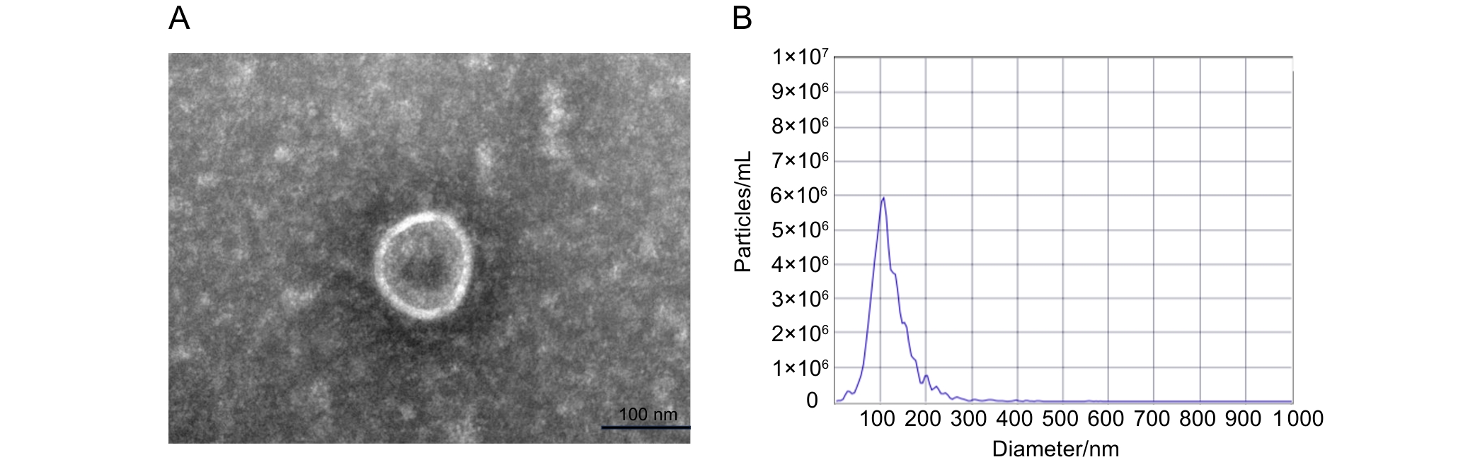
Figure 1 Electron microscopic observation and particle size analysis of exosomes
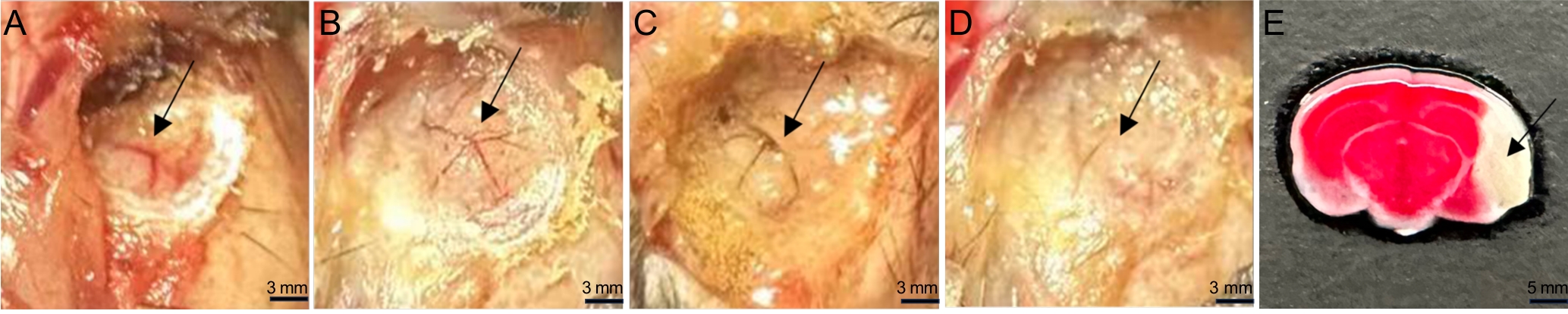
Figure 2 MCA blood flow changes during MCAO model establishment and TTC staining of the cerebral infarction area
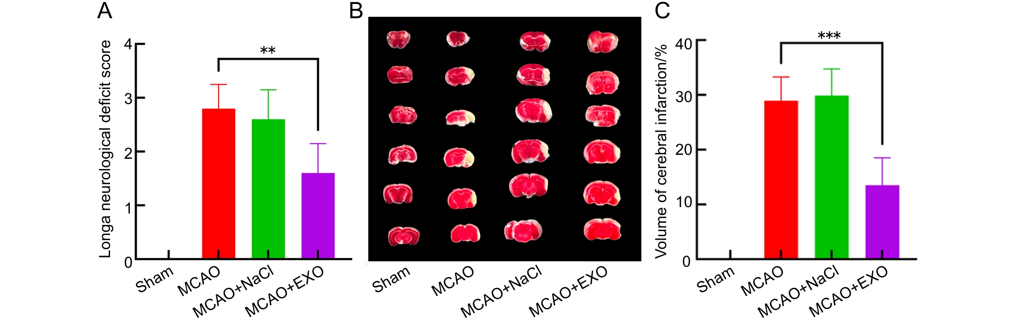
Figure 3 Analysis of neurological deficit scores and the percentages of cerebral infarction volume in each group of mice
Note:A, Neurological Longa scores of each group of mice; B, 2,3,5-triphenyltetrazolium chloride (TTC) staining results of brain sections in each group of mice, each column contains six slices representing the complete brain of one mouse, normal brain tissue was red, while brain tissue in the infarcted area was white; C, The percentages of brain infarction volume in each group of mice. The sham operation group (Sham) only exposed the middle cerebral artery without electrocoagulation of the vessel; the model group (MCAO) used high-frequency surgical forceps to electrocoagulate the middle cerebral artery at a power of 8 W; the model + normal saline group (MCAO+NaCl) first injected 100 μL of normal saline through the tail vein, and then performed electrocoagulation modeling; the model + exosome group (MCAO+EXO) first injected 100 μL of exosomes (9.5×10¹¹ particles per mL) from the culture supernatant of human amniotic mesenchymal stem cells (hAMSCs) through the tail vein, and then performed electrocoagulation modeling. There were eight mice in each group. **P<0.01, ***P<0.001.
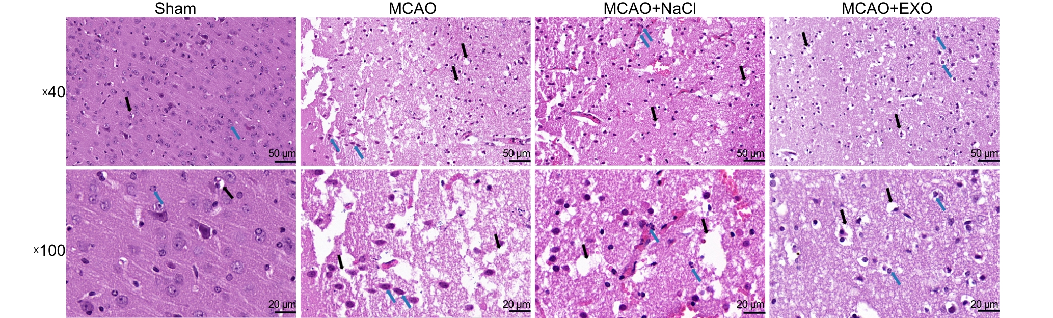
Figure 4 HE staining of brain tissue sections from each group of mice to observe neuronal morphology
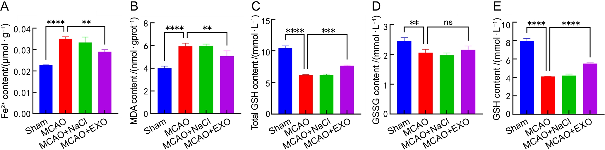
Figure 5 Microcolorimetric assays of Fe2+, MDA, total GSH, GSSG and GSH contents in the brains of each group of mice
Note:A-E, Contents of Fe2+, malondialdehyde (MDA), total glutathione (total GSH), oxidized glutathione (GSSG), and reduced glutathione (GSH) in the brains of mice from each group. Reduced GSH was calculated as the difference between the contents of total GSH and GSSG. The sham operation group (Sham) only exposed the middle cerebral artery without electrocoagulation of the vessel; the model group (MCAO) used high-frequency surgical forceps to electrocoagulate the middle cerebral artery at a power of 8 W; the model + normal saline group (MCAO+NaCl) first injected 100 μL of normal saline through the tail vein, and then performed electrocoagulation modeling; the model + exosome group (MCAO+EXO) first injected 100 μL of exosomes (9.5×10¹¹ particles per mL) from the culture supernatant of human amniotic mesenchymal stem cells (hAMSCs) through the tail vein, and then performed electrocoagulation modeling. There were eight mice in each group. nsP >0.05, **P<0.01, ***P<0.001, ****P<0.000 1.
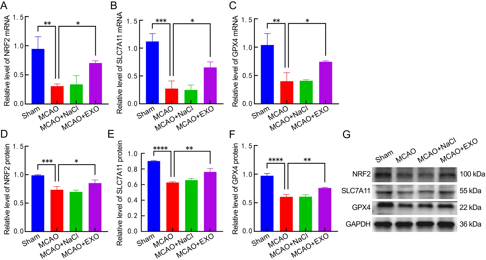
Figure 6 Detection and analysis of mRNA and protein expression of NRF2, SLC7A11 and GPX4 in the brains of each group of mice
Note:A-C, mRNA transcription levels of nuclear factor-erythroid 2-related factor 2 (NRF2), solute carrier family 7 member 11 (SLC7A11), and glutathione peroxidase 4 (GPX4) in the brains of mice from each group. D-F, Relative protein expression levels of NRF2, SLC7A11, and GPX4 in the brains of mice from each group. G, Western blotting images of NRF2, SLC7A11, GPX4, and GAPDH. The sham operation group (Sham) only exposed the middle cerebral artery without electrocoagulation of the vessel; the model group (MCAO) used high-frequency surgical forceps to electrocoagulate the middle cerebral artery at a power of 8 W; the model + normal saline group (MCAO+NaCl) first injected 100 μL of normal saline through the tail vein, and then performed electrocoagulation modeling; the model + exosome group (MCAO+EXO) first injected 100 μL of exosomes (9.5×10¹¹ particles per mL) from the culture supernatant of human amniotic mesenchymal stem cells (hAMSCs) through the tail vein, and then performed electrocoagulation modeling. There were eight mice in each group. *P<0.05, **P<0.01, ***P<0.001, ****P<0.000 1.
| [1] | ZHU Z B, SHI M Y, YU Q, et al. Burden and risk factors of stroke worldwide and in China: An analysis from the Global Burden of Disease Study 2021[J]. Chin Med J (Engl), 2025,138(20):2588-2595. DOI: 10.1097/CM9.0000000000003778 . |
| [2] | GREEN T L, MCNAIR N D, HINKLE J L, et al. Care of the patient with acute ischemic stroke (posthyperacute and prehospital discharge): update to 2009 comprehensive nursing care scientific statement: a scientific statement from the American heart association[J]. Stroke, 2021, 52(5): e179-e197. DOI:10.1161/STR.0000000000000357 . |
| [3] | QIN C, YANG S, CHU Y H, et al. Signaling pathways involved in ischemic stroke: molecular mechanisms and therapeutic interventions[J]. Signal Transduct Target Ther, 2022, 7(1):215. DOI:10.1038/s41392-022-01064-1 . |
| [4] | TUO Q Z, LEI P. Ferroptosis in ischemic stroke: Animal models and mechanisms[J]. Zool Res, 2024, 45(6):1235-1248. DOI:10.24272/j.issn.2095-8137.2024.239 . |
| [5] | ZHANG Y F, LU X Y, TAI B, et al. Ferroptosis and its multifaceted roles in cerebral stroke[J]. Front Cell Neurosci, 2021, 15:615372. DOI: 10.3389/fncel.2021.615372 . |
| [6] | JIANG X J, STOCKWELL B R, CONRAD M. Ferroptosis: mechanisms, biology and role in disease[J]. Nat Rev Mol Cell Biol, 2021, 22(4):266-282. DOI:10.1038/s41580-020-00324-8 . |
| [7] | YANG Y P, NICOL C J B, CHIANG M C. A review of the neuroprotective properties of exosomes derived from stem cells and exosome-coated nanoparticles for treating neurodegenerative diseases and stroke[J]. Int J Mol Sci, 2025, 26(8):3915. DOI: 10.3390/ijms26083915 . |
| [8] | SONG Y, ZHANG T, SHI P, et al. Exosomes derived from human amniotic mesenchymal stem cells promotes angiogenesis in hUVECs by delivering novel miRNA N-194[J]. Mol Med, 2025, 31:173. DOI:10.1186/s10020-025-01192-8 . |
| [9] | SUN J, YUAN Q, GUO L,et al. Brain microvascular endothelial cell-derived exosomes protect neurons from ischemia-reperfusion injury in mice[J]. Pharmaceuticals (Basel), 2022, 15(10):1287. DOI:10.3390/ph15101287 . |
| [10] | QIN J, ZHOU L, YU L, et al. Exosomes derived from HUVECs alleviate ischemia-reperfusion induced inflammation in neural cells by upregulating KLF14 expression[J]. Front Pharmacol, 2024, 15:1365928. DOI: 10.3389/fphar.2024. 1365928 . |
| [11] | CHEN Y X, LI B F, QUAN J, et al. Inhibition of ferroptosis by mesenchymal stem cell-derived exosomes in acute spinal cord injury: role of Nrf2/GCH1/BH4 axis[J]. Neurospine, 2024, 21(2):642-655. DOI:10.14245/ns.2448038.019 . |
| [12] | LI X, ZHANG X, LIU Y J, et al. Exosomes derived from mesenchyml stem cells ameliorate oxygen-glucose deprivation/reoxygenation-induced neuronal injury via transferring microRNA-194 and targeting Bach1[J]. Tissue Cell, 2021, 73:101651. DOI:10.1016/j.tice.2021.101651 . |
| [13] | HUO S Z, SHI P, PANG X N. Culture and identification of human amniotic mesenchymal stem cells[J]. Chin Med Sci J, 25(4):211-214. DOI: 10.1016/s1001-9294(11)60004-7 . |
| [14] | WANG M M, BAI Y, CHI H T, et al. miR-451 protects against ischemic stroke by targeting Phd3[J]. Exp Neurol, 2021, 343:113777. DOI:10.1016/j.expneurol.2021.113777 . |
| [15] | LONGA E Z, WEINSTEIN P R, CARLSON S, et al. Reversible middle cerebral artery occlusion without craniectomy in rats[J]. Stroke, 1989, 20(1): 84-91. DOI: 10.1161/01.str.20.1.84 . |
| [16] | SWANSON R A, MORTON M T, TSAO-WU G, et al. A semiautomated method for measuring brain infarct volume[J]. J Cereb Blood Flow Metab, 1990, 10(2): 290-293. DOI:10.1038/jcbfm.1990.47 . |
| [17] | LI C, LUO Y P, LI S G, et al. Mechanistic insights of neuronal death and neuroprotective therapeutic approaches in stroke[J]. Neural Regen Res, 2025, 21(3):869-886. DOI:10.4103/NRR.NRR-D-24-01324 . |
| [18] | TANG D L, CHEN X, KANG R, et al. Ferroptosis: molecular mechanisms and health implications[J]. Cell Res, 2021, 31(2):107-125. DOI:10.1038/s41422-020-00441-1 . |
| [19] | SHEN K, WANG X J, WANG Y W, et al. miR-125b-5p in adipose derived stem cells exosome alleviates pulmonary microvascular endothelial cells ferroptosis via Keap1/Nrf2/GPX4 in sepsis lung injury[J]. Redox Biol, 2023, 62:102655. DOI:10.1016/j.redox.2023.102655 . |
| [20] | LIN F Y, CHEN W Y, ZHOU J H, et al. Mesenchymal stem cells protect against ferroptosis via exosome-mediated stabilization of SLC7A11 in acute liver injury[J]. Cell Death Dis, 2022, 13(3):271. DOI:10.1038/s41419-022-04708-w . |
| [21] | 邢凤英, 周颖, 马政文, 等. 线栓法大鼠大脑中动脉脑缺血模型的评价方法初探[J]. 实验动物与比较医学, 2013, 33(5): 339-346. DOI: 10.3969/j.issn.1674-5817.2013.05.003 . |
| XING F Y, ZHOU Y, MA Z W, et al. Preliminary study for evaluation system of rat permanent cerebral artery occlusion model by intraluminal suture method[J]. Lab Anim Comp Med, 2013, 33(5): 339-346. DOI: 10.3969/j.issn.1674-5817.2013.05.003 . | |
| [22] | SUN Y, YANG X, XU L, et al. The role of Nrf2 in relieving cerebral ischemia-reperfusion injury[J]. Curr Neuropharmacol, 2023, 21(6):1405-1420. DOI: 10.2174/1570159X21666221129100308 . |
| [23] | VILLAVICENCIO-TEJO F, OLESEN M A, ARÁNGUIZ A, et al. Activation of the Nrf2 pathway prevents mitochondrial dysfunction induced by caspase-3 cleaved Tau: implications for Alzheimer's disease[J]. Antioxidants (Basel), 2022,11(3):515. DOI: 10.3390/antiox11030515 . |
| [24] | LIU H, ZHANG T A, ZHANG W Y, et al. Rhein attenuates cerebral ischemia-reperfusion injury via inhibition of ferroptosis through NRF2/SLC7A11/GPX4 pathway[J]. Exp Neurol, 2023, 369:114541. DOI:10.1016/j.expneurol.2023.114541 . |
| [25] | FU C, WU Y F, LIU S J, et al. Rehmannioside A improves cognitive impairment and alleviates ferroptosis via activating PI3K/AKT/Nrf2 and SLC7A11/GPX4 signaling pathway after ischemia[J]. J Ethnopharmacol, 2022, 289:115021. DOI:10.1016/j.jep.2022.115021 . |
| [1] | PAN Linqin, DENG Xiangliang, LUO Yunxia. Advances in Integrative Translational Research on Animal Models of Ischemic Stroke in Traditional Chinese and Western Medicine [J]. Laboratory Animal and Comparative Medicine, 2026, 46(3): 344-356. |
| [2] | WANG Juan, XU Jiahui, TIAN Yunyuan, ZHANG Mengmeng, LI Min, WANG Siwang, LI Yao. Comparison and Behavioral Observation of Two Female Mice Models of Ulcerative Colitis [J]. Laboratory Animal and Comparative Medicine, 2026, 46(3): 332-343. |
| [3] | AI Xiufeng, ZHANG Lizong, FANG Mingsun, LÜ Dongying, CHEN Chu, CAI Zhaowei, WANG Dejun. Analysis of Differences in the Intestinal Flora of Rats and Mice after Drinking Chlorinated Water Based on 16S rRNA Sequencing [J]. Laboratory Animal and Comparative Medicine, 2026, 46(3): 437-445. |
| [4] | RONG Wenshuang, NIU Yuanfei, LIU Meiting, YANG Mengyuan, CUI Shuang, MA Lina, FU Yao, WANG Lianmei, CAO Junling. Influence of Antigen Type on the Establishment of an Induced Sjögren Syndrome Mouse Model [J]. Laboratory Animal and Comparative Medicine, 2026, 46(2): 178-190. |
| [5] | WU Xianwen, LIU Lili, CHEN Ye, XU Guoheng. Optimization of Cage-Changing Intervals and Wood Shavings Usage for Mice During the Growth Phase in Breeding Systems [J]. Laboratory Animal and Comparative Medicine, 2026, 46(2): 251-260. |
| [6] | YANG Yunrong, WU Wenyu, TAN Yue, YAN Guofeng, LI Yao, LU Jin. A Review of Methods for Establishing and Evaluating Animal Models of Stroke [J]. Laboratory Animal and Comparative Medicine, 2026, 46(1): 94-106. |
| [7] | Yisu ZHANG, Xinru LIU, Ruojie WU, Rui LIU, Hong OUYANG, Xiaohong LI. Establishment and Evaluation of Mouse Model of Pregnancy Pain-depression Comorbidity Induced by Chronic Unpredictable Stress, Complete Freund's Adjuvant and Formalin [J]. Laboratory Animal and Comparative Medicine, 2024, 44(3): 259-269. |
| [8] | Jia LIU, Yanrong YE, Yun SHEN, Qiying TANG, Meiqing CHEN, Kehui YI, Shaozhuang CHEN. Ginkgolide B Promotes Neural Function Recovery of Ischemic Stroke Mice by Regulating Characteristics of Brain T Cells and Their Interactions with Glial Cells [J]. Laboratory Animal and Comparative Medicine, 2024, 44(2): 139-148. |
| [9] | Min LIANG, Yang GUO, Jinjin WANG, Mengyan ZHU, Jun CHI, Yanjuan CHEN, Chengji WANG, Zhilan YU, Ruling SHEN. Construction of Dmd Gene Mutant Mice and Phenotype Verification in Muscle and Immune Systems [J]. Laboratory Animal and Comparative Medicine, 2024, 44(1): 42-51. |
| [10] | Han LI, Xiaorui ZHANG, Chengfang ZHANG. Mechanism of Intermittent Fasting in Improving Olanzapine-induced Metabolic Disorders in Mice [J]. Laboratory Animal and Comparative Medicine, 2023, 43(1): 3-10. |
| [11] | Yanbing ZHU, Fan BAI, Shaoxin TAO, Yuhualei PAN, Huan WANG, Yushang ZHAO, Song WANG, Yan YU. Inhibition of Phospholipase D1 Activity Improves the Recovery of Neurological Function in Mice with Ischemic Stroke [J]. Laboratory Animal and Comparative Medicine, 2022, 42(4): 322-332. |
| [12] | Bo DONG, Jiaxin LIU, Wei XIONG, Songqi TANG, Wei HUANG. Progress in Animal Models of Ischemic Stroke [J]. Laboratory Animal and Comparative Medicine, 2022, 42(1): 54-61. |
| [13] | LI Zifa, ZHANG Hao, REN Meng, XU Kaiyong, HU Minghui, ZHOU Miaomiao, WANG Kezhou. Protective Effect of Quercetin on Lipid Metabolism Disorder in Mice Livers Caused by Cadmium [J]. Laboratory Animal and Comparative Medicine, 2021, 41(4): 305-312. |
| [14] | FENG Yan, WU Wenqing, ZHANG Jingyuan, LI Yu, LI Leichen, YUAN Zheng, CUI Shufang. Isolation and Identification of Exosomes from Skin Fibroblasts of Naked Mole Rats [J]. Laboratory Animal and Comparative Medicine, 2020, 40(6): 506-512. |
| [15] | LEI Shan, LIU Qiang, HUANG Wei-jin, WANG You-chun. Influence of Strain, Gender and Hair Coat of Mice on Establishing Bioluminescent Imaging Pseudovirus Mouse Model [J]. Laboratory Animal and Comparative Medicine, 2019, 39(6): 423-428. |
| Viewed | ||||||
|
Full text |
|
|||||
|
Abstract |
|
|||||