
Laboratory Animal and Comparative Medicine ›› 2023, Vol. 43 ›› Issue (4): 440-445.DOI: 10.12300/j.issn.1674-5817.2022.173
• Case Report • Previous Articles Next Articles
Shanshan ZHAI1( ), Liang LIANG1, Yingying CAO1, Zhuxin LI1, Qing WANG1, Junyu TAO1,2, Chenxia YUN1,3, Jing LENG1,2,3(
 )(), Haibo TANG1,2()()
Received:2022-11-08
Revised:2023-04-20
Online:2023-08-25
Published:2023-08-25
Contact:
Jing LENG, Haibo TANG
CLC Number:
Shanshan ZHAI,Liang LIANG,Yingying CAO,et al. Diagnosis of Trichoepithelioma in a Tree Shrew and Observation of Cell Biological Characteristics[J]. Laboratory Animal and Comparative Medicine, 2023, 43(4): 440-445. DOI: 10.12300/j.issn.1674-5817.2022.173.
Add to citation manager EndNote|Ris|BibTeX
URL: https://www.slarc.org.cn/dwyx/EN/10.12300/j.issn.1674-5817.2022.173
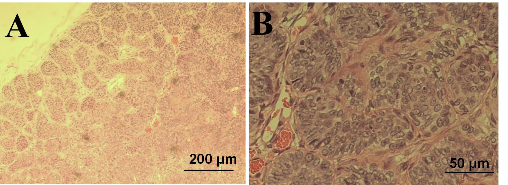
Figure 1 Histopathological changes of primary tumor in the tree shrews (HE staining)Note: A, The edge of the tumor tissue was clearly outlined and the tumor cells were arranged in a striated and palisade pattern (×50); B, HE-stained tumor cells were basal cell-like with deep nuclear staining and minimal cytoplasm (×200).
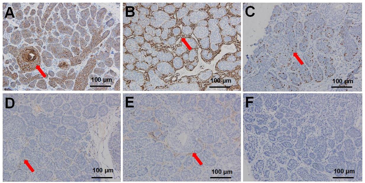
Figure 2 Immunohistochemistry of trichoepithelioma from a tree shrew(DAB staining,×100)Note: A, Strongly positive expression of CK in tumor cells (As indicated by the red arrow); B, Strongly positive expression of vimentin in tumor cell mesenchyme (as indicated by the red arrow); C, Low index expression of ki-67 in tumor cells (as indicated by the red arrow); D, Low positive expression of Bcl2 in tumor cell mesenchyme (as indicated by the red arrow); E, Low proportion expression of CD10 in tumor cell mesenchyme (as indicated by the red arrow) ; F, Negative expression of CD34 in tumor cells.
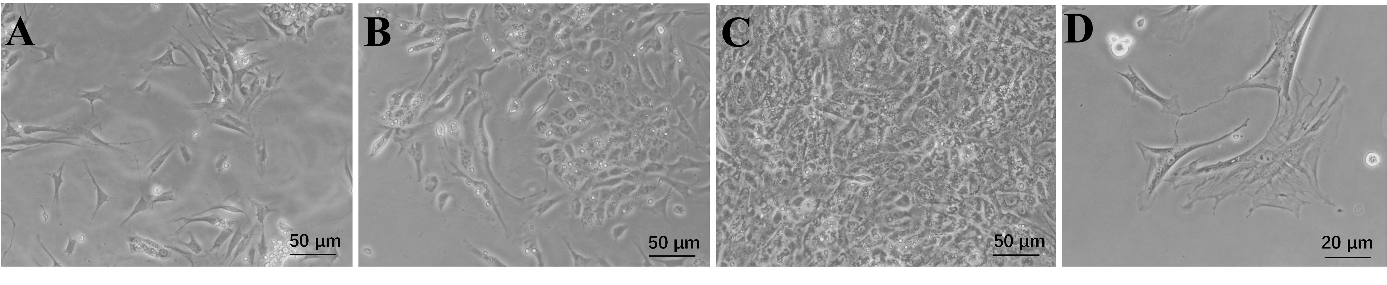
Figure 3 Culture of primary tumor cells from a tree shrew trichoepithelioma and morphological observationNote:A, Day 1 after tumor cell isolation and culture (×100); B, Day 2 after tumor cell isolation and culture (×100); C, Day 5 after tumor cell isolation and culture (×100). D, Tumor cells were pleomorphic, irregular, and tentacled (×400).
| 1 | KARIMZADEH I, NAMAZI M R, KARIMZADEH A. Trichoepithelioma: a comprehensive review[J]. Acta Dermatovenerol Croat, 2018, 26(2):162-165. |
| 2 | ALSAAD K O, OBAIDAT N A, GHAZARIAN D. Skin adnexal neoplasms: part 1: an approach to tumours of the pilosebaceous unit[J]. J Clin Pathol, 2007, 60(2):129-144. DOI: 10.1136/jcp.2006.040337 . |
| 3 | SMOLLER B R. Lever's histopathology of the skin, 10th edition[J]. J Cutan Pathol, 2009, 36(5):605. DOI: 10.1111/j.1600-0560.2008.01213.x . |
| 4 | YE M S, ZHANG J Y, YU D D, et al. Comprehensive annotation of the Chinese tree shrew genome by large-scale RNA sequencing and long-read isoform sequencing[J]. Zool Res, 2021, 42(6):692-709. DOI: 10.24272/j.issn.2095-8137.2021.272 . |
| 5 | 曾雯, 雷玲, 赵铖. 树鼩用于构建自身免疫性疾病动物模型展望[J]. 中国免疫学杂志, 2022, 38(15):1918-1921. DOI: 10.3969/j.issn.1000-484X.2022.15.024 . |
| ZENG W, LEI L, ZHAO C. Prospects of tree shrews used to establish animal models of autoimmune diseases[J]. Chin J Immunol, 2022, 38(15):1918-1921. DOI: 10.3969/j.issn.1000-484X.2022.15.024 . | |
| 6 | LI R F, ZANIN M, XIA X S, et al. The tree shrew as a model for infectious diseases research[J]. J Thorac Dis, 2018, 10(S9): S2272-S2279. DOI: 10.21037/jtd.2017.12.121 . |
| 7 | 贾杰, 代解杰. 树鼩在生物医学研究中的优势与挑战[J]. 实验动物与比较医学, 2019, 39(1):3-8. DOI: 10.3969/j.issn.1674-5817.2019.01.002 . |
| JIA J, DAI J J. Advantages and challenges of tree shrews in biomedical research[J]. Lab An Comp Med, 2019, 39(1): 3-8. DOI: 10.3969/j.issn.1674-5817.2019.01.002 . | |
| 8 | 徐新峰. 犬乳腺肿瘤细胞的分离、培养及鉴定[D]. 石河子: 石河子大学, 2014. |
| XU X F. Isolation, culture and identification of canine breast tumor cells[D]. Shihezi: Shihezi University, 2014. | |
| 9 | MIGDEN M R, CHANG A L S, DIRIX L, et al. Emerging trends in the treatment of advanced basal cell carcinoma[J]. Cancer Treat Rev, 2018, 64:1-10. DOI: 10.1016/j.ctrv.2017.12.009 . |
| 10 | PATTERSON J W. Weedon's skin pathology[M]. 4th ed. Amsterdam: Elsevier, 2015. |
| 11 | LEAR J T, CORNER C, DZIEWULSKI P, et al. Challenges and new horizons in the management of advanced basal cell carcinoma: a UK perspective[J]. Br J Cancer, 2014, 111(8):1476-1481. DOI: 10.1038/bjc.2014.270 . |
| 12 | 孙彩虹, 张彩萍, 丁克云, 等. 基底细胞癌72例临床特征分析[J]. 皮肤性病诊疗学杂志, 2015, 22(2): 99-101. DOI: 10.3969/j.issn.1674-8468.2015.02.004 . |
| SUN C H, ZHANG C P, DING K Y, et al. Analysis of the clinical feature of 72 cases with basal cell carcinoma[J]. J Diagn Ther Derm Venereol, 2015, 22(2): 99-101. DOI: 10.3969/j.issn.1674-8468.2015.02.004 . | |
| 13 | 吴海建. BCL-2和CK15在毛发上皮瘤和基底细胞癌中的表达及其意义[D]. 石家庄: 河北医科大学, 2015. |
| WU H J. Expression and significance of BCL-2 and CK15 in trichoepithelioma and basa cell carcinoma[D]. Shijiazhuang: Hebei Medical University, 2015. | |
| 14 | CARRASQUILLO O Y, CRUZVAL-O'REILLY E, SÁNCHEZ J E, et al. Differentiation of basal cell carcinoma and trichoepithelioma: an immunohistochemical study[J]. Am J Dermatopathol, 2020, 43(3):191-197. DOI: 10.1097/dad.0000000000001783 . |
| 15 | SMOLLER B R, VAN DE RIJN M, LEBRUN D, et al. Bcl-2 expression reliably distinguishes trichoepitheliomas from basal cell carcinomas[J]. Br J Dermatol, 1994, 131(1):28-31. DOI: 10.1111/j.1365-2133.1994.tb08453.x . |
| 16 | SWANSON P E, FITZPATRICK M M, RITTER J H, et al. Immunohistologic differential diagnosis of basal cell carcinoma, squamous cell carcinoma, and trichoepithelioma in small cutaneous biopsy specimens[J]. J Cutan Pathol, 1998, 25(3):153-159. DOI: 10.1111/j.1600-0560.1998.tb01708.x . |
| 17 | ARITS A H M M, PARREN L J M T, VAN MARION A M W, et al. Basal cell carcinoma and trichoepithelioma: a possible matter of confusion[J]. Int J Dermatol, 2008, 47():13-17. DOI: 10.1111/j.1365-4632.2008.03951.x . |
| 18 | 刘金丽, 张弛, 薛浩伟, 等. 127例皮肤基底细胞癌患者的临床观察[J]. 安徽医科大学学报, 2021, 56(4):659-662. DOI: 10.19405/j.cnki.issn1000-1492.2021.04.031 . |
| LIU J L, ZHANG C, XUE H W, et al. Analysis of clinical feature of basal cell carcinoma in 127 patients[J]. Acta Univ Med Anhui, 2021, 56(4):659-662. DOI: 10.19405/j.cnki.issn1000-1492.2021.04.031 . | |
| 19 | SARI ASLANI F, AKBARZADEH-JAHROMI M, JOWKAR F. Value of CD10 expression in differentiating cutaneous basal from squamous cell carcinomas and basal cell carcinoma from trichoepithelioma[J]. Iran J Med Sci, 2013, 38(2):100-106. |
| 20 | ASTARCI H M, GURBUZ G A, SENGUL D, et al. Significance of androgen receptor and CD10 expression in cutaneous basal cell carcinoma and trichoepithelioma[J]. Oncol Lett, 2015, 10(6):3466-3470. DOI: 10.3892/ol.2015.3804 . |
| [1] | KONG Zhihao, WEI Xiaofeng, YU Lingzhi, FENG Liping, ZHU Qi, SHI Guojun, WANG Chen. Isolation and Identification of Staphylococcus xylosus in Nude Mice with Squamous Skin Scurfs [J]. Laboratory Animal and Comparative Medicine, 2025, 45(3): 368-375. |
| [2] | LIU Xin, QI Mengdi, WANG Wenguang, HAN Yuanyuan, LU Meili, LI Na, DAI Jiejie, LU Caixia. Study on Susceptibility and Infection Characteristics of Dengue Virus in Cells Sourced from Different Tissues of Tree Shrews [J]. Laboratory Animal and Comparative Medicine, 2025, 45(2): 229-238. |
| [3] | MENG Yu, LIANG Dongli, ZHENG Linlin, ZHOU Yuanyuan, WANG Zhaoxia. Optimization and Evaluation of Conditions for Orthotopic Nude Mouse Models of Human Liver Tumor Cells [J]. Laboratory Animal and Comparative Medicine, 2024, 44(5): 511-522. |
| [4] | Minbo HOU, Tiantian CUI, Naying SU, Miaomiao ZHANG, Yongmin JIAO, Jianyan YAN, Xijie WANG, Ohira TOKO. Pathologic Diagnosis of a Pituicytoma in a Han-Wistar Rat [J]. Laboratory Animal and Comparative Medicine, 2023, 43(6): 654-658. |
| [5] | Zhuxin LI, Liang LIANG, Yingying CAO, Shanshan ZHAI, Yinhan DAI, Xia HE, Junyu TAO, Jing LENG, Haibo TANG. Diagnosis of a Primary Pleomorphic Liposarcoma in a Tree Shrew(Tupaia belangeri subsp. yaoshanensis) [J]. Laboratory Animal and Comparative Medicine, 2023, 43(6): 647-653. |
| [6] | SHI Meiyan, WANG Xuan, WANG Wenguang, RUAN Leiying, DAI Jiejie. Isolation and Culture of Spinal Microvascular Endothelial Cells of Tree Shrews and Experimental Study on Infection with Enterovirus 71 [J]. Laboratory Animal and Comparative Medicine, 2021, 41(2): 148-154. |
| [7] | LI Xiaofei, SUN Xiaomei, WANG Wenguang, KUANG Dexuan, LU Caixia, TONG Pinfen, HAN Yuanyuan, LI Na, DAI Jiejie. Study on Gene Cloning and Preliminary Function of Junction Adhesion Molecule A in Tree Shrew#br# [J]. Laboratory Animal and Comparative Medicine, 2020, 40(3): 196-. |
| [8] | JIA Huan-huan, ZENG Ye-wen, LUO Ting, GONG Bao-yong, MAI Dong-mei, PAN Ying-chun, ZHAO Wei-bo. Effects of Different Concentrations of Chromium-containing Bedding Materials on Toxity of Blood and Organs in BALB/c Nude Mice and KM Mice [J]. Laboratory Animal and Comparative Medicine, 2019, 39(6): 454-461. |
| [9] | TU Jue, ZHOU Wei-min, Cai Yue-qin, Ling Yun, Chen Min-li. Expression of Phospholipase A2 in Different Xenografted Tumor Models of Nude Mice [J]. Laboratory Animal and Comparative Medicine, 2019, 39(4): 298-304. |
| [10] | WANG Wen-guang, KUANG De-xuan, LI Na, LU Cai-xia, HAN Yuan-yuan, TONG Pin-fen, SUN Xiao-mei, DAI Jie-jie. Cloning and Analysis of Tree Shrew Mfsd2a Gene and Detection of Its Expression in Different Tissues [J]. Laboratory Animal and Comparative Medicine, 2019, 39(3): 178-186. |
| [11] | WANG Wen-guang, KUANG De-xuan, LU Cai-xia, HAN Yuan-yuan, LI Na, TONG Pin-fen, SUN Xiao-mei, DAI Jie-jie. Isolation, Identification and Subculture of Pulmonary Fibroblasts in Tree Shrew [J]. Laboratory Animal and Comparative Medicine, 2019, 39(2): 105-110. |
| [12] | JIA Jie, DAI Jie-jie. Advantages and Challenges of Tree Shrews in Biomedical Research [J]. Laboratory Animal and Comparative Medicine, 2019, 39(1): 3-8. |
| [13] | ZHANG Zhi-cheng, SONG Qing-kai, LI Xiao-hui, LI Na, YUAN Yuan, HUANG Xin, WANG Xuan, LI Ming-xue, DAI Jie-jie. Behavior Detection Method of MPTP Induced Tree Shrew Parkinson’s Disease Models [J]. Laboratory Animal and Comparative Medicine, 2019, 39(1): 9-14. |
| [14] | WANG Xuan, WANG Wen-guang, LI Na, YUAN Yuan, ZHANG Zhi-cheng, SUN Xiao-mei. Isolation and Identification of Spinal Astrocytes from Tree Shrews [J]. Laboratory Animal and Comparative Medicine, 2019, 39(1): 15-20. |
| [15] | SONG Deng-peng, RAO Hong, HAN An-yan, WU Fu-yun, CHEN De-sen. Effects of Icariin on Human BALB/c-nu Prostate Cancer Model in Nude Mice [J]. Laboratory Animal and Comparative Medicine, 2018, 38(6): 428-433. |
| Viewed | ||||||
|
Full text |
|
|||||
|
Abstract |
|
|||||