Laboratory Animal and Comparative Medicine ›› 2023, Vol. 43 ›› Issue (6): 647-653.DOI: 10.12300/j.issn.1674-5817.2023.058
• Case Reports • Previous Articles Next Articles
Zhuxin LI1, Liang LIANG1( ), Yingying CAO1, Shanshan ZHAI1, Yinhan DAI1, Xia HE1, Junyu TAO1,2, Jing LENG1,2,3, Haibo TANG1,2(
 )(
)( )
)
Received:2023-05-06
Revised:2023-08-08
Online:2023-12-25
Published:2023-12-25
Contact:
Haibo TANG
CLC Number:
Zhuxin LI,Liang LIANG,Yingying CAO,et al. Diagnosis of a Primary Pleomorphic Liposarcoma in a Tree Shrew(Tupaia belangeri subsp. yaoshanensis)[J]. Laboratory Animal and Comparative Medicine, 2023, 43(6): 647-653. DOI: 10.12300/j.issn.1674-5817.2023.058.
Add to citation manager EndNote|Ris|BibTeX
URL: https://www.slarc.org.cn/dwyx/EN/10.12300/j.issn.1674-5817.2023.058

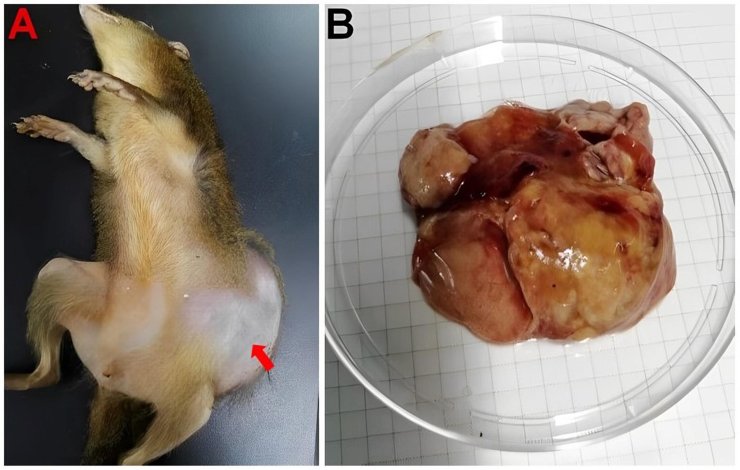
Figure 1 The overall morphology and autopsy of Tupaia belangeri subsp.yaoshanensisNote:A, Overall morphology and tumor location of tree shrew (as indicated by the red arrow); B, Exfoliated tumor form.

Figure 2 Histopathological changes of primary tumor in Tupaia belangeri subsp. yaoshanensis (HE staining)Note:A, Pleomorphic sarcoma region, epithelioid cells, obvious cell atypia, coarse nucleoli (as indicated by the red arrow) (×200). B, Adipocytes have different sizes, and there are many vesicles in the cytoplasm (as indicated by the red arrow). Visible pleomorphic adipocytes display large cell size, large nuclei, coarse granular chromatin and coarse nucleoli (×400). C, Adipocytes and pleomorphic adipocytes. The nuclei of pleomorphic adipocytes are vacuolarly squeezed by cytoplasmic lipid droplets, showing indentation (as indicated by the red arrow) (×400).

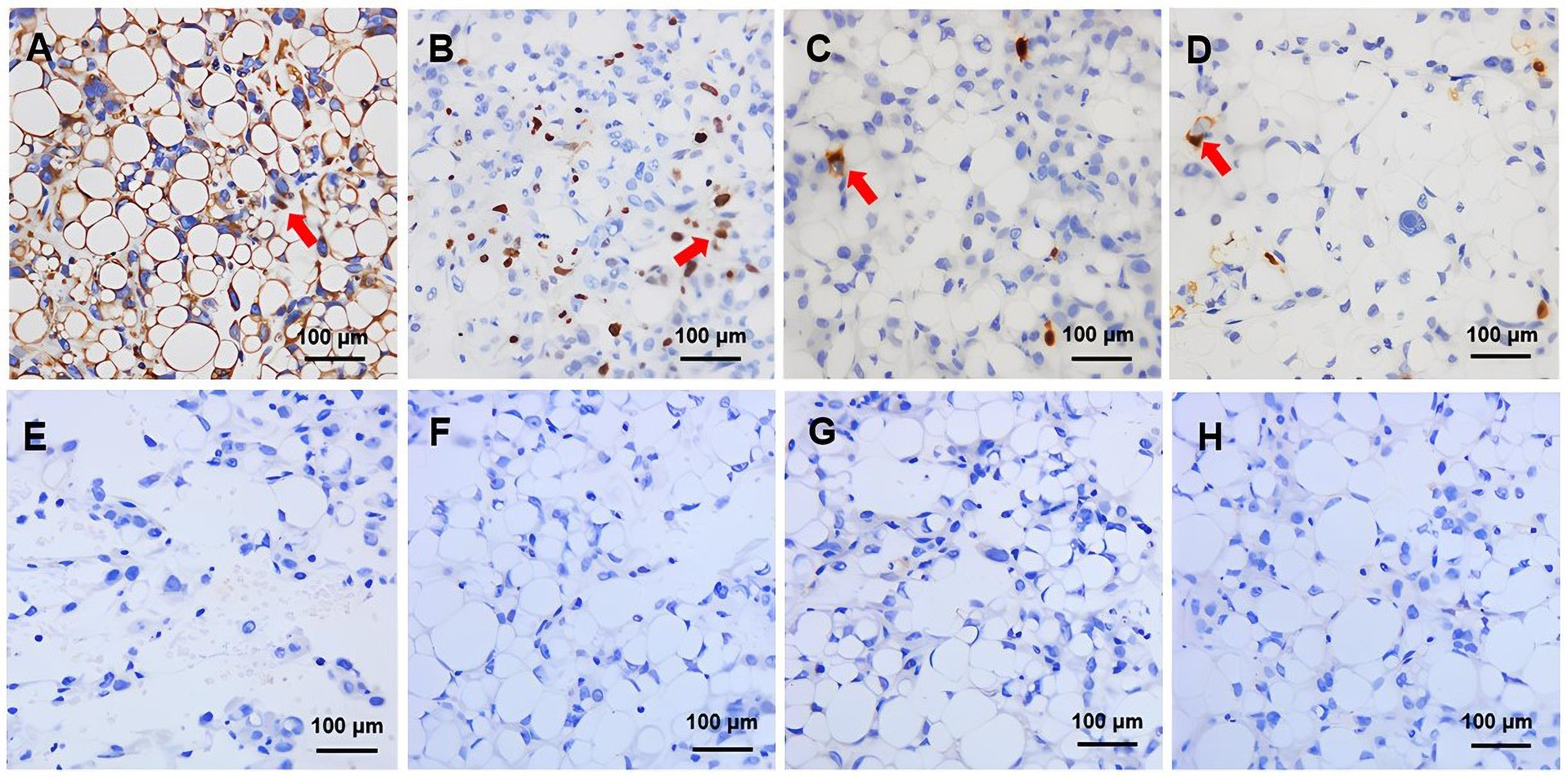
Figure 3 Immunohistochemical detection of primary tumors suggestive of pleomorphic liposarcoma in the Tupaia belangeri subsp.yaoshanensis (DAB stain)Note:A, Diffuse positive expression of vimentin in tumor cells (as indicated by the red arrow)(×400); B, High expression of Ki-67 in small focal nuclei of tumor cells (as indicated by the red arrow)(×400); C/D, Positive expression of S-100 in small tumor cells (as indicated by the red arrow) (×400); E, Negative expression of cytokeratin (CK) in tumor cells (×400); F, Negative expression of gross cystic disease fluid protein-15 (GCDFP-15) in tumor cells (×400); G, Negative expression of estrogen receptor (ER) in tumor cells (×400); H, Negative expression of GATA Binding Protein 3 (GATA) in tumor cells (×400).
| 1 | DEI TOS A P. Classification of pleomorphic sarcomas: where are we now?[J]. Histopathology, 2006, 48(1):51-62. DOI: 10.1111/j.1365-2559.2005.02289.x . |
| 2 | WANG L, REN W M, ZHOU X Y, et al. Pleomorphic liposarcoma: a clinicopathological, immunohistochemical and molecular cytogenetic study of 32 additional cases[J]. Pathol Int, 2013, 63(11):523-531. DOI: 10.1111/pin.12104 . |
| 3 | WANG L W, LUO R L, XIONG Z M, et al. Pleomorphic liposarcoma[J]. Medicine, 2018, 97(8): e9986. DOI: 10.1097/md.0000000000009986 . |
| 4 | 宋亚宁, 曹永宽. 胆囊三角区巨大脂肪肉瘤一例[J]. 中华临床医师杂志(电子版), 2012, 6(5):1368. |
| SONG Y N, CAO Y K. A case of giant liposarcoma in gallbladder triangle[J]. Chin J Clin Electron Ed, 2012, 6(5):1368. | |
| 5 | 季旸, 秦杰, 李霞, 等. 肢体不同病理亚型脂肪肉瘤超声学表现[J]. 医学影像学杂志, 2022, 32(7): 1215-1218. DOI: 10.3969/j.issn.1001-7399.2010.01.024 . |
| JI Y, QIN J, LI X, et al. The ultrasound performance of liposarcoma in different subtypes of pathology[J]. J Med Imaging, 2022, 32(7): 1215-1218. DOI: 10.3969/j.issn.1001-7399.2010.01.024 . | |
| 6 | 李元歌, 陈武标, 郁成, 等. 脂肪肉瘤CT及MRI的影像特征与病理分型的对照分析[J]. 医学影像学杂志, 2020, 30(2): 303-307. DOI: 10.13315/j.cnki.cjcep.2018.06.024 . |
| LI Y G, CHEN W B, YU C, et al. Comparative analysis of CT and MRI imaging features and pathological classification of liposarcoma[J]. J Med Imaging, 2020, 30(2): 303-307. DOI: 10.13315/j.cnki.cjcep.2018.06.024 . | |
| 7 | ANDERSON W J, JO V Y. Pleomorphic liposarcoma: Updates and current differential diagnosis[J]. Semin Diagn Pathol, 2019, 36(2):122-128. DOI: 10.1053/j.semdp.2019.02.007 . |
| 8 | APOSTOLOU G, BITELI M, CHATZIPANTELIS P. Cytopathological diagnosis of metastatic pleomorphic liposarcoma in the lung: a report of a case correlated with the histopathology of the primary tumour[J]. Diagn Cytopathol, 2009, 37(9):667-670. DOI: 10.1002/dc.21085 . |
| 9 | VON MEHREN M, KANE J M, BUI M M, et al. NCCN guidelines insights: soft tissue sarcoma, version 1.2021[J]. J Natl Compr Canc Netw, 2020, 18(12):1604-1612. DOI: 10.6004/jnccn.2020.0058 . |
| 10 | EWING E. Soft tissue and visceral sarcomas: ESMO Clinical Practice Guidelines for diagnosis, treatment and follow-up[J]. Annals of Oncology, 2012, 23(): vii92–vii99.doi:10.1093/annonc/mds253 . |
| 11 | 中华医学会肿瘤学分会, 中华医学会杂志社, 中国医师协会肛肠医师分会腹膜后疾病专业委员会, 等. 中国腹膜后肿瘤诊治专家共识( 2019版)[J]. 中华肿瘤杂志, 2019, 41(10):728-733. DOI:10.3760/cma.j.issn.0253-3766.2019.10.002 . |
| Chinese Medical Association, Cancer Society of Chinese Medical Association, Journal of Chinese Medical Association, et al. Expert consensus on treatment of Retroperitoneal tumors in China (Edition 2019)[J]. Chin J Oncol, 2019, 41(10):728-733. DOI: 10.3760/cma.j.issn.0253-3766.2019.10.002 . | |
| 12 | WILSON D E, REEDER D M. Mammal species of the World: a taxonomic and geographic reference[M]. 3rd ed. Maryland: Johns Hopkins University Press, 2005:104-9. |
| 13 | LI R F, ZANIN M, XIA X S, et al. The tree shrew as a model for infectious diseases research[J]. J Thorac Dis, 2018, 10(S9): S2272-S2279. DOI: 10.21037/jtd.2017.12.121 . |
| 14 | 夏巍, 赖永静, 杜龙, 等. 树鼩在人类肿瘤疾病动物模型中的应用进展[J]. 动物医学进展, 2019, 40(3): 109-113. DOI: 10.3969/j.issn.1007-5038.2019.03.022 . |
| XIA W, LAI Y J, DU L, et al. Application of tree shrew in animal models of human tumor diseases[J]. Prog Vet Med, 2019, 40(3): 109-113. DOI: 10.3969/j.issn.1007-5038.2019.03.022 . | |
| 15 | 唐海波, 梁亮, 曹颖颖, 等. 一例瑶山亚种树鼩自发隆突性皮肤纤维肉瘤的诊断[J]. 动物医学进展, 2021, 42(11):140-144. DOI: 10.3969/j.issn.1007-5038.2021.11.028 . |
| TANG H B, LIANG L, CAO Y Y, et al. Diagnosis of a spontaneous dermatofibrosarcoma protuberans from tree shrew (Tupaia belangeri subsp.yaoshanensis)[J]. Prog Vet Med, 2021, 42(11):140-144. DOI: 10.3969/j.issn.1007-5038.2021.11.028 . | |
| 16 | 张圆圆, 蒋艳玲, 文容,等. 反射式共聚焦显微镜在儿童毛发上皮瘤的诊断价值[J]. 实用皮肤病学杂志, 2021, 14(2):81-83, 87. DOI: 10.11786/sypfbxzz.1674-1293.20210205 . |
| ZHANG Y Y, JIANG Y L, WEN R, et al. The diagnostic value of reflective confocal microscopy in children with tricho-epithelioma[J]. Prac J Dermat, 2021, 14(2):81-83, 87. DOI:10.11786/sypfbxzz.1674-1293.20210205 . | |
| 17 | SBARAGLIA M, BELLAN E, DEI TOS A P. The 2020 WHO classification of soft tissue tumours: news and perspectives[J]. Pathologica, 2021, 113(2):70-84. DOI: 10.32074/1591-951X-213 . |
| 18 | LEE A T J, THWAY K, HUANG P H, et al. Clinical and molecular spectrum of liposarcoma[J]. J Clin Oncol, 2018, 36(2):151-159. DOI: 10.1200/JCO.2017.74.9598 . |
| 19 | GHADIMI M P, LIU P, PENG T S, et al. Pleomorphic liposarcoma: clinical observations and molecular variables[J]. Cancer, 2011, 117(23):5359-5369. DOI: 10.1002/cncr.26195 . |
| 20 | DOWNES K A, GOLDBLUM J R, MONTGOMERY E A, et al. Pleomorphic liposarcoma: a clinicopathologic analysis of 19 cases[J]. Mod Pathol, 2001, 14(3):179-184. DOI: 10.1038/modpathol.3880280 . |
| 21 | MIETTINEN M, ENZINGER F M. Epithelioid variant of pleomorphic liposarcoma: a study of 12 cases of a distinctive variant of high-grade liposarcoma[J]. Mod Pathol, 1999, 12(7):722-728. |
| 22 | 刘喆, 杨婕, 朱萌, 等. 多形性脂肪肉瘤4例临床病理分析[J]. 中国处方药, 2022, 20(12): 14-16. DOI: 10.3969/j.issn.1671-945X.2022.12.005 . |
| LIU Z, YANG J, ZHU M, et al. Pleomorphic liposarcoma: a clinicopathologic analysis of 4 cases[J]. J China Prescr Drug, 2022, 20(12): 14-16. DOI: 10.3969/j.issn.1671-945X.2022.12.005 . | |
| 23 | MANGHAM D. World Health Organisation classification of tumours: pathology and genetics of tumours of soft tissue and bone[M]. Lyon, France: IARC Press, 2002:427. |
| 24 | HORNICK J L, BOSENBERG M W, MENTZEL T, et al. Pleomorphic liposarcoma[J]. Am J Surg Pathol, 2004, 28(10):1257-1267. DOI: 10.1097/01.pas.0000135524.73447.4a . |
| 25 | YAN P, SUN M L, SUN Y P, et al. Effective apatinib treatment of pleomorphic liposarcoma[J]. Medicine, 2017, 96(33): e7771. DOI: 10.1097/md.0000000000007771 . |
| 26 | GEBHARD S, COINDRE J M, MICHELS J J, et al. Pleomorphic liposarcoma: clinicopathologic, immunohistochemical, and follow-up analysis of 63 cases: a study from the French Federation of Cancer Centers Sarcoma Group[J]. Am J Surg Pathol, 2002, 26(5):601-616. DOI: 10.1097/00000478-200205000-00006 . |
| 27 | NASCIMENTO A F, RAUT C P. Diagnosis and management of pleomorphic sarcomas (so-called MFH) in adults[J]. J Surg Oncol, 2008, 97(4):330-339. DOI: 10.1002/jso.20972 . |
| 28 | CRAGO A M, DICKSON M A. Liposarcoma[J]. Surg Oncol Clin N Am, 2016, 25(4):761-773. DOI: 10.1016/j.soc.2016.05.007 . |
| 29 | 施继鼎, 王占兴, 陈振声, 等. 膀胱原发多形性脂肪肉瘤1例报告[J]. 福建医药杂志, 2021, 43(3): 179-180. DOI: 10.3969/j.issn.1002-2600.2021.03.073 . |
| SHI J D, WANG Z X, CHEN Z S, et al. Primary liposarcoma pleomorphic bladder: a case report[J]. Fujian Med J, 2021, 43(3): 179-180. DOI: 10.3969/j.issn.1002-2600.2021.03.073 . | |
| 30 | CHOI J H, RO J Y. The 2020 WHO classification of tumors of soft tissue: selected changes and new entities[J]. Adv Anat Pathol, 2021, 28(1):44-58. DOI: 10.1097/PAP.0000000000000284 . |
| [1] | SUN Xiaorong, SU Dan, GUI Wenjuan, CHEN Yue. Establishment and Evaluation of a Moderate-to-Severe Knee Osteoarthritis Model in Rats Induced by Surgery [J]. Laboratory Animal and Comparative Medicine, 2024, 44(6): 597-604. |
| [2] | Minbo HOU, Tiantian CUI, Naying SU, Miaomiao ZHANG, Yongmin JIAO, Jianyan YAN, Xijie WANG, Ohira TOKO. Pathologic Diagnosis of a Pituicytoma in a Han-Wistar Rat [J]. Laboratory Animal and Comparative Medicine, 2023, 43(6): 654-658. |
| [3] | Shanshan ZHAI, Liang LIANG, Yingying CAO, Zhuxin LI, Qing WANG, Junyu TAO, Chenxia YUN, Jing LENG, Haibo TANG. Diagnosis of Trichoepithelioma in a Tree Shrew and Observation of Cell Biological Characteristics [J]. Laboratory Animal and Comparative Medicine, 2023, 43(4): 440-445. |
| [4] | Ying TAN, Wenping LIAO, Qilong GAO, Yong LI, Xinhui SHI, Jingkun WANG. Physiological Indexes and Histopathology Analysis of Sodium Iodate-Induced Retinitis Pigmentosa in Rats [J]. Laboratory Animal and Comparative Medicine, 2023, 43(2): 124-135. |
| [5] | WEN Fuli. Establishment of Eimeria stiedai Infected Rabbit Model and Nested PCR Assay [J]. Laboratory Animal and Comparative Medicine, 2020, 40(6): 477-482. |
| [6] | HUANG Jisheng, WU Shuyi, ZHAN Jinhe, NI Qingchun. Analysis of Incidence of Spontaneous Histopathological Lesions in Young SD Rats [J]. Laboratory Animal and Comparative Medicine, 2020, 40(2): 128-135. |
| [7] | XIAO Kunlin, ZHANG Rui, Sun Hong, XIAO Kuntai, MA Jianbing. Evaluation of Monoiodoacetic Acid-induced Knee Osteoarthritis SD Rats with Diseasse Progression at Different Time Points [J]. Laboratory Animal and Comparative Medicine, 2020, 40(1): 47-52. |
| [8] | ZHOU Fei, WANG Hao-an, CHEN Tao, QIU Shuang, CHEN Ke, CEN Xiao-bo. Case Report of Geniposide Induced Bilirubin Pigmentation in Sprague-Dawley Rat [J]. Laboratory Animal and Comparative Medicine, 2018, 38(4): 284-287. |
| [9] | HUANG Chao, GONG Yi-juan, TIAN Fang, WANG Yu-zhu, SUN Bing, ZHI Rui-na, IA Min-jie, DING Xun-cheng, ZHOU Xin-chu, LI Wei-hua. Establishment of Vaginal Mucosal Immuno-inflammatory Response Model in Minipig [J]. Laboratory Animal and Comparative Medicine, 2016, 36(2): 81-86. |
| [10] | LI Wei-hua1,2,WANG Hai-lan3,Gaku Ichihara4,DING Xun-cheng2,ZHOU Zhi-jun1. Comparative Study of 1-Bromopropane Induced Toxic Effects in Fisher 344/NSIc and Wistar NWN Rats [J]. Laboratory Animal and Comparative Medicine, 2010, 30(2): 105-108. |
| [11] | ZHOU Xiao-mm, BA Cai-feng, FENG Hui-quan . Experimental Study on Mouse Model Infected with Mycoplasma suis [J]. Laboratory Animal and Comparative Medicine, 2008, 28(6): 372-376. |
| [12] | XU Xiao-Ping, XU Jian-Qin, CHEN Min-Li, CHEN Fang-Ming, CHEN Liang, PENG Ding-Guo, ZHOU Wei-Min. Establishment of Rabbit Model of Allergic Rhinitis and Histological Observation of Nasal Mucosa [J]. Laboratory Animal and Comparative Medicine, 2006, 26(2): 80-82. |
| [13] | LIU Yi-1, ZHANG Chi-1, XIAO Jun-Xia-2, XI Shou-Min-1, YIN Wei-Dong-1, WANG Zong-Bao-1. Disorders of Glucose and Lipid Metabolism and Pathological Changes of Pancreas, Liver and Kidney in New Zealand White Rabbits Induced by High Sucrose,High Fat and High Cholesterol Diet [J]. Laboratory Animal and Comparative Medicine, 2005, 25(3): 151-156. |
| [14] | CHENG Shu-jun, HUANG Ren, QING Yao, TAN Wen-ya. Histopathological Observation on Spontaneous Lesion of Digestive System in Macaca mulatta [J]. Laboratory Animal and Comparative Medicine, 2003, 23(3): 131-134. |
| [15] | ZHOU Zhong-xin1,WANG Jie2, LUO Bao-ming2. Functional and Histopathological Changes of Liver in Cirrhosis Model of Mini-pigs [J]. Laboratory Animal and Comparative Medicine, 2002, 22(2): 102-105. |
| Viewed | ||||||
|
Full text |
|
|||||
|
Abstract |
|
|||||