

Laboratory Animal and Comparative Medicine ›› 2026, Vol. 46 ›› Issue (5): 650-659.DOI: 10.12300/j.issn.1674-5817.2026.010
• Laboratory Animals for Traditional Chinese Medicine Research • Previous Articles Next Articles
XU Haochen, LE Qingqing, HE Xiwen, CAI Bing, YANG Longhe( )(
)( )
)
Received:2026-01-08
Revised:2026-06-16
Online:2026-10-25
Published:2026-10-16
Correspondence to:
YANG Longhe
CLC Number:
XU Haochen,LE Qingqing,HE Xiwen,et al. Evaluation of the Efficacy of an Eye-Protective Compound Formula on the Retina in a Mouse Model of Blue Light-Induced Damage[J]. Laboratory Animal and Comparative Medicine, 2026, 46(5): 650-659. DOI: 10.12300/j.issn.1674-5817.2026.010.
Add to citation manager EndNote|Ris|BibTeX
URL: https://www.slarc.org.cn/dwyx/EN/10.12300/j.issn.1674-5817.2026.010
The sequences of primers used in real-time fluorescent quantitative reverse transcription PCR
| 基因名称 Gene name | 引物序列 Primer sequences (5' - 3') |
|---|---|
| IL-1β | F-GAAATGCCACCTTTTGACAGTG R-TGGATGCTCTCATCAGGACAG |
| IL-6 | F-AATTAAGCCTCCGACTTGTGAAG R-CTTCCATCCAGTTGCCTTCTTG |
| TNF-α | F-GTAGCAAACCACCAAGTGGAGG R-CAGCCTTGTCCCTTGAAGAGAA |
| GAPDH | F-GGTGAAGGTCGGTGTGAACG R-CTCGCTCCTGGAAGATGGTG |
Table 1
The sequences of primers used in real-time fluorescent quantitative reverse transcription PCR
| 基因名称 Gene name | 引物序列 Primer sequences (5' - 3') |
|---|---|
| IL-1β | F-GAAATGCCACCTTTTGACAGTG R-TGGATGCTCTCATCAGGACAG |
| IL-6 | F-AATTAAGCCTCCGACTTGTGAAG R-CTTCCATCCAGTTGCCTTCTTG |
| TNF-α | F-GTAGCAAACCACCAAGTGGAGG R-CAGCCTTGTCCCTTGAAGAGAA |
| GAPDH | F-GGTGAAGGTCGGTGTGAACG R-CTCGCTCCTGGAAGATGGTG |
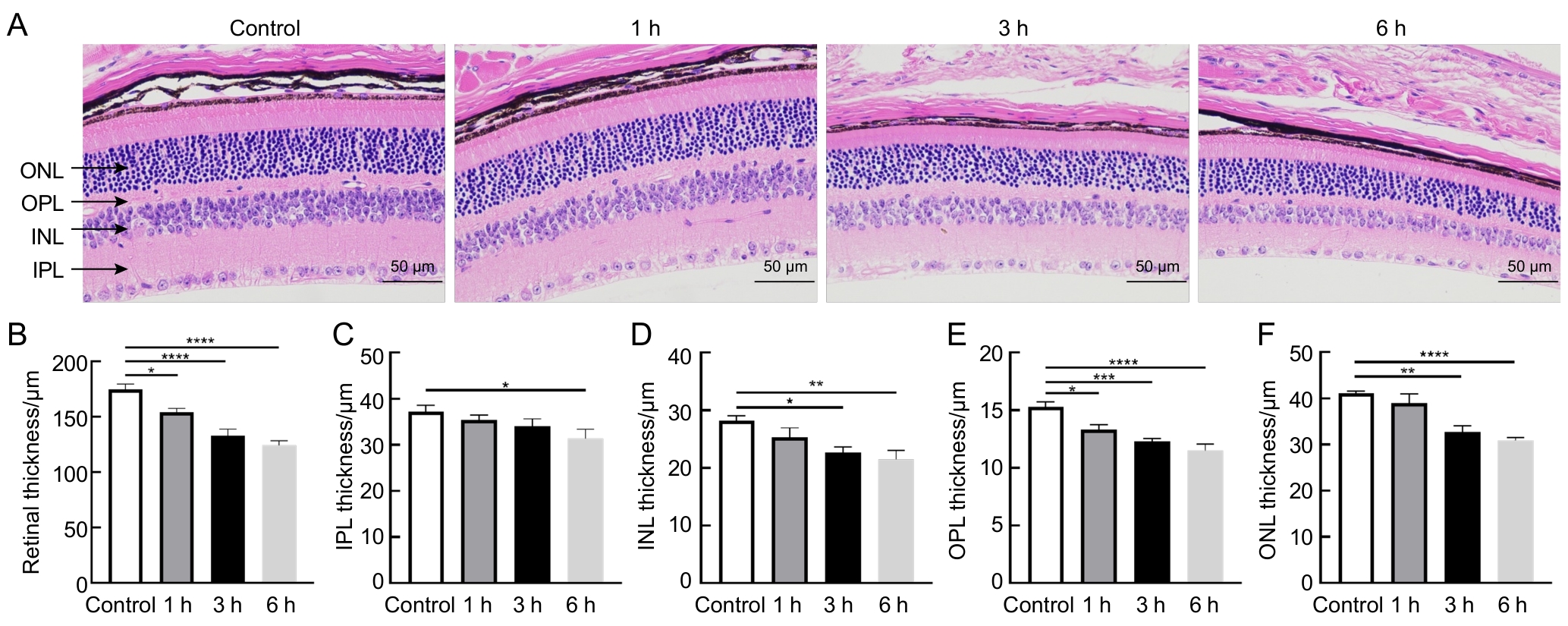
Figure 1
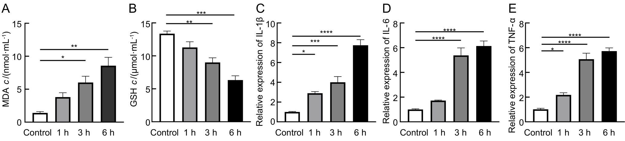
Figure 2
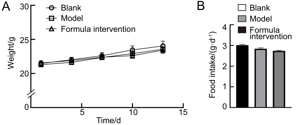
Figure 3
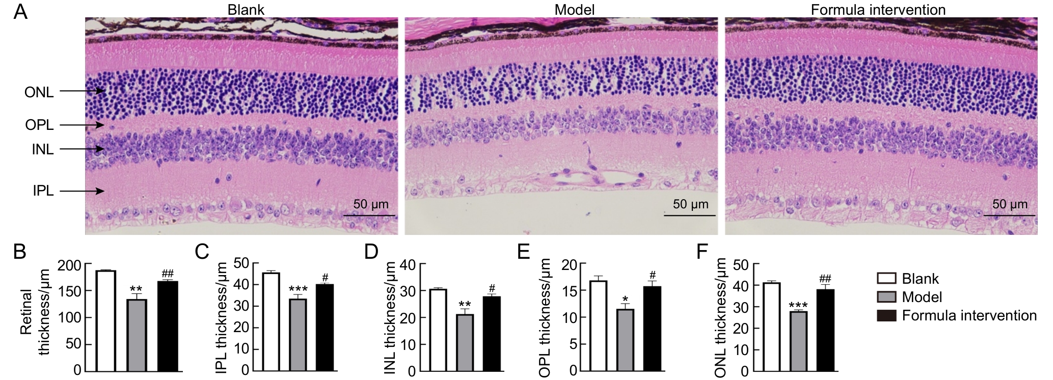
Figure 4

Figure 5
| [1] |
ZHOU Z X, LI S Z, YANG Q S, et al. BlingLife®-marigold extract alleviates blue light-induced retinal mitochondria oxidative stress and senescence by activating NRF2/HO-1 signaling[J]. J Microbiol Biotechnol, 2025, 35: e2411037. DOI: 10.4014/jmb. 2411.11037.
|
| [2] |
CHAKRAVARTHY H, GEORGYEV V, WAGEN C, et al. Blue light-induced phototoxicity in retinal cells: implications in age-related macular degeneration[J]. Front Aging Neurosci, 2024, 16: 1509434. DOI: 10.3389/fnagi.2024.1509434.
|
| [3] |
YEH W J, CHIEN P T, WEN Y T, et al. A comprehensive review of experimental models for investigating blue light-induced ocular damage: Insights into parameters, limitations, and new opportunities[J]. Exp Eye Res, 2024, 249: 110142. DOI: 10. 1016/j.exer.2024.110142.
|
| [4] |
ZHANG S Y, REN J P, GHAI R T. Global burden of low vision and blindness due to age-related macular degeneration from 1990 to 2021 and projections for 2050[J]. BMC Public Health, 2024, 24(1): 3510. DOI: 10.1186/s12889-024-21047-x.
|
| [5] |
THOMAS C J, MIRZA R G, GILL M K. Age-related macular degeneration[J]. Med Clin North Am, 2021, 105(3): 473-491. DOI: 10.1016/j.mcna.2021.01.003.
|
| [6] |
GARCÍA-ROMERA M C, PONCE-GARCÍA V, TORRES-PAREJO Ú, et al. Effect of exposure to blue light from electronic devices and the Mediterranean diet on macular pigment[J]. J Clin Med, 2024, 13(24): 7688. DOI: 10.3390/jcm13247688.
|
| [7] |
KIM S H, KIM Y K, SHIN Y I, et al. Nighttime outdoor artificial light and risk of age-related macular degeneration[J]. JAMA Netw Open, 2024, 7(1): e2351650. DOI: 10.1001/jamanet workopen.2023.51650.
|
| [8] |
SINGH S, KELLER P R, BUSIJA L, et al. Blue-light filtering spectacle lenses for visual performance, sleep, and macular health in adults[J]. Cochrane Database Syst Rev, 2023(8): CD013244. DOI: 10.1002/14651858.cd013244.pub2.
|
| [9] |
MOISEEV R V, MORRISON P W J, STEELE F, et al. Penetration enhancers in ocular drug delivery[J]. Pharmaceutics, 2019, 11(7): 321. DOI: 10.3390/pharmaceutics11070321.
|
| [10] |
YEH W J, YAN C, WU C H. Photoprotective effects of phytochemicals on blue light-induced retinal damage: current evidence and future perspectives[J]. Nutrients, 2025, 17(2): 331. DOI: 10.3390/nu17020331.
|
| [11] |
WON J H, SITNIKOV D, HONG J N. Protective effects of carotenoids against blue light induced-cellular damage in human retinal pigment epithelium[J]. Food Sci Biotechnol, 2025, 34(8): 1713-1723. DOI: 10.1007/s10068-024-01757-z.
|
| [12] |
ACAR N. The implication of omega-3 polyunsaturated fatty acids in retinal physiology[J]. Oléagineux Corps Gras Lipides, 2007, 14(3-4): 194-197. DOI: 10.1051/ocl.2007.0121.
|
| [13] |
LIU Y X, ZHANG D, WU Y P, et al. Docosahexaenoic acid aggravates photooxidative damage in retinal pigment epithelial cells via lipid peroxidation[J]. J Photochem Photobiol B, 2014, 140: 85-93. DOI: 10.1016/j.jphotobiol.2014. 07.016.
|
| [14] |
LI P, LI Z G, SUN Q X, et al. Protective effect and mechanism of Lycium ruthenicum Murray anthocyanins against retinal damage induced by blue light exposure[J]. J Food Sci, 2024, 89(8): 5113-5129. DOI: 10.1111/1750-3841.17184.
|
| [15] |
庞雨菲. 黑果枸杞多酚对丙烯酰胺诱导大鼠纹状体氧化应激损伤的保护作用及机制[D]. 银川: 宁夏医科大学, 2023. DOI: 10.27258/d.cnki.gnxyc.2023.000305.
|
|
PANG Y F. Protective effect and mechanism of polyphenols from Lycium ruthenicum Murray on acrylamide-induced oxidative stress injury in rat striatum [D]. Yinchuan: Ningxia Medical University, 2023. DOI: 10.27258/d.cnki.gnxyc.2023.000305.
|
|
| [16] |
OUYANG X L, YANG J, HONG Z X, et al. Mechanisms of blue light-induced eye hazard and protective measures: a review[J]. Biomed Pharmacother, 2020, 130: 110577. DOI: 10.1016/j.biopha.2020.110577.
|
| [17] |
娄解南, 谭书明, 谢国芳, 等. 刺梨多酚对丙烯酰胺致肝脏氧化损伤的保护作用[J]. 食品安全质量检测学报, 2023, 14(17): 122-128. DOI: 10.19812/j.cnki.jfsq11-5956/ts.2023.17.012.
|
|
LOU J N, TAN S M, XIE G F, et al. Protective effect of Rosa roxburghii Tratt.polyphenols against acrylamide-induced hepatic oxidative damage[J]. J Food Saf Qual, 2023, 14(17): 122-128. DOI: 10.19812/j.cnki.jfsq11-5956/ts.2023.17.012.
|
|
| [18] |
张丹, 葛颖颖, 杨琼琼, 等. 刺梨果抗氧化活性成分与体外抗氧化活性研究[C]//中国食品科学技术学会第十五届年会论文摘要集. 2018: 283-284.
|
|
ZHANG D, GE Y Y, YANG Q Q, et al. Study on antioxidant active components and in vitro antioxidant activity of Rosa roxburghii fruit[C]//Abstracts of the 15th Annual Meeting of Chinese Institute of Food Science and Technology. 2018: 283-284.
|
|
| [19] |
SKIBSTED L H. Anthocyanidins regenerating xanthophylls: a quantum mechanical approach to eye health[J]. Curr Opin Food Sci, 2018, 20: 24-29. DOI: 10.1016/j.cofs.2018.02.014.
|
| [20] |
TÄHTIVAARA L, THAPA R, PARTANEN P, et al. Comparison of blue light-induced damage (BLD) in albino and pigmented mice[J]. Invest Ophthalmol Vis Sci, 2025, 66(8): 2528.
|
| [21] |
KING A, GOTTLIEB E, BROOKS D G, et al. Mitochondria-derived reactive oxygen species mediate blue light-induced death of retinal pigment epithelial cells[J]. Photochem Photobiol, 2004, 79(5): 470-475. DOI: 10.1111/j.1751-1097.2004.tb00036.x.
|
| [22] |
KAYNEZHAD P, FOSBURY R, HOGG C, et al. Near infrared spectroscopy reveals instability in retinal mitochondrial metabolism and haemodynamics with blue light exposure at environmental levels[J]. J Biophotonics, 2022, 15(4): e202100283. DOI: 10.1002/jbio.202100283.
|
| [23] |
ROEHLECKE C, SCHUMANN U, ADER M, et al. Stress reaction in outer segments of photoreceptors after blue light irradiation[J]. PLoS One, 2013, 8(9): e71570. DOI: 10.1371/journal.pone.0071570.
|
| [24] |
马修彬, 陈松. 蓝光对视网膜色素上皮细胞的损伤机制[J]. 国际眼科纵览, 2007, 31(6): 415-419. DOI: 10.3760/cma.j.issn.1673-5803.2007.06.015.
|
|
MA X B, CHEN S. Mechanism of blue-light-induced damage to the retinal pigment epithelium cells[J]. International Rev Ophthalmol, 2007, 31(6): 415-419. DOI: 10.3760/cma.j.issn.1673-5803.2007.06.015.
|
|
| [25] |
NAKAMURA M, KUSE Y, TSURUMA K, et al. The involvement of the oxidative stress in murine blue LED light-induced retinal damage model[J]. Biol Pharm Bull, 2017, 40(8): 1219-1225. DOI: 10.1248/bpb.b16-01008.
|
| [26] |
KAIDZU S, OKUNO T, TANITO M, et al. Structural and functional change in albino rat retina induced by various visible light wavelengths[J]. Int J Mol Sci, 2022, 23(1): 309. DOI: 10.3390/ijms23010309.
|
| [27] |
JI Z Z, ZHANG D C, WANG Y M, et al. The role of the SIRT1/NF-κB/NLRP3 pathway in the pyroptosis of lens epithelial cells under shortwave blue light radiation[J]. Exp Eye Res, 2024, 246: 110019. DOI: 10.1016/j.exer.2024.110019.
|
| [28] |
YANG Y C, YANG R, DENG F Q, et al. Immunoactivation by cutaneous blue light irradiation inhibits remote tumor growth and metastasis[J]. ACS Pharmacol Transl Sci, 2024, 7(4): 1055-1068. DOI: 10.1021/acsptsci.3c00355.
|
| [29] |
GRIMM C, WENZEL A, WILLIAMS T, et al. Rhodopsin-mediated blue-light damage to the rat retina: effect of photoreversal of bleaching[J]. Invest Ophthalmol Vis Sci, 2001,42(2): 497-505.
|
| [30] |
KAMOSHITA M, TODA E, OSADA H, et al. Lutein acts via multiple antioxidant pathways in the photo-stressed retina[J]. Sci Rep, 2016, 6: 30226. DOI: 10.1038/srep30226.
|
| [31] |
KIM J, CHO K, CHOUNG S Y. Protective effect of Prunella vulgaris var. L extract against blue light induced damages in ARPE-19 cells and mouse retina[J]. Free Radic Biol Med, 2020, 152: 622-631. DOI: 10.1016/j.freeradbiomed.2019.12.003.
|
| [32] |
ORGANISCIAK D T, VAUGHAN D K. Retinal light damage: Mechanisms and protection[J]. Prog Retin Eye Res, 2010, 29(2): 113-134. DOI: 10.1016/j.preteyeres.2009.11.004.
|
| [33] |
JAADANE I, BOULENGUEZ P, CHAHORY S, et al. Retinal damage induced by commercial light emitting diodes (LEDs)[J]. Free Radic Biol Med, 2015, 84: 373-384. DOI: 10.1016/j.freeradbiomed.2015.03.034.
|
| [34] |
SHANG Y M, WANG G S, SLINEY D, et al. White light–emitting diodes (LEDs) at domestic lighting levels and retinal injury in a rat model[J]. Environ Health Perspect, 2014, 122(3): 269-276. DOI: 10.1289/ehp.1307294.
|
| [35] |
OGAWA K, KUSE Y, TSURUMA K, et al. Protective effects of bilberry and lingonberry extracts against blue light-emitting diode light-induced retinal photoreceptor cell damage in vitro[J]. BMC Complement Altern Med, 2014, 14: 120. DOI: 10.1186/1472-6882-14-120.
|
| [36] |
WU J M, SEREGARD S, ALGVERE P V. Photochemical damage of the retina[J]. Surv Ophthalmol, 2006, 51(5): 461-481. DOI: 10.1016/j.survophthal.2006.06.009.
|
| [37] |
DABOUZ R, CHENG C W H, ABRAM P, et al. An allosteric interleukin-1 receptor modulator mitigates inflammation and photoreceptor toxicity in a model of retinal degeneration[J]. J Neuroinflammation, 2020, 17(1): 359. DOI: 10.1186/s12974-020-02032-8.
|
| [38] |
朱恒杏, 邵孟茹, 洪天畅, 等. 黑枸杞花青素、EGCG对中波紫外线诱导HaCaT细胞氧化损伤的协同保护作用[J]. 现代食品科技, 2024, 40(8): 106-115. DOI: 10.13982/j.mfst.1673-9078.2024.8.0918.
|
|
ZHU H X, SHAO M R, HONG T C, et al. Synergistic protective effects of the anthocyanins from black wolfberry and EGCG against the UVB-induced oxidative damage in HaCaT cells[J]. Mod Food Sci Technol, 2024, 40(8): 106-115. DOI: 10.13982/j.mfst.1673-9078.2024.8.0918.
|
|
| [39] |
邓楷, 欧阳健, 胡娜, 等. 黑果枸杞花青素结构差异对其稳定性及细胞抗氧化活性的影响[J]. 天然产物研究与开发, 2022, 34(2): 213-219. DOI: 10.16333/j.1001-6880.2022.2.005.
|
|
DENG K, OUYANG J, HU N, et al. Effects of structural difference on stability and cellular antioxidative activity of anthocyanins from Lycium ruthenicum Murr[J]. Nat Prod Res Dev, 2022, 34(2): 213-219. DOI: 10.16333/j.1001-6880.2022.2.005.
|
|
| [40] |
李婧, 苏蓉, 刘振华, 等. 黑果枸杞果实中抗炎活性成分的筛选及作用机制研究[J]. 中国野生植物资源, 2023, 42(5): 9-15, 38. DOI: 10.3969/j.issn.1006-9690.2023.05.002.
|
|
LI J, SU R, LIU Z H, et al. Research on the screening and mechanism of active ingredients for anti-inflammatory in Lycium ruthenicum Murr. fruit[J]. Chin Wild Plant Resour, 2023, 42(5): 9-15, 38. DOI: 10.3969/j.issn.1006-9690.2023.05.002.
|
|
| [41] |
薛才华, 武强, 王梦杰, 等. 黑枸杞花青素对巨噬细胞RAW264.7炎性与自噬交互作用的影响[J]. 中国畜牧杂志, 2021, 57(12): 244-249. DOI: 10.19556/j.0258-7033.20210220-06.
|
|
XUE C H, WU Q, WANG M J, et al. Effect of L. ruthenicum on the interaction between inflammation and autophagy of macrophage RAW264.7[J]. Chin J Anim Sci, 2021, 57(12): 244-249. DOI: 10.19556/j.0258-7033.20210220-06.
|
|
| [42] |
BERNSTEIN P S. The role of lutein and zeaxanthin in protection against age-related macular degeneration[J]. Acta Hortic, 2015(1106): 153-160. DOI: 10.17660/ActaHortic.2015. 1106.24.
|
| [43] |
ZHANG H J, LIU X B, CHEN X M, et al. Lutein delays photoreceptor degeneration in a mouse model of retinitis pigmentosa[J]. Neural Regen Res, 2022, 17(7): 1596-1603. DOI: 10.4103/1673-5374.330622.
|
| [44] |
BRAND A, BAUER N G, HALLOTT A, et al. Membrane lipid modification by polyunsaturated fatty acids sensitizes oligodendroglial OLN-93 cells against oxidative stress and promotes up-regulation of heme oxygenase-1 (HSP32)[J]. J Neurochem, 2010, 113(2): 465-476. DOI: 10.1111/j.1471-4159.2010.06611.x.
|
| [45] |
EGGERS B, STEPIEN J, REKER A K, et al. The protective effect of docosahexaenoic acid on mitochondria in SH-SY5Y model of rotenone-induced toxicity[J]. Metabolites, 2025, 15(1): 29. DOI: 10.3390/metabo15010029.
|
| [46] |
BORGONOVI S M, IAMETTI S, DI NUNZIO M. Docosahexaenoic acid as master regulator of cellular antioxidant defenses: a systematic review[J]. Antioxidants, 2023, 12(6): 1283. DOI: 10.3390/antiox12061283.
|
| [1] | JIANG Miaomiao, FANG Jinping, HUANG Zongxuan, LI Jiamin, LIAO Huan, ZHANG Wenqiang, YU Jiaying, YANG Yanyan, LIAN Dawei. Study on the Mechanism of Gegen Qinlian Decoction in Improving Diabetic Myocardial Injury [J]. Laboratory Animal and Comparative Medicine, 2026, 46(5): 629-639. |
| [2] | LI Yanping, HUANG Shan, YAO Jengyuan, SHI Yimin, SUI Huaxiu, HUANG Houtan, YU Yanping, MI Yan, LIN Tahui. Protective Effect and Mechanism of Artemitin on Ozone-Induced Pulmonary Inflammation in Mice [J]. Laboratory Animal and Comparative Medicine, 2026, 46(5): 640-649. |
| [3] | ZHANG Jiaoling, LIN Aping, XU Shiying, PAN Dequan, CAI Yijun. Optimization and Application of Mouse Embryo Transfer Technology [J]. Laboratory Animal and Comparative Medicine, 2026, 46(5): 750-758. |
| [4] | ZHONG Qiuming, QIAN Liping, CAI Yuanyuan, LIU Deqiang, LIN Wei, YU Chunying, XIE Jindong, WANG Xunli, WU Sheng, CHEN Biyin, HUANG Zhen. Comparative Analysis of Sex Differences Between Cyclophosphamide- and Paclitaxel-Induced Mouse Models of Chemotherapy-Induced Alopecia [J]. Laboratory Animal and Comparative Medicine, 2026, 46(5): 677-688. |
| [5] | WANG Juan, XU Jiahui, TIAN Yunyuan, ZHANG Mengmeng, LI Min, WANG Siwang, LI Yao. Comparison and Behavioral Observation of Two Female Mice Models of Ulcerative Colitis [J]. Laboratory Animal and Comparative Medicine, 2026, 46(3): 332-343. |
| [6] | LI Jiafei, ZHANG Zhenhao, WANG Shuo, TIAN Ge, WEN Shuang, YAN Yuxue, CUI Ran, YE Zhen, CUI Yongchun. Effects of Autonomic Neuromodulators on Atrial Electrical Remodeling and Histopathological Changes in a Rat Model of Atrial Fibrillation [J]. Laboratory Animal and Comparative Medicine, 2026, 46(3): 321-331. |
| [7] | LI Longxue, WAN Chongfan, ZHANG Qi, LEI Ruting, WANG Xiaoyue, CHENG Leyan, LAI Qi, LIU Ronghua, LIU Xuan, XU Tielong. Molecular Mechanisms of Qingfei Paidu Decoction in the Prevention and Treatment of Acute Lung Injury in Mice Based on miRNA Sequencing [J]. Laboratory Animal and Comparative Medicine, 2026, 46(3): 311-320. |
| [8] | AI Xiufeng, ZHANG Lizong, FANG Mingsun, LÜ Dongying, CHEN Chu, CAI Zhaowei, WANG Dejun. Analysis of Differences in the Intestinal Flora of Rats and Mice after Drinking Chlorinated Water Based on 16S rRNA Sequencing [J]. Laboratory Animal and Comparative Medicine, 2026, 46(3): 437-445. |
| [9] | TANG Xiaohang, GU Yingmin, LÜ Yangyang, HUANG Mingshu, TIAN Xuesong. Evaluation of the Histological Staining Performance of Rat Eyeball Sections Prepared Using a Self-Developed Fixative [J]. Laboratory Animal and Comparative Medicine, 2026, 46(2): 261-270. |
| [10] | WU Xianwen, LIU Lili, CHEN Ye, XU Guoheng. Optimization of Cage-Changing Intervals and Wood Shavings Usage for Mice During the Growth Phase in Breeding Systems [J]. Laboratory Animal and Comparative Medicine, 2026, 46(2): 251-260. |
| [11] | RONG Wenshuang, NIU Yuanfei, LIU Meiting, YANG Mengyuan, CUI Shuang, MA Lina, FU Yao, WANG Lianmei, CAO Junling. Influence of Antigen Type on the Establishment of an Induced Sjögren Syndrome Mouse Model [J]. Laboratory Animal and Comparative Medicine, 2026, 46(2): 178-190. |
| [12] | XU Yingtao, WANG Mengmeng, LIN Ping, CHI Haitao, WANG Yi, BAI Ying. Exosomes Treat Ischemic Stroke by Regulation of Ferroptosis Through the NRF2/SLC7A11/GPX4 Pathway in Mice [J]. Laboratory Animal and Comparative Medicine, 2026, 46(1): 20-31. |
| [13] | LIU Chang, XIANG Xuesong, HE Huihuang, CHEN Xiaoqing, QIU Wenhong. Establishment and Evaluation of an Oxidative Stress Model of Atopic Dermatitis Induced by 2,4-dinitrofluorobenzene [J]. Laboratory Animal and Comparative Medicine, 2026, 46(1): 46-54. |
| [14] | LIU Zhiwei, YANG Ran, LIAN Hao, ZHANG Yu, JIN Lilun. Cartilage Protection and Anti-Inflammatory Effects of Fraxetin on Monosodium Iodoacetate-Induced Rat Model of Osteoarthritis [J]. Laboratory Animal and Comparative Medicine, 2025, 45(3): 259-268. |
| [15] | WU Haifeng, ZHOU Xiaojiang, LI Chenjiang, LI Huaiyin, GAO Ming. Comparison of the Fixation Effects of Six Composite Fixatives on Retinal Tissue of Golden Hamsters [J]. Laboratory Animal and Comparative Medicine, 2024, 44(6): 675-681. |
| Viewed | ||||||
|
Full text |
|
|||||
|
Abstract |
|
|||||
