
Laboratory Animal and Comparative Medicine ›› 2025, Vol. 45 ›› Issue (6): 738-751.DOI: 10.12300/j.issn.1674-5817.2025.134
• Invertebrate Laboratory Animal: Nematode • Previous Articles Next Articles
SUN Han1( ), GUO Peng2, YU Xinhe1, ZHANG Junqiao3, YAO Ying4, YANG Wen1()(
 )
)
Received:2025-08-12
Revised:2025-12-05
Online:2025-12-25
Published:2025-12-19
Correspondence to:
YANG Wen
CLC Number:
SUN Han,GUO Peng,YU Xinhe,et al. Progress in Caenorhabditis elegans as a Degenerative Disease Model for Molecular Pathways Studying[J]. Laboratory Animal and Comparative Medicine, 2025, 45(6): 738-751. DOI: 10.12300/j.issn.1674-5817.2025.134.
Add to citation manager EndNote|Ris|BibTeX
URL: https://www.slarc.org.cn/dwyx/EN/10.12300/j.issn.1674-5817.2025.134
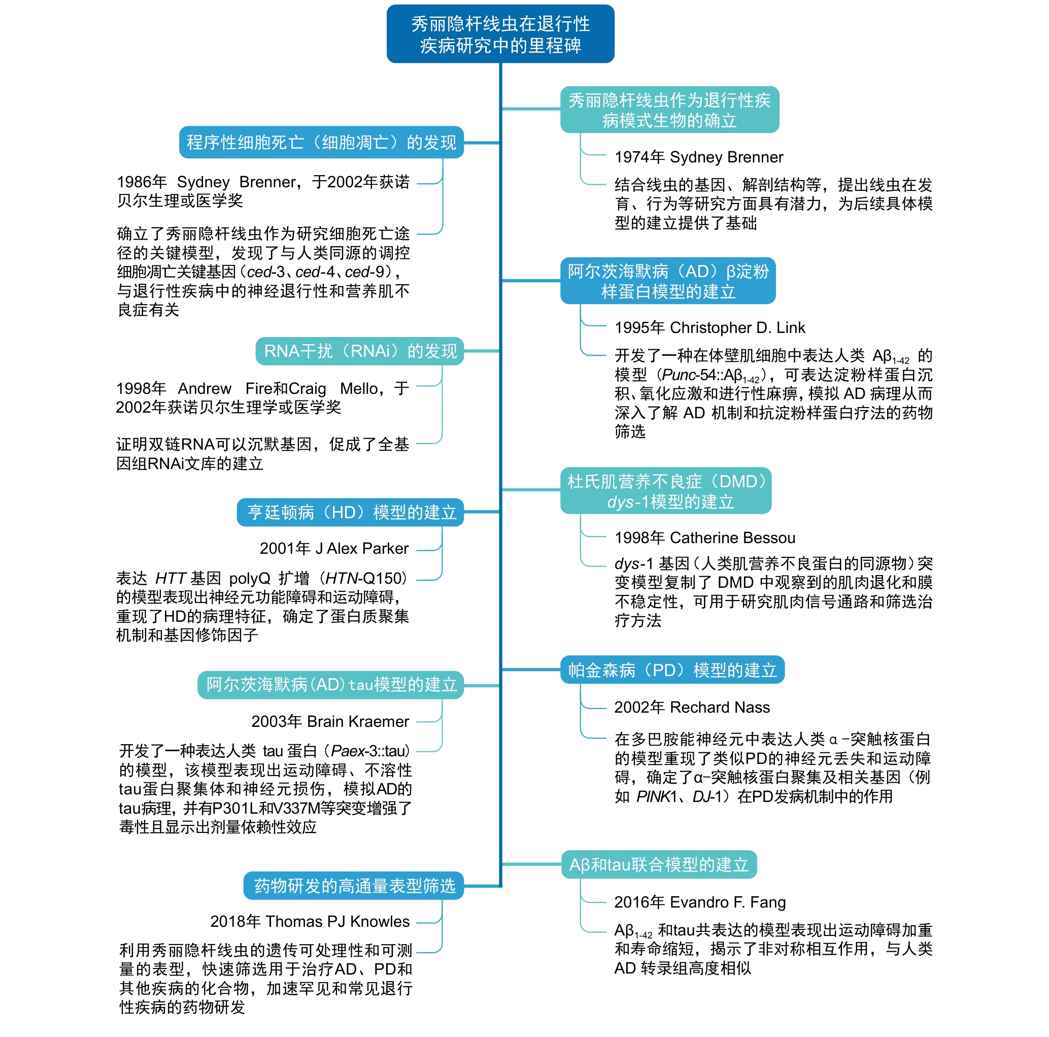
Figure 1 Major milestones of Caenorhabditis elegans in degenerative disease research
疾病 Diseases | 定义/特征 Definition/Characteristics | 秀丽隐杆线虫模型 Caenorhabditis elegans models |
|---|---|---|
杜氏肌营养不良症 Duchenne muscular dystrophy (DMD) | X染色体连锁隐性遗传,肌肉纤维逐渐坏死和假性肥大 | dys-1; ace-1[ |
肢带型肌营养不良 Limb-girdle muscular dystrophy (LGMD) | 累及肩胛带与骨盆带,表现为对称性近端肌无力 | LS587; giv otIs117[ |
肌少症 Sarcopenia | 衰老引起的骨骼肌质量显著减少,力量减弱 | PD2856; PD2859; PD4092[ |
线粒体肌病 Mitochondrial myopathies | 线粒体功能障碍引起的骨骼肌病变 | dct-1; allo-1[ |
糖原累积病 Glycogen storage diseases | 酶缺陷导致糖原异常积聚,常见于婴幼儿 | RT258;VK2882; pwIs50[ |
视网膜色素变性 Retinitis pigmentosa (RP) | 感光细胞退化,以夜盲和视野缩小为早期表现 | snrp-200; prp-8[ |
骨质疏松症 Osteoporosis | 骨密度下降,骨小梁破坏,易致脆性骨折 | 无 |
马凡综合征 Marfan syndrome | FBN1基因突变致结缔组织发育异常,累及骨、心血管、眼等系统 | 无 |
家族黑蒙性痴呆症 Tay-Sachs disease | HEXA基因突变,溶酶体中GM2神经节苷脂异常积聚 | 无 |
脊髓小脑性共济失调 Spinocerebellar ataxia | 以共济失调为主要表现的常染色体显性遗传病 | 无 |
尼曼-皮克病 Niemann-Pick disease | 脂类代谢障碍致肝脾大和神经系统损害 | 无 |
阿尔茨海默病 Alzheimer's disease (AD) | β淀粉样蛋白沉积和tau蛋白异常磷酸化引起的认知功能持续退化 | |
帕金森病 Parkinson's disease (PD) | 黑质多巴胺能神经元丢失,表现为震颤、运动迟缓等 | P301L;V337M |
亨廷顿病 Huntington's disease (HD) | HTT基因CAG重复扩增引起基底节神经元退化,表现为舞蹈样运动和精神症状 | HTN-Q150 |
肌萎缩侧索硬化 Amyotrophic lateral sclerosis (ALS) | 上下运动神经元退化,导致肌肉萎缩、瘫痪 | G93A SOD1 |
进行性核上性麻痹 Progressive supranuclear palsy | 以垂直眼动障碍、姿势不稳和额叶认知障碍为特征的tau蛋白病 | 无 |
多发性硬化症 Multiple sclerosis | 中枢神经系统白质脱髓鞘,伴随神经功能缺失 | 无 |
Table 1 Common clinical degenerative diseases and their corresponding Caenorhabditis elegans models
疾病 Diseases | 定义/特征 Definition/Characteristics | 秀丽隐杆线虫模型 Caenorhabditis elegans models |
|---|---|---|
杜氏肌营养不良症 Duchenne muscular dystrophy (DMD) | X染色体连锁隐性遗传,肌肉纤维逐渐坏死和假性肥大 | dys-1; ace-1[ |
肢带型肌营养不良 Limb-girdle muscular dystrophy (LGMD) | 累及肩胛带与骨盆带,表现为对称性近端肌无力 | LS587; giv otIs117[ |
肌少症 Sarcopenia | 衰老引起的骨骼肌质量显著减少,力量减弱 | PD2856; PD2859; PD4092[ |
线粒体肌病 Mitochondrial myopathies | 线粒体功能障碍引起的骨骼肌病变 | dct-1; allo-1[ |
糖原累积病 Glycogen storage diseases | 酶缺陷导致糖原异常积聚,常见于婴幼儿 | RT258;VK2882; pwIs50[ |
视网膜色素变性 Retinitis pigmentosa (RP) | 感光细胞退化,以夜盲和视野缩小为早期表现 | snrp-200; prp-8[ |
骨质疏松症 Osteoporosis | 骨密度下降,骨小梁破坏,易致脆性骨折 | 无 |
马凡综合征 Marfan syndrome | FBN1基因突变致结缔组织发育异常,累及骨、心血管、眼等系统 | 无 |
家族黑蒙性痴呆症 Tay-Sachs disease | HEXA基因突变,溶酶体中GM2神经节苷脂异常积聚 | 无 |
脊髓小脑性共济失调 Spinocerebellar ataxia | 以共济失调为主要表现的常染色体显性遗传病 | 无 |
尼曼-皮克病 Niemann-Pick disease | 脂类代谢障碍致肝脾大和神经系统损害 | 无 |
阿尔茨海默病 Alzheimer's disease (AD) | β淀粉样蛋白沉积和tau蛋白异常磷酸化引起的认知功能持续退化 | |
帕金森病 Parkinson's disease (PD) | 黑质多巴胺能神经元丢失,表现为震颤、运动迟缓等 | P301L;V337M |
亨廷顿病 Huntington's disease (HD) | HTT基因CAG重复扩增引起基底节神经元退化,表现为舞蹈样运动和精神症状 | HTN-Q150 |
肌萎缩侧索硬化 Amyotrophic lateral sclerosis (ALS) | 上下运动神经元退化,导致肌肉萎缩、瘫痪 | G93A SOD1 |
进行性核上性麻痹 Progressive supranuclear palsy | 以垂直眼动障碍、姿势不稳和额叶认知障碍为特征的tau蛋白病 | 无 |
多发性硬化症 Multiple sclerosis | 中枢神经系统白质脱髓鞘,伴随神经功能缺失 | 无 |
品系名称 Strain names | CGC名称 CGC names | CGC编号 CGC numbers | 疾病模型Disease models | 主要蛋白 Key proteins | 应用 Applications | 主要机制 Key mechanisms |
|---|---|---|---|---|---|---|
| 抗肌萎缩蛋白(Dystrophin) 缺失;乙酰胆碱酯酶(AChE)活性下降 | 模拟人类 DMD 的 dys-1 突变线虫,表现运动过度与胆碱酯酶活性降低,用于研究肌营养不良的致病机制 | |||||
LS587 | 抗肌萎缩蛋白(Dystrophin)与 MyoD(MyoD)双突变(加重肌纤维退化) | dys-1(cx18); hlh-1(cc561ts) 双突变诱导重度肌纤维退变,更接近 LGMD 表型,用于深入研究肌肉退行机制 | ||||
BA1 | Dysferlin 蛋白缺失(膜修复缺陷) | fer-1 突变部分模拟 LGMD2B 的肌膜修复缺陷,用于阐释dysferlinopathy 的病理机制 | ||||
cc2856 | 肌丝绿色荧光蛋白(GFP)报告基因(muscle filament GFP reporter)用于体壁肌监测 | UNC-54::GFP 标记肌丝结构,可随龄追踪非病理性肌肉衰老变化 | ||||
cc2859 | 内源性 unc-54::GFP(UNC-54::GFP)标记肌球蛋白重链 | CRISPR 标记的肌球蛋白重链,用于量化年龄相关的肌肉蛋白聚集与结构衰退 | ||||
cc4092 | non-stop mRNA 降解报告(non-stop mRNA decay reporter) | unc-54::gfp 无终止密码子模型,用于遗传筛选影响肌肉蛋白稳态的调控基因 | ||||
WJA780 | srf-780 (Δnonu-1) | mRNA 监测缺陷(defective mRNA surveillance;non-stop 降解受抑) | 在 nonu-1 缺失背景缓解 non-stop 肌肉缺陷,用于解析 mRNA 质量控制与肌肉衰老的联系 | |||
| dct-1 功能缺失导致线粒体堆积,用于研究线粒体肌病与选择性自噬机制 | ||||||
| allo-1 缺失使父源线粒体无法清除,用于研究异常胞器累积导致的肌病机制 | ||||||
RT258 | pwIs50 | 溶酶体膜蛋白 LMP-1::GFP(LMP-1::GFP)标记 | 用于监测 Pompe 病中溶酶体体积异常并进行药物高通量筛选 | |||
VK2882 | vkIs2882 | 肠道双标:GLO-1::CemOr-ange(GLO-1)+ LMP-1::GFP(LMP-1::GFP) | 用于多参数表型成像,分析 Pompe 病中糖原/溶酶体积聚并提高筛选精度 | |||
cer23 | 剪接因子 (SNRNP200) V676L 突变 | 模拟视网膜退化相关剪接因子突变,用于化合物筛选与疾病修饰子分析 | ||||
cer14 | 剪接体核心蛋白 (PRP-8)H2302 缺失突变 | 模拟 PRP8 致病突变,重现光感受细胞退行,用于药物与剪接机制研究 | ||||
CL2006 | dvIs2 | β-淀粉样肽1–42(Aβ₁₋₄₂)在肌肉持续表达 | CL2006 产生年龄依赖性 Aβ 斑块与瘫痪,用于筛选抗 Aβ 聚集药物 | |||
| CL4176 | β-淀粉样肽1–42(Aβ₁₋₄₂)温度诱导表达 | CL4176 升温急性致瘫,用于快速评估抗 Aβ 毒性候选物 | ||||
CL2355 | β-淀粉样肽1–42(Aβ₁₋₄₂)在神经元泛神经表达 | CL2355 表现学习、嗅觉缺陷,用于研究 Aβ 对神经回路功能的影响 | ||||
| CK10 (extrachr.) | 人源突变 Tau 蛋白 P301L(Tau P301L) | 再现 Tau 异常磷酸化与神经退变,用于机制研究和抗 Tau 药物筛选 | ||||
Strain w/ bkIs10 | bkIs10 | 人源突变 Tau 蛋白 V337M(Tau V337M) | 导致严重的神经退行与瘫痪,可用于分析不同 Tau 突变毒性差异及药物筛选 | |||
| UA196 (Pdat-1::α-Syn) | α-突触核蛋白(α-synuclein)A53T 突变 | 诱导多巴胺神经元退变和路易体样聚集,用于筛选保护黑质神经元的药物 | ||||
| 亨廷顿蛋白 Exon1 携带 150Q(Htt Exon1–150Q) | 呈现 polyQ 依赖毒性与行为缺陷,用于筛选减轻蛋白聚集与神经毒性的分子 | |||||
| 超氧化物歧化酶1(SOD1)G93A 突变 | 形成致病聚集并诱发运动障碍,用于研究自噬清除机制和药物筛选 | |||||
TDP-43 M337V 突变 | 再现 TDP-43 致病聚集与运动神经退行,用于修饰子与抗聚集筛选 | |||||
| TDP-43 A315T 在 GABA 能神经元中表达 | 表现后肢麻痹与神经元退变,用于筛选改善运动功能的候选化合物 | |||||
| FUS 蛋白(FUS)截短型或 P525L 突变 | 诱导 FUS 异常聚集与轴突断裂,用于研究 FUS 相关 ALS 的分子机制及药物筛选 |
Table 2 Summary of disease strains in Caenorhabditis elegans
品系名称 Strain names | CGC名称 CGC names | CGC编号 CGC numbers | 疾病模型Disease models | 主要蛋白 Key proteins | 应用 Applications | 主要机制 Key mechanisms |
|---|---|---|---|---|---|---|
| 抗肌萎缩蛋白(Dystrophin) 缺失;乙酰胆碱酯酶(AChE)活性下降 | 模拟人类 DMD 的 dys-1 突变线虫,表现运动过度与胆碱酯酶活性降低,用于研究肌营养不良的致病机制 | |||||
LS587 | 抗肌萎缩蛋白(Dystrophin)与 MyoD(MyoD)双突变(加重肌纤维退化) | dys-1(cx18); hlh-1(cc561ts) 双突变诱导重度肌纤维退变,更接近 LGMD 表型,用于深入研究肌肉退行机制 | ||||
BA1 | Dysferlin 蛋白缺失(膜修复缺陷) | fer-1 突变部分模拟 LGMD2B 的肌膜修复缺陷,用于阐释dysferlinopathy 的病理机制 | ||||
cc2856 | 肌丝绿色荧光蛋白(GFP)报告基因(muscle filament GFP reporter)用于体壁肌监测 | UNC-54::GFP 标记肌丝结构,可随龄追踪非病理性肌肉衰老变化 | ||||
cc2859 | 内源性 unc-54::GFP(UNC-54::GFP)标记肌球蛋白重链 | CRISPR 标记的肌球蛋白重链,用于量化年龄相关的肌肉蛋白聚集与结构衰退 | ||||
cc4092 | non-stop mRNA 降解报告(non-stop mRNA decay reporter) | unc-54::gfp 无终止密码子模型,用于遗传筛选影响肌肉蛋白稳态的调控基因 | ||||
WJA780 | srf-780 (Δnonu-1) | mRNA 监测缺陷(defective mRNA surveillance;non-stop 降解受抑) | 在 nonu-1 缺失背景缓解 non-stop 肌肉缺陷,用于解析 mRNA 质量控制与肌肉衰老的联系 | |||
| dct-1 功能缺失导致线粒体堆积,用于研究线粒体肌病与选择性自噬机制 | ||||||
| allo-1 缺失使父源线粒体无法清除,用于研究异常胞器累积导致的肌病机制 | ||||||
RT258 | pwIs50 | 溶酶体膜蛋白 LMP-1::GFP(LMP-1::GFP)标记 | 用于监测 Pompe 病中溶酶体体积异常并进行药物高通量筛选 | |||
VK2882 | vkIs2882 | 肠道双标:GLO-1::CemOr-ange(GLO-1)+ LMP-1::GFP(LMP-1::GFP) | 用于多参数表型成像,分析 Pompe 病中糖原/溶酶体积聚并提高筛选精度 | |||
cer23 | 剪接因子 (SNRNP200) V676L 突变 | 模拟视网膜退化相关剪接因子突变,用于化合物筛选与疾病修饰子分析 | ||||
cer14 | 剪接体核心蛋白 (PRP-8)H2302 缺失突变 | 模拟 PRP8 致病突变,重现光感受细胞退行,用于药物与剪接机制研究 | ||||
CL2006 | dvIs2 | β-淀粉样肽1–42(Aβ₁₋₄₂)在肌肉持续表达 | CL2006 产生年龄依赖性 Aβ 斑块与瘫痪,用于筛选抗 Aβ 聚集药物 | |||
| CL4176 | β-淀粉样肽1–42(Aβ₁₋₄₂)温度诱导表达 | CL4176 升温急性致瘫,用于快速评估抗 Aβ 毒性候选物 | ||||
CL2355 | β-淀粉样肽1–42(Aβ₁₋₄₂)在神经元泛神经表达 | CL2355 表现学习、嗅觉缺陷,用于研究 Aβ 对神经回路功能的影响 | ||||
| CK10 (extrachr.) | 人源突变 Tau 蛋白 P301L(Tau P301L) | 再现 Tau 异常磷酸化与神经退变,用于机制研究和抗 Tau 药物筛选 | ||||
Strain w/ bkIs10 | bkIs10 | 人源突变 Tau 蛋白 V337M(Tau V337M) | 导致严重的神经退行与瘫痪,可用于分析不同 Tau 突变毒性差异及药物筛选 | |||
| UA196 (Pdat-1::α-Syn) | α-突触核蛋白(α-synuclein)A53T 突变 | 诱导多巴胺神经元退变和路易体样聚集,用于筛选保护黑质神经元的药物 | ||||
| 亨廷顿蛋白 Exon1 携带 150Q(Htt Exon1–150Q) | 呈现 polyQ 依赖毒性与行为缺陷,用于筛选减轻蛋白聚集与神经毒性的分子 | |||||
| 超氧化物歧化酶1(SOD1)G93A 突变 | 形成致病聚集并诱发运动障碍,用于研究自噬清除机制和药物筛选 | |||||
TDP-43 M337V 突变 | 再现 TDP-43 致病聚集与运动神经退行,用于修饰子与抗聚集筛选 | |||||
| TDP-43 A315T 在 GABA 能神经元中表达 | 表现后肢麻痹与神经元退变,用于筛选改善运动功能的候选化合物 | |||||
| FUS 蛋白(FUS)截短型或 P525L 突变 | 诱导 FUS 异常聚集与轴突断裂,用于研究 FUS 相关 ALS 的分子机制及药物筛选 |
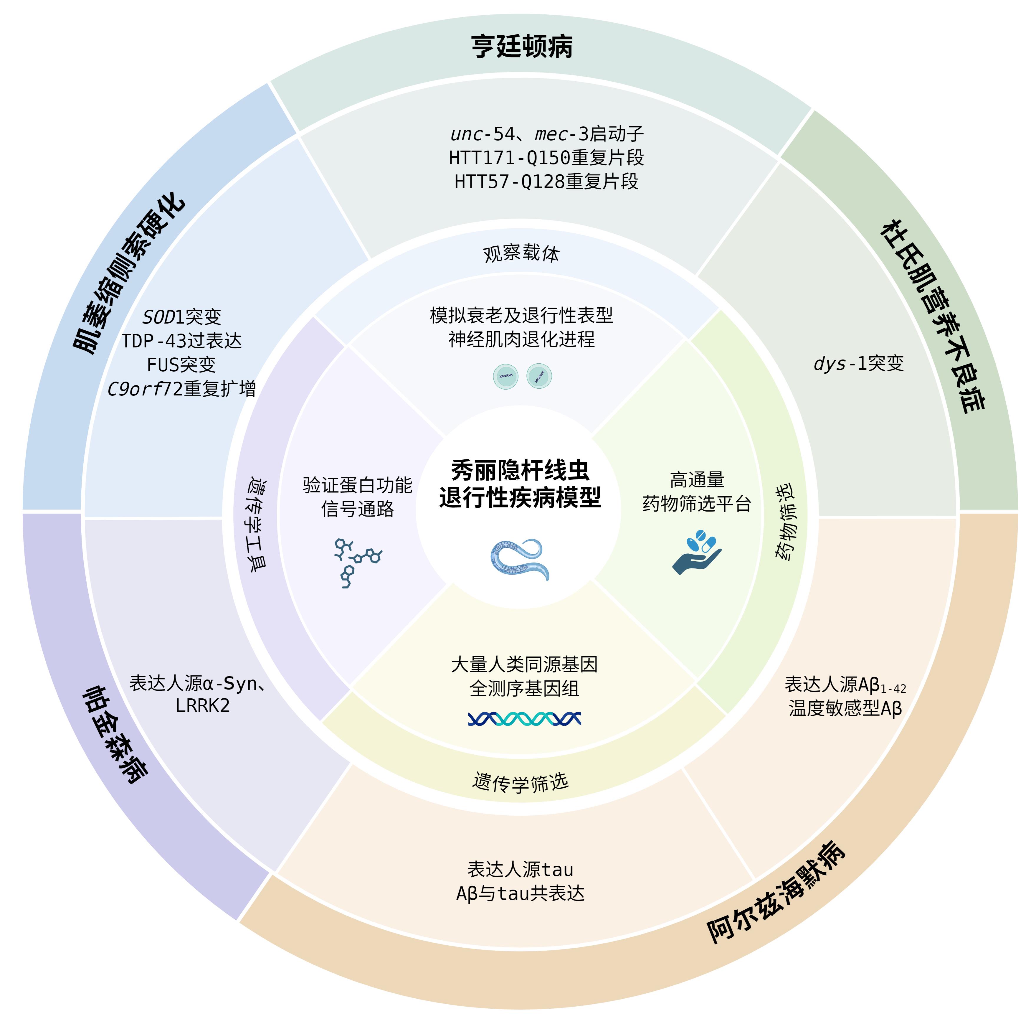
Figure 2 Overview of applications of Caenorhabditis elegans in various degenerative disease models
| [1] | HOU Y, DAN X, BABBAR M, WEI Y, HASSELBALCH S G, CROTEAU D L, BOHR V A. Ageing as a risk factor for neurodegenerative disease[J]. Nat Rev Neurol, 2019, 15(10): 565–581. DOI: 10.1038/s41582-019-0244-7 . |
| [2] | THE C. ELEGANS SEQUENCING CONSORTIUM. Genome sequence of the nematode C. elegans: a platform for investigating biology[J]. Science, 1998, 282(5396): 2012-2018. DOI: 10.1126/science.282.5396.2012 . |
| [3] | CULETTO E, SATTELLE D B. A role for Caenorhabditis elegans in understanding the function and interactions of human disease genes[J]. Hum Mol Genet, 2000, 9(6): 869-877. DOI: 10.1093/hmg/9.6.869 . |
| [4] | SHAYE D D, GREENWALD I. OrthoList: a compendium of C. elegans genes with human orthologs[J]. PLoS One, 2011, 6(5): 1-12. DOI: 10.1371/journal.pone.0020085 . |
| [5] | BARGMANN C I, HORVITZ H R. Control of larval development by chemosensory neurons in Caenorhabditis elegans [J]. Science, 1991, 251(4998): 1243-1246. DOI: 10.1126/science. 2006412 . |
| [6] | GRAY J M, KAROW D S, LU H, et al. Oxygen sensation and social feeding mediated by a C. elegans guanylate cyclase homologue[J]. Nature, 2004, 430(6997): 317-322. DOI: 10.1038/nature02714 . |
| [7] | GILES A C, ROSE J K, RANKIN C H. Investigations of learning and memory in Caenorhabditis elegans [J]. Int Rev Neurobiol, 2006, 69: 37-71. DOI: 10.1016/S0074-7742(05)69002-2 . |
| [8] | HERMS J, ANLIKER B, HEBER S, et al. Cortical dysplasia resembling human type 2 lissencephaly in mice lacking all three APP family members[J]. EMBO J, 2004, 23(20): 4106-4115. DOI: 10.1038/sj.emboj.7600390 . |
| [9] | DUAN D S, GOEMANS N, TAKEDA S, et al. Duchenne muscular dystrophy[J]. Nat Rev Dis Primers, 2021, 7: 13. DOI: 10.1038/s41572-021-00248-3 . |
| [10] | HAENGGI T, FRITSCHY J M. Role of dystrophin and utrophin for assembly and function of the dystrophin glycoprotein complex in non-muscle tissue[J]. Cell Mol Life Sci, 2006, 63(14): 1614-1631. DOI: 10.1007/s00018-005-5461-0 . |
| [11] | BRENNER S. The genetics of Caenorhabditis elegans [J]. Genetics, 1974, 77(1): 71-94. DOI: 10.1093/genetics/77.1.71 . |
| [12] | GIUGIA J, GIESELER K, ARPAGAUS M, et al. Mutations in the dystrophin-like dys-1 gene of Caenorhabditis elegans result in reduced acetylcholinesterase activity[J]. FEBS Lett, 1999, 463(3): 270-272. DOI: 10.1016/s0014-5793(99)01651-8 . |
| [13] | KRAJACIC P, HERMANOWSKI J, LOZYNSKA O, et al. C. elegans dysferlin homolog fer-1 is expressed in muscle, and fer-1 mutations initiate altered gene expression of muscle enriched genes[J]. Physiol Genomics, 2009, 40(1): 8-14. DOI: 10.1152/physiolgenomics.00106.2009 . |
| [14] | ARRIBERE J A, FIRE A Z. Nonsense mRNA suppression via nonstop decay[J]. eLife, 2018, 7: 1-23. DOI: 10.7554/eLife.33292 . |
| [15] | GLOVER M L, BURROUGHS A M, MONEM P C, et al. NONU-1 encodes a conserved endonuclease required for mRNA translation surveillance[J]. Cell Rep, 2020, 30(13): 4321-4331.e4. DOI: 10.1016/j.celrep.2020.03.023 . |
| [16] | PALIKARAS K, LIONAKI E, TAVERNARAKIS N. Coordination of mitophagy and mitochondrial biogenesis during ageing in C. elegans [J]. Nature, 2015, 521(7553): 525-528. DOI: 10.1038/nature14300 . |
| [17] | HUYNH J M, DANG H, FARES H. Measurement of lysosomal size and lysosomal marker intensities in adult Caenorhabditis elegans [J]. Bio Protoc, 2018, 8(3): 1-12. DOI: 10.21769/BioProtoc.2724 . |
| [18] | KUKHTAR D, RUBIO-PEÑA K, SERRAT X, et al. Mimicking of splicing-related retinitis pigmentosa mutations in C. elegans allow drug screens and identification of disease modifiers[J]. Hum Mol Genet, 2020, 29(5): 756-765. DOI: 10.1093/hmg/ddz315 . |
| [19] | LINK C D. Expression of human beta-amyloid peptide in transgenic Caenorhabditis elegans [J]. Proc Natl Acad Sci USA, 1995, 92(20): 9368-9372. DOI: 10.1073/pnas.92.20.9368 . |
| [20] | KRAEMER B C, ZHANG B, LEVERENZ J B, et al. Neurodegeneration and defective neurotransmission in a Caenorhabditis elegans model of tauopathy[J]. Proc Natl Acad Sci USA, 2003, 100(17): 9980-9985. DOI: 10.1073/pnas.1533448100 . |
| [21] | LAKSO M, VARTIAINEN S, MOILANEN A M, et al. Dopaminergic neuronal loss and motor deficits in Caenorhabditis elegans overexpressing human alpha-synuclein[J]. J Neurochem, 2003, 86(1): 165-172. DOI: 10.1046/j.1471-4159.2003.01809.x . |
| [22] | FABER P W, ALTER J R, MACDONALD M E, et al. Polyglutamine-mediated dysfunction and apoptotic death of a Caenorhabditis elegans sensory neuron[J]. Proc Natl Acad Sci USA, 1999, 96(1): 179-184. DOI: 10.1073/pnas.96.1.179 . |
| [23] | LI J, HUANG K X, LE W D. Establishing a novel C. elegans model to investigate the role of autophagy in amyotrophic lateral sclerosis[J]. Acta Pharmacol Sin, 2013, 34(5): 644-650. DOI: 10.1038/aps.2012.190 . |
| [24] | RING S, WEYER S W, KILIAN S B, et al. The secreted beta-amyloid precursor protein ectodomain APPs alpha is sufficient to rescue the anatomical, behavioral, and electrophysiological abnormalities of APP-deficient mice[J]. J Neurosci, 2007, 27(29): 7817-7826. DOI: 10.1523/JNEUROSCI.1026-07.2007 . |
| [25] | WASCO W, BUPP K, MAGENDANTZ M, et al. Identification of a mouse brain cDNA that encodes a protein related to the Alzheimer disease-associated amyloid beta protein precursor[J]. Proc Natl Acad Sci USA, 1992, 89(22): 10758-10762. DOI: 10.1073/pnas.89.22.10758 . |
| [26] | DAIGLE I, LI C. Apl-1, a Caenorhabditis elegans gene encoding a protein related to the human beta-amyloid protein precursor[J]. Proc Natl Acad Sci USA, 1993, 90(24): 12045-12049. DOI: 10.1073/pnas.90.24.12045 . |
| [27] | LEE V M, OTVOS L Jr, CARDEN M J, et al. Identification of the major multiphosphorylation site in mammalian neurofilaments[J]. Proc Natl Acad Sci USA, 1988, 85(6): 1998-2002. DOI: 10.1073/pnas.85.6.1998 . |
| [28] | MCCOLL G, ROBERTS B R, GUNN A P, et al. The Caenorhabditis elegans Aβ1-42 model of Alzheimer disease predominantly expresses Aβ3-42 [J]. J Biol Chem, 2009, 284(34): 22697-22702. DOI: 10.1074/jbc.C109.028514 . |
| [29] | MANGO S E. Stop making nonSense: the C. elegans smg genes[J]. Trends Genet, 2001, 17(11): 646-653. DOI: 10.1016/s0168-9525(01)02479-9 . |
| [30] | LINK C D, TAFT A, KAPULKIN V, et al. Gene expression analysis in a transgenic Caenorhabditis elegans Alzheimer's disease model[J]. Neurobiol Aging, 2003, 24(3): 397-413. DOI: 10.1016/s0197-4580(02)00224-5 . |
| [31] | LU T, ARON L, ZULLO J, et al. REST and stress resistance in ageing and Alzheimer's disease[J]. Nature, 2014, 507(7493): 448-454. DOI: 10.1038/nature13163 . |
| [32] | SIDDIQUI A A, MERQUIOL E, BRUCK-HAIMSON R, et al. Cathepsin B promotes Aβ proteotoxicity by modulating aging regulating mechanisms[J]. Nat Commun, 2024, 15(1): 8564. DOI: 10.1038/s41467-024-52540-x . |
| [33] | MARIANI E, POLIDORI M C, CHERUBINI A, et al. Oxidative stress in brain aging, neurodegenerative and vascular diseases: an overview[J]. J Chromatogr B Analyt Technol Biomed Life Sci, 2005, 827(1): 65-75. DOI: 10.1016/j.jchromb. 2005.04.023 . |
| [34] | KRAEMER B C, BURGESS J K, CHEN J H, et al. Molecular pathways that influence human tau-induced pathology in Caenorhabditis elegans [J]. Hum Mol Genet, 2006, 15(9): 1483-1496. DOI: 10.1093/hmg/ddl067 . |
| [35] | Diomede L., et al. Expression of A2V-mutated Aβ in Caenorhabditis elegans results in oligomer formation and toxicity[J]. Neurobiol Dis, 2014. 62(100): p. 521-32. |
| [36] | Huang A, Stultz CM. The effect of a DeltaK280 mutation on the unfolded state of a microtubule-binding repeat in Tau[J]. PLoS Comput Biol. 2008 Aug 22;4(8):e1000155. |
| [37] | Tiwari V, Buvarp E, Borbolis F, et al. Loss of DNA glycosylases improves health and cognitive function in a C. elegans model of human tauopathy[J]. Nucleic Acids Res. 2024 Oct 14;52(18):10965-10985. |
| [38] | FANG E F, HOU Y J, PALIKARAS K, et al. Mitophagy inhibits amyloid-β and tau pathology and reverses cognitive deficits in models of Alzheimer's disease[J]. Nat Neurosci, 2019, 22(3): 401-412. DOI: 10.1038/s41593-018-0332-9 . |
| [39] | MACDONALD M. A novel gene containing a trinucleotide repeat that is expanded and unstable on Huntington's disease chromosomes[J]. Cell, 1993, 72(6): 971-983. DOI: 10.1016/0092-8674(93)90585-e . |
| [40] | COSTA M D, MACIEL P. Modifier pathways in polyglutamine (PolyQ) diseases: from genetic screens to drug targets[J]. Cell Mol Life Sci, 2022, 79(5): 1-31. DOI: 10.1007/s00018-022-04280-8 . |
| [41] | ADAMLA F, IGNATOVA Z. Somatic expression of unc-54 and vha-6 mRNAs declines but not pan-neuronal rgef-1 and unc-119 expression in aging Caenorhabditis elegans [J]. Sci Rep, 2015, 5: 1-10. DOI: 10.1038/srep10692 . |
| [42] | MARCH Z M, MACK K L, SHORTER J. AAA+ protein-based technologies to counter neurodegenerative disease[J]. Biophys J, 2019, 116(8): 1380-1385. DOI: 10.1016/j.bpj. 2019. 03.007 . |
| [43] | MOUCHIROUD L, HOUTKOOPER R H, MOULLAN N, et al. The NAD+/sirtuin pathway modulates longevity through activation of mitochondrial UPR and FOXO signaling[J]. Cell, 2013, 154(2): 430-441. DOI: 10.1016/j.cell.2013.06.016 . |
| [44] | SHIN B H, LIM Y, OH H J, et al. Pharmacological activation of Sirt1 ameliorates polyglutamine-induced toxicity through the regulation of autophagy[J]. PLoS One, 2013, 8(6): 1-10. DOI: 10.1371/journal.pone.0064953 . |
| [45] | GAETA A L, CALDWELL K A, CALDWELL G A. Found in translation: the utility of C. elegans alpha-synuclein models of Parkinson's disease[J]. Brain Sci, 2019, 9(4): 1-15. DOI: 10.3390/brainsci9040073 . |
| [46] | BLIEDERHAEUSER C, GROZDANOV V, SPEIDEL A, et al. Age-dependent defects of alpha-synuclein oligomer uptake in microglia and monocytes[J]. Acta Neuropathol, 2016, 131(3): 379-391. DOI: 10.1007/s00401-015-1504-2 . |
| [47] | BELLOMO G, PACIOTTI S, GATTICCHI L, et al. The vicious cycle between α-synuclein aggregation and autophagic-lysosomal dysfunction[J]. Mov Disord, 2020, 35(1): 34-44. DOI: 10.1002/mds.27895 . |
| [48] | MAO X B, OU M T, KARUPPAGOUNDER S S, et al. Pathological α-synuclein transmission initiated by binding lymphocyte-activation gene 3[J]. Science, 2016, 353(6307): 1-33. DOI: 10.1126/science.aah3374 . |
| [49] | PRICE A, MANZONI C, COOKSON M R, et al. The LRRK2 signalling system[J]. Cell Tissue Res, 2018, 373(1): 39-50. DOI: 10.1007/s00441-017-2759-9 . |
| [50] | TULLET J M A, ARAIZ C, SANDERS M J, et al. DAF-16/FoxO directly regulates an atypical AMP-activated protein kinase gamma isoform to mediate the effects of insulin/IGF-1 signaling on aging in Caenorhabditis elegans [J]. PLoS Genet, 2014, 10(2): 1-16. DOI: 10.1371/journal.pgen.1004109 . |
| [51] | DHONDT I, PETYUK V A, CAI H H, et al. FOXO/DAF-16 activation slows down turnover of the majority of proteins in C. elegans [J]. Cell Rep, 2016, 16(11): 3028-3040. DOI: 10.1016/j.celrep.2016.07.088 . |
| [52] | SALAŠOVÁ A, YOKOTA C, POTĚŠIL D, et al. A proteomic analysis of LRRK2 binding partners reveals interactions with multiple signaling components of the WNT/PCP pathway[J]. Mol Neurodegener, 2017, 12(1): 1-19. DOI: 10.1186/s13024-017-0193-9 . |
| [53] | ARBO B D, ANDRÉ-MIRAL C, NASRE-NASSER R G, et al. Resveratrol derivatives as potential treatments for Alzheimer's and Parkinson's disease[J]. Front Aging Neurosci, 2020, 12: 1-15. DOI: 10.3389/fnagi.2020.00103 . |
| [54] | CHEN L L, ZHANG S M, LIU S, et al. Amyotrophic lateral sclerosis mechanism: insights from the Caenorhabditis elegans models[J]. Cells, 2024, 13(1): 1-18. DOI: 10.3390/cells13010099 . |
| [55] | WANG J O, FARR G W, HALL D H, et al. An ALS-linked mutant SOD1 produces a locomotor defect associated with aggregation and synaptic dysfunction when expressed in neurons of Caenorhabditis elegans [J]. PLoS Genet, 2009, 5(1): 1-14. DOI: 10.1371/journal.pgen.1000350 . |
| [56] | ZHONG Y W, WANG J O, HENDERSON M J, et al. Nuclear export of misfolded SOD1 mediated by a normally buried NES-like sequence reduces proteotoxicity in the nucleus[J]. eLife, 2017, 6: 1-23. DOI: 10.7554/eLife.23759 . |
| [57] | OGAWA M, SHIDARA H, OKA K, et al. Cysteine residues in Cu, Zn-superoxide dismutase are essential to toxicity in Caenorhabditis elegans model of amyotrophic lateral sclerosis[J]. Biochem Biophys Res Commun, 2015, 463(4): 1196-1202. DOI: 10.1016/j.bbrc.2015.06.084 . |
| [58] | SILVA M C, FOX S, BEAM M, et al. A genetic screening strategy identifies novel regulators of the proteostasis network[J]. PLoS Genet, 2011, 7(12): 1-15. DOI: 10.1371/journal.pgen.1002438 . |
| [59] | ZHANG T, PERIZ G, LU Y N, et al. USP7 regulates ALS-associated proteotoxicity and quality control through the NEDD4L-SMAD pathway[J]. Proc Natl Acad Sci USA, 2020, 117(45): 28114-28125. DOI: 10.1073/pnas.2014349117 . |
| [60] | XU H, JIA C C, CHENG C, et al. Activation of autophagy attenuates motor deficits and extends lifespan in a C. elegans model of ALS[J]. Free Radic Biol Med, 2022, 181: 52-61. DOI: 10.1016/j.freeradbiomed.2022.01.030 . |
| [61] | ZHANG T, MULLANE P C, PERIZ G, et al. TDP-43 neurotoxicity and protein aggregation modulated by heat shock factor and insulin/IGF-1 signaling[J]. Hum Mol Genet, 2011, 20(10): 1952-1965. DOI: 10.1093/hmg/ddr076 . |
| [62] | ASH P E A, ZHANG Y J, ROBERTS C M, et al. Neurotoxic effects of TDP-43 overexpression in C. elegans [J]. Hum Mol Genet, 2010, 19(16): 3206-3218. DOI: 10.1093/hmg/ddq230 . |
| [63] | VACCARO A, TAUFFENBERGER A, AGGAD D, et al. Mutant TDP-43 and FUS cause age-dependent paralysis and neurodegeneration in C. elegans [J]. PLoS One, 2012, 7(2): 1-10. DOI: 10.1371/journal.pone.0031321 . |
| [64] | LIACHKO N F, GUTHRIE C R, KRAEMER B C. Phosphorylation promotes neurotoxicity in a Caenorhabditis elegans model of TDP-43 proteinopathy[J]. J Neurosci, 2010, 30(48): 16208-16219. DOI: 10.1523/JNEUROSCI.2911-10.2010 . |
| [65] | LIACHKO N F, MCMILLAN P J, GUTHRIE C R, et al. CDC7 inhibition blocks pathological TDP-43 phosphorylation and neurodegeneration[J]. Ann Neurol, 2013, 74(1): 39-52. DOI: 10.1002/ana.23870 . |
| [66] | AARON C, BEAUDRY G, PARKER J A, et al. Maple syrup decreases TDP-43 proteotoxicity in a Caenorhabditis elegans model of amyotrophic lateral sclerosis (ALS)[J]. J Agric Food Chem, 2016, 64(17): 3338-3344. DOI: 10.1021/acs.jafc.5b05432 . |
| [67] | MURAKAMI T, YANG S P, XIE L, et al. ALS mutations in FUS cause neuronal dysfunction and death in Caenorhabditis elegans by a dominant gain-of-function mechanism[J]. Hum Mol Genet, 2012, 21(1): 1-9. DOI: 10.1093/hmg/ddr417 . |
| [68] | VÉRIÈPE J, FOSSOUO L, PARKER J A. Neurodegeneration in C. elegans models of ALS requires TIR-1/Sarm1 immune pathway activation in neurons[J]. Nat Commun, 2015, 6: 1-9. DOI: 10.1038/ncomms8319 . |
| [69] | TOSSING G, LIVERNOCHE R, MAIOS C, et al. Genetic and pharmacological PARP inhibition reduces axonal degeneration in C. elegans models of ALS[J]. Hum Mol Genet, 2022, 31(19): 3313-3324. DOI: 10.1093/hmg/ddac116 . |
| [70] | LABARRE A, GUITARD E, TOSSING G, et al. Fatty acids derived from the probiotic Lacticaseibacillus rhamnosus HA-114 suppress age-dependent neurodegeneration[J]. Commun Biol, 2022, 5(1): 1-19. DOI: 10.1038/s42003-022-04295-8 . |
| [71] | WANG X, HAO L M, SAUR T, et al. Forward genetic screen in Caenorhabditis elegans suggests F57A10.2 and acp-4 as suppressors of C9ORF72 related phenotypes[J]. Front Mol Neurosci, 2016, 9: 1-10. DOI: 10.3389/fnmol.2016.00113 . |
| [72] | SNOZNIK C, MEDVEDEVA V, MOJSILOVIC-PETROVIC J, et al. The nuclear ubiquitin ligase adaptor SPOP is a conserved regulator of C9orf72 dipeptide toxicity[J]. Proc Natl Acad Sci USA, 2021, 118(40): 1-10. DOI: 10.1073/pnas.2104664118 . |
| [73] | PULEO N, LAMITINA T. The conserved multi-functional nuclear protein dss-1/Sem1 is required for C9orf72-associated ALS/FTD dipeptide toxicity[J]. MicroPubl Biol, 2020: 1-5. DOI: 10.17912/micropub.biology.000262 . |
| [74] | CHANG M Y, CAI Y, GAO Z H, et al. Duchenne muscular dystrophy: pathogenesis and promising therapies[J]. J Neurol, 2023, 270(8): 3733-3749. DOI: 10.1007/s00415-023-11796-x . |
| [75] | WU X T, NAGASAWA S, MUTO K, et al. Mitochonic acid 5 improves Duchenne muscular dystrophy and Parkinson's disease model of Caenorhabditis elegans [J]. Int J Mol Sci, 2022, 23(17): 1-15. DOI: 10.3390/ijms23179572 . |
| [76] | YOSHINA S, IZUHARA L, MASHIMA R, et al. Febuxostat ameliorates muscle degeneration and movement disorder of the dystrophin mutant model in Caenorhabditis elegans [J]. J Physiol Sci, 2023, 73(1): 1-9. DOI: 10.1186/s12576-023-00888-y . |
| [77] | CARRE-PIERRAT M, MARIOL M C, CHAMBONNIER L, et al. Blocking of striated muscle degeneration by serotonin in C. elegans [J]. J Muscle Res Cell Motil, 2006, 27(3-4): 253-258. DOI: 10.1007/s10974-006-9070-9 . |
| [78] | NYAMSUREN O, FAGGIONATO D, LOCH W, et al. A mutation in CHN-1/CHIP suppresses muscle degeneration in Caenorhabditis elegans [J]. Dev Biol, 2007, 312(1): 193-202. DOI: 10.1016/j.ydbio.2007.09.033 . |
| [79] | ELLWOOD R A, SLADE L, LEWIS J, et al. Sulfur amino acid supplementation displays therapeutic potential in a C. elegans model of Duchenne muscular dystrophy[J]. Commun Biol, 2022, 5: 1-12. DOI: 10.1038/s42003-022-04212-z . |
| [1] | SONG Mengjiao, SHEN Yidong. Approaches and Application Examples for Studying Mitochondrial Morphology and Function in Caenorhabditis elegans [J]. Laboratory Animal and Comparative Medicine, 2025, 45(6): 726-737. |
| [2] | LUO Lianlian, YUAN Yanchun, WANG Junling, SHI Guangsen. Advances in Mouse Models of Amyotrophic Lateral Sclerosis [J]. Laboratory Animal and Comparative Medicine, 2025, 45(3): 290-299. |
| [3] | Ruiqi LI, Han DUAN, Luo GAN, Yuan ZHENG, Wen YANG. Advantages of Ciona intestinalis as a Model Organism and Its Applications [J]. Laboratory Animal and Comparative Medicine, 2024, 44(2): 162-179. |
| [4] | Hui CHENG, Fei FANG, Jiahao SHI, Hua YANG, Mengjie ZHANG, Ping YANG, Jian FEI. H1 Linker Histone Gene Regulates Lifespan via Dietary Restriction Pathways in Caenorhabditis elegans [J]. Laboratory Animal and Comparative Medicine, 2023, 43(3): 271-281. |
| Viewed | ||||||
|
Full text |
|
|||||
|
Abstract |
|
|||||