




Laboratory Animal and Comparative Medicine ›› 2025, Vol. 45 ›› Issue (6): 726-737.DOI: 10.12300/j.issn.1674-5817.2025.119
• Invertebrate Laboratory Animal: Nematode • Previous Articles Next Articles
SONG Mengjiao( ), SHEN Yidong()(
 )
)
Received:2025-07-16
Revised:2025-10-17
Online:2025-12-25
Published:2025-12-19
Correspondence to:
SHEN Yidong
CLC Number:
SONG Mengjiao,SHEN Yidong. Approaches and Application Examples for Studying Mitochondrial Morphology and Function in Caenorhabditis elegans[J]. Laboratory Animal and Comparative Medicine, 2025, 45(6): 726-737. DOI: 10.12300/j.issn.1674-5817.2025.119.
Add to citation manager EndNote|Ris|BibTeX
URL: https://www.slarc.org.cn/dwyx/EN/10.12300/j.issn.1674-5817.2025.119
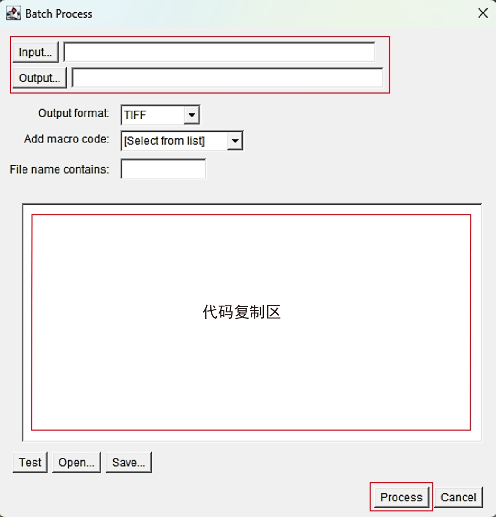
Figure 1 Introduction to Batch Process operation interface
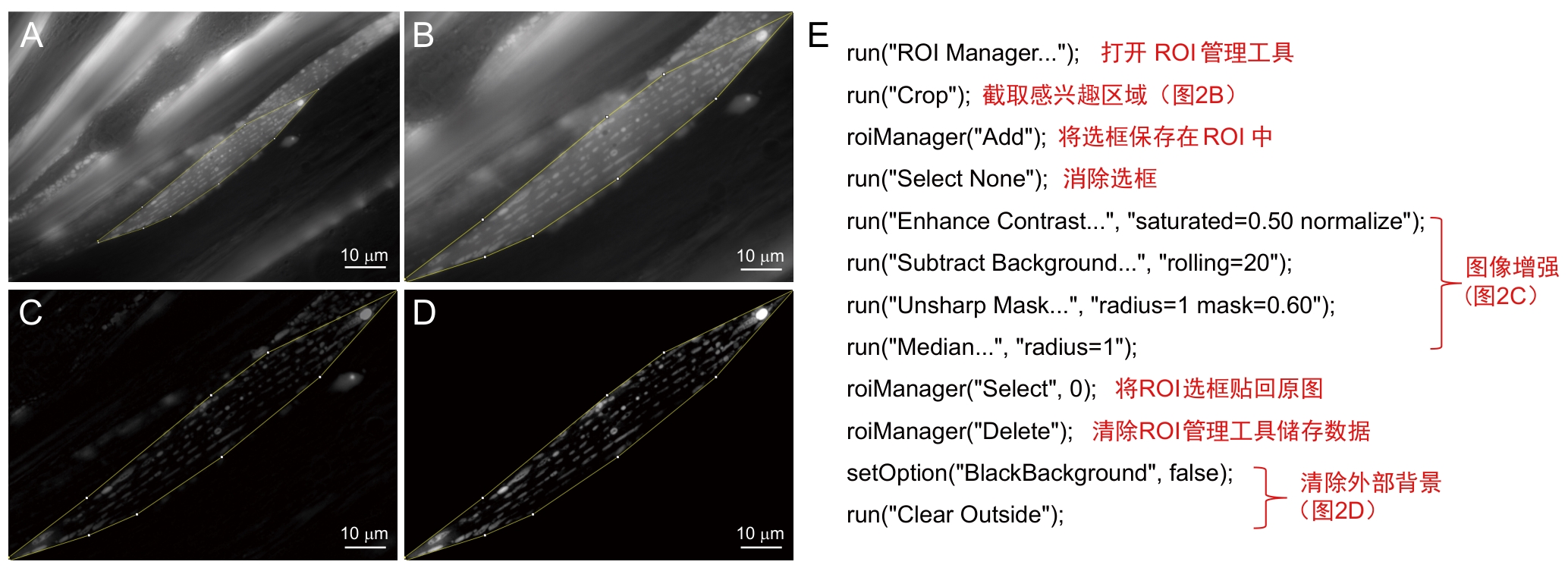
Figure 2 Preprocessing procedures and annotations
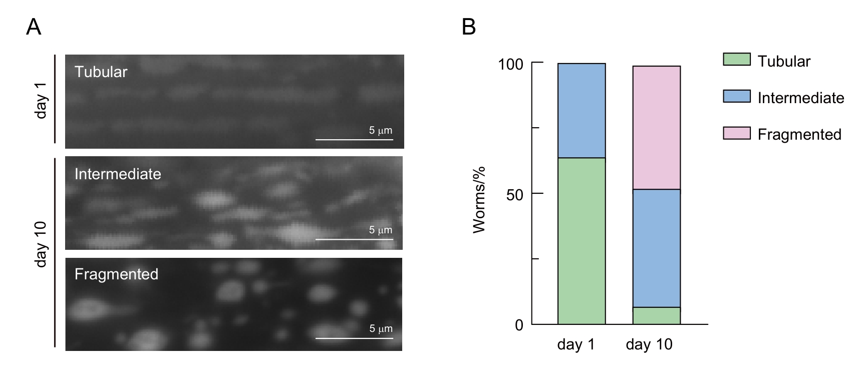
Figure 3 Classification of mitochondrial morphology in C. elegans muscle cells
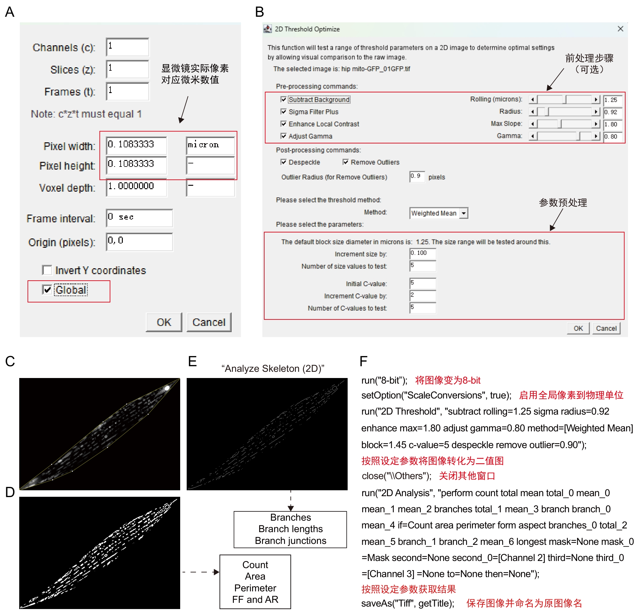
Figure 4 Procedures and annotations of Mitochondria Analyzer plugin
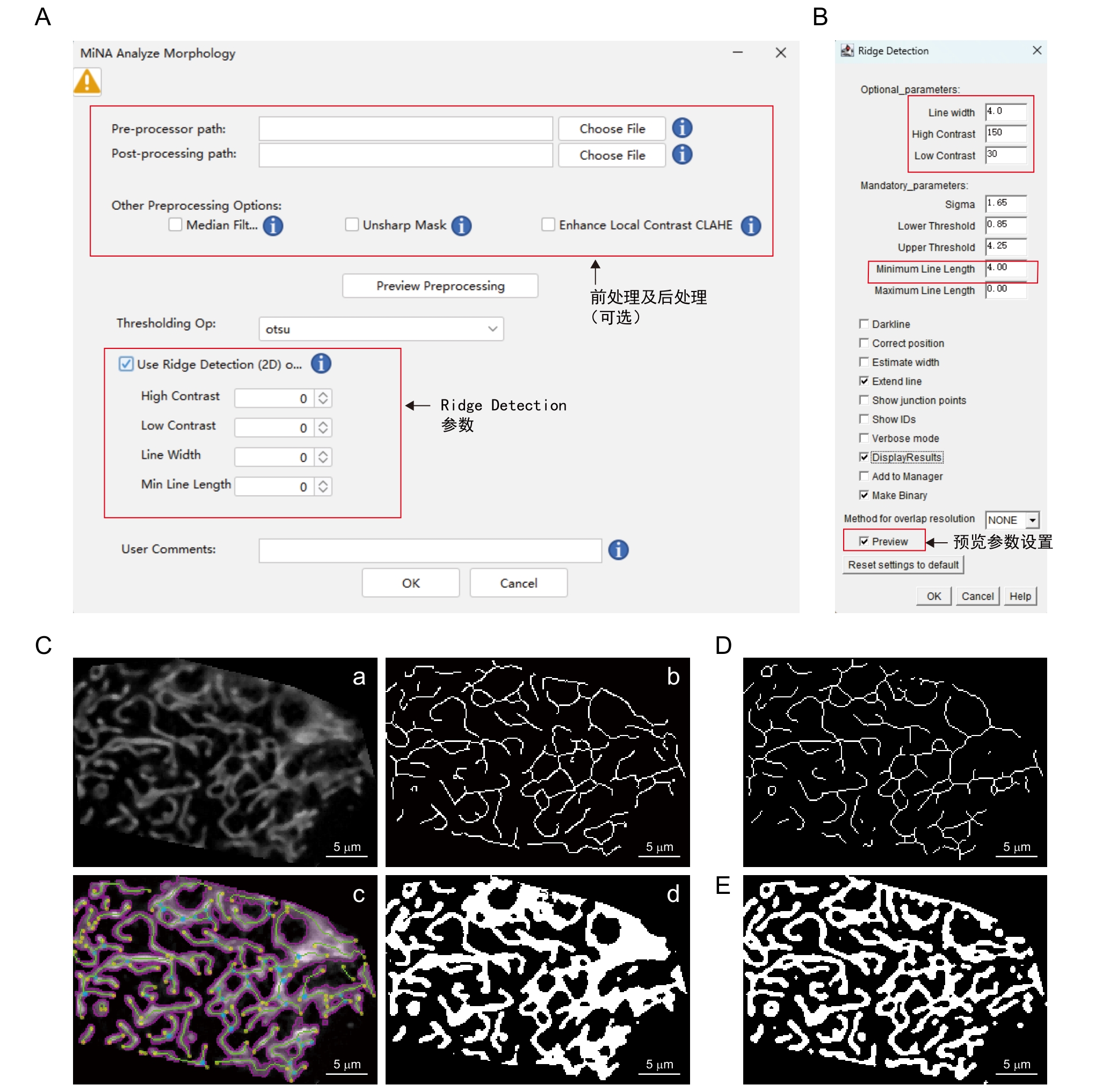
Figure 5 Procedures and annotations of MiNA - Mitochondrial Network Analysis plugin
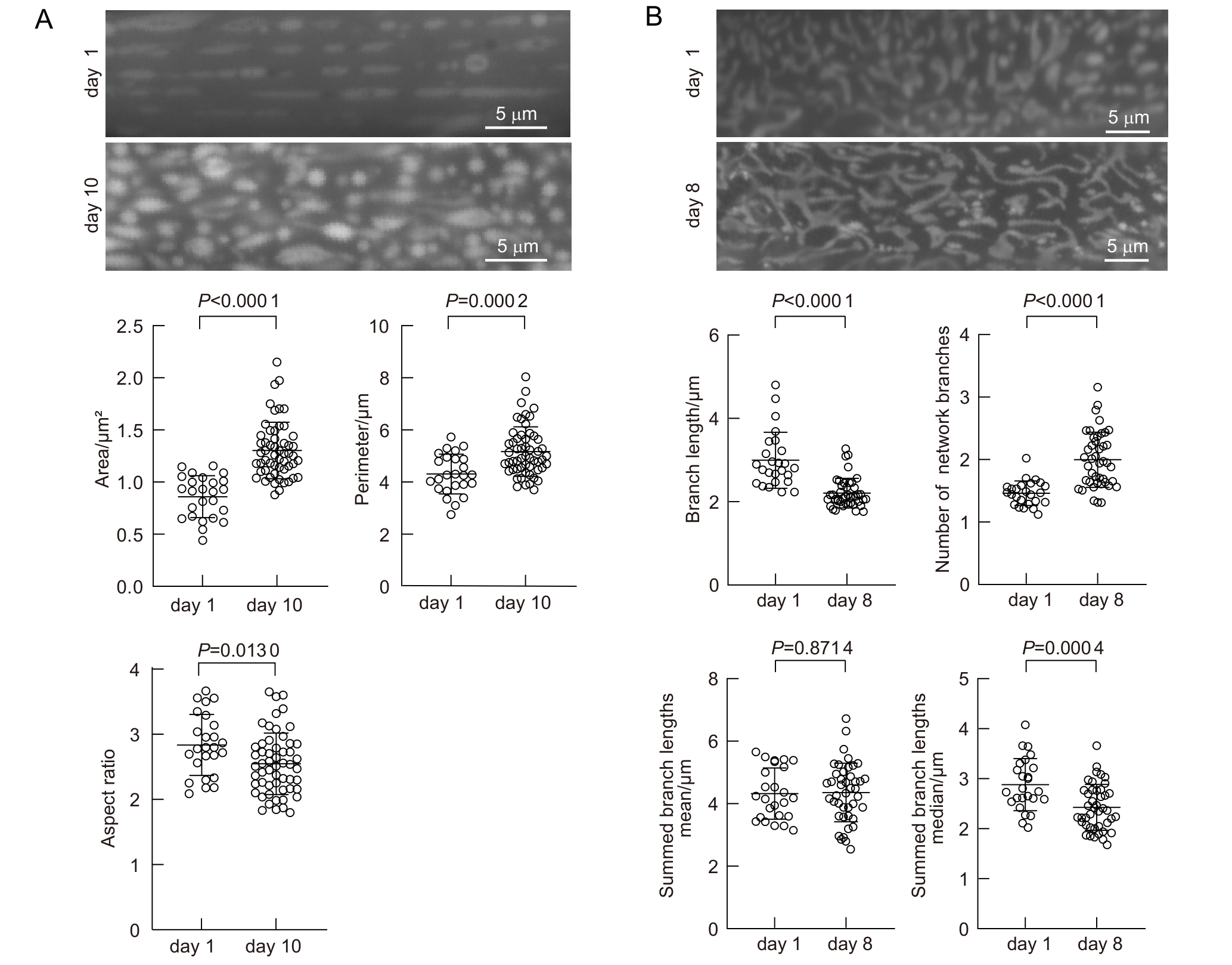
Figure 6 Age-dependent mitochondrial morphology changes in C. elegans
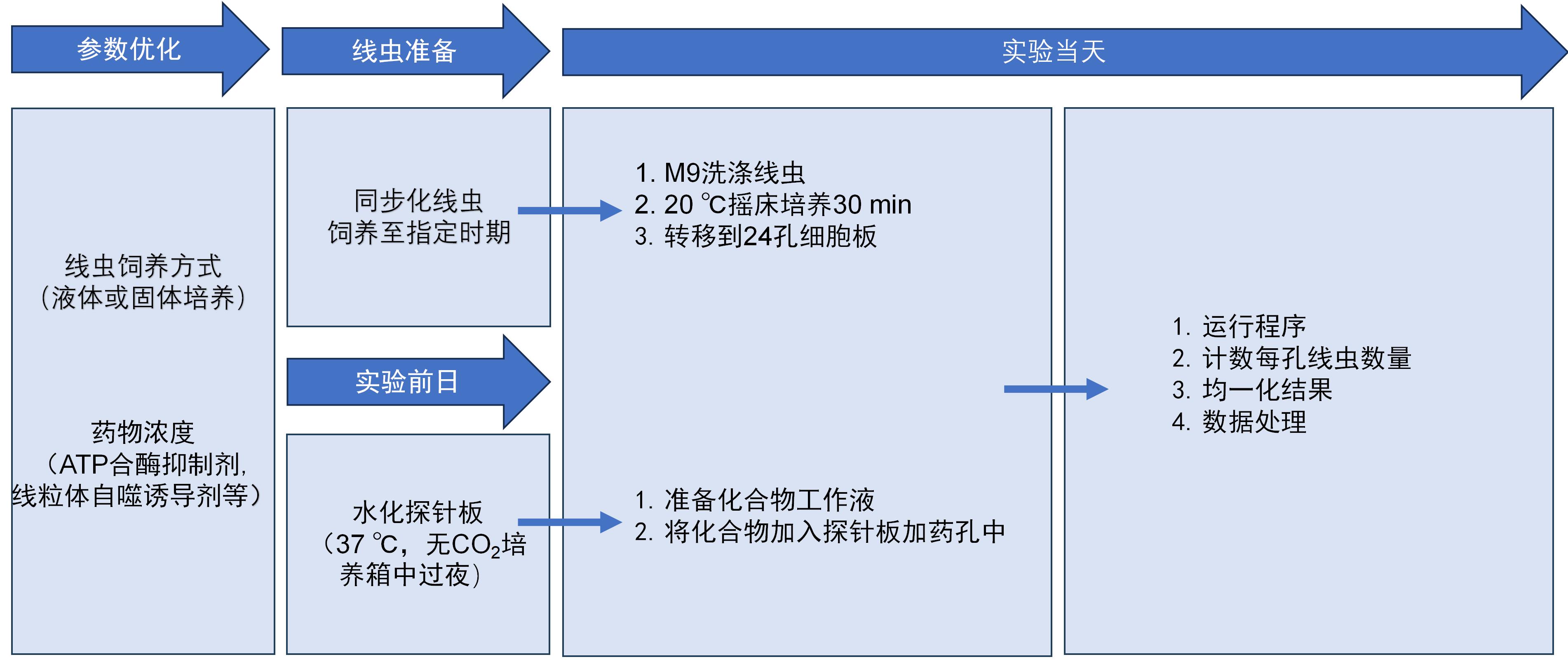
Figure 7 Seahorse workflow
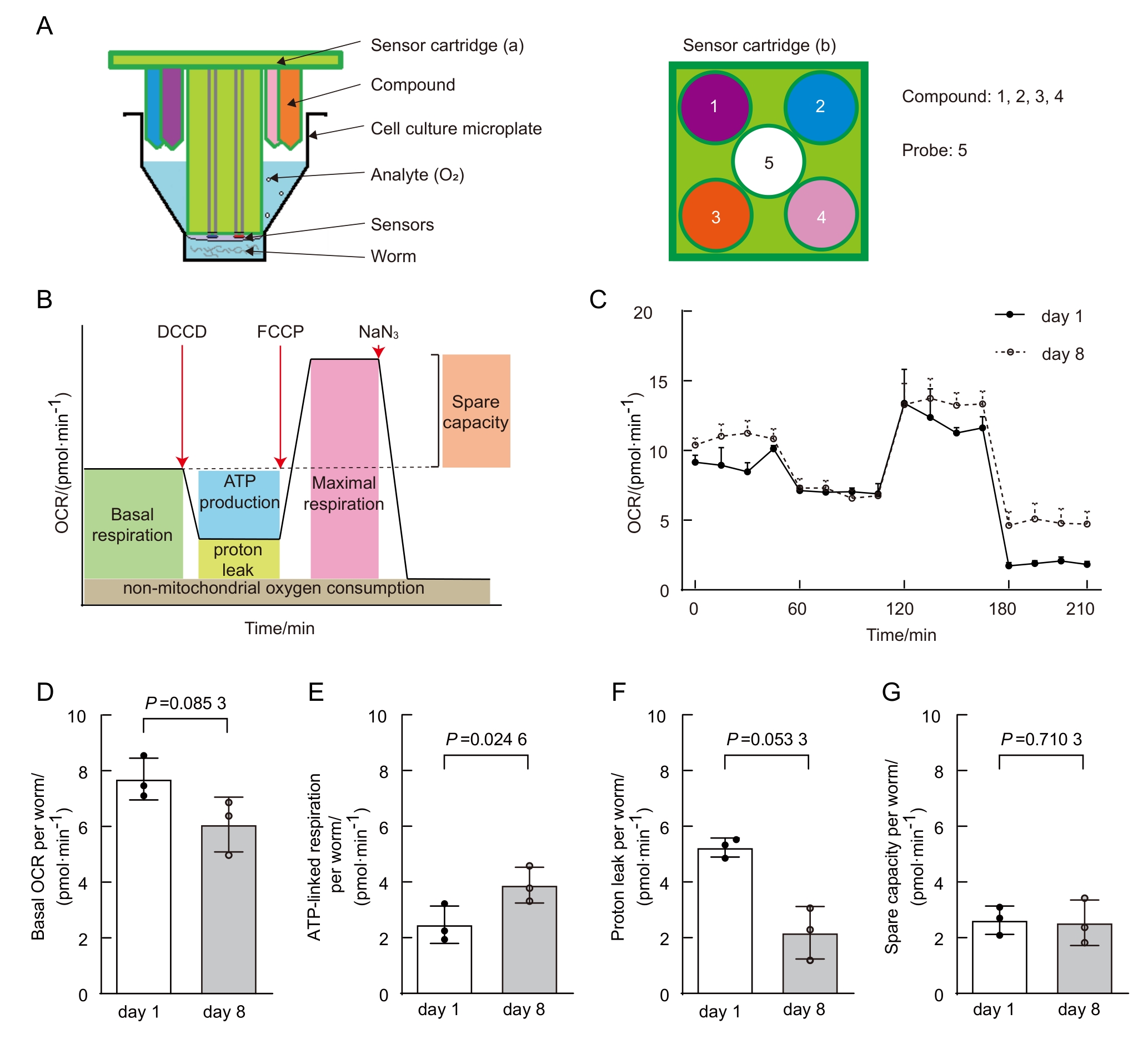
Figure 8 Age-dependent mitochondrial function changes in C.elegans (data from published paper of our lab [
| [1] | MUKHERJEE I, GHOSH M, MEINECKE M. MICOS and the mitochondrial inner membrane morphology–when things get out of shape[J]. FEBS Lett, 2021, 595(8):1159-1183. DOI:10.1002/1873-3468.14089 . |
| [2] | OKAMOTO K, SHAW J M. Mitochondrial morphology and dynamics in yeast and multicellular eukaryotes[J]. Annu Rev Genet, 2005, 39:503-536. DOI:10.1146/annurev.genet.38. 072902.093019 . |
| [3] | LÓPEZ-OTÍN C, BLASCO M A, PARTRIDGE L, et al. Hallmarks of aging: an expanding universe[J]. Cell, 2023, 186(2):243-278. DOI:10.1016/j.cell.2022.11.001 . |
| [4] | YAN C J, DUANMU X Y, ZENG L, et al. Mitochondrial DNA: distribution, mutations, and elimination[J]. Cells, 2019, 8(4):379. DOI:10.3390/cells8040379 . |
| [5] | KLIONSKY D J, ABDELMOHSEN K, ABE A, et al. Guidelines for the use and interpretation of assays for monitoring autophagy (3rd edition)[J]. Autophagy, 2016, 12(1):1-222. DOI:10.1080/15548627.2015.1100356 . |
| [6] | MACK H I D, HEIMBUCHER T, MURPHY C T. The nematode Caenorhabditis elegans as a model for aging research[J]. Drug Discov Today Dis Models, 2018, 27:3-13. DOI:10.1016/j.ddmod.2018.11.001 . |
| [7] | JI T, ZHANG X L, XIN Z L, et al. Does perturbation in the mitochondrial protein folding pave the way for neurodegeneration diseases?[J]. Ageing Res Rev, 2020, 57:100997. DOI:10.1016/j.arr.2019.100997 . |
| [8] | SOHRABI S, MOR D E, KALETSKY R, et al. High-throughput behavioral screen in C. elegans reveals Parkinson's disease drug candidates[J]. Commun Biol, 2021, 4(1):203. DOI:10.1038/s42003-021-01731-z . |
| [9] | YE S W, SONG S D, LIU X J, et al. A small-molecule screen identifies novel aging modulators by targeting 5-HT/DA signaling pathway[J]. Aging Cell, 2025, 24(3): 1-13. DOI:10.1111/acel.14411 . |
| [10] | PANDA M, FAKITSA M, MARKAKI M, et al. Caenorhabditis elegans as an emerging high throughput chronotherapeutic drug screening platform for human neurodegenerative disorders[J]. Adv Drug Deliv Rev, 2025, 224:115655. DOI:10.1016/j.addr.2025.115655 . |
| [11] | YOO I, AHN I, LEE J, et al. Extracellular flux assay (Seahorse assay): Diverse applications in metabolic research across biological disciplines[J]. Mol Cells, 2024, 47(8):100095. DOI:10.1016/j.mocell.2024.100095 . |
| [12] | STIERNAGLE T. Maintenance of C. elegans [J]. WormBook, 2006:1-11. DOI:10.1895/wormbook.1.101.1 . |
| [13] | MALLICK A, RANAWADE A, VAN DEN BERG W, et al. Axin-mediated regulation of lifespan and muscle health in C. elegans requires AMPK-FOXO signaling[J]. iScience, 2020, 23(12):101843. DOI:10.1016/j.isci.2020.101843 . |
| [14] | REGMI S G, ROLLAND S G, CONRADT B. Age-dependent changes in mitochondrial morphology and volume are not predictors of lifespan[J]. Aging (Albany NY), 2014, 6(2):118-30. DOI: 10.18632/aging.100639 . |
| [15] | XIA Q, LI P L, CASAS-MARTINEZ J C, et al. Peroxiredoxin 2 regulates DAF-16/FOXO mediated mitochondrial remodelling in response to exercise that is disrupted in ageing[J]. Mol Metab, 2024, 88:102003. DOI: 10.1016/j.molmet.2024.102003 . |
| [16] | CHAUDHRY A, SHI R, LUCIANI D S. A pipeline for multidimensional confocal analysis of mitochondrial morphology, function, and dynamics in pancreatic β-cells[J]. Am J Physiol Endocrinol Metab, 2020, 318(2):E87-E101. DOI:10.1152/ajpendo.00457.2019 . |
| [17] | Mitochondria Analyzer[Z/OL]. [2025-07-16]. . |
| [18] | VALENTE A J, MADDALENA L A, ROBB E L, et al. A simple ImageJ macro tool for analyzing mitochondrial network morphology in mammalian cell culture[J]. Acta Histochem, 2017, 119(3):315-326. DOI:10.1016/j.acthis.2017.03.001 . |
| [19] | MiNA - Mitochondrial Network Analysis[Z/OL]. [2025-07-16]. . |
| [20] | NG L F, GRUBER J. Measurement of respiration rate in live Caenorhabditis elegans [J]. Bio Protoc, 2019, 9(10):1-12. DOI:10.21769/BioProtoc.3243 . |
| [21] | HAROON S, VERMULST M. Oxygen consumption measurements in Caenorhabditis elegans using the seahorse XF24[J]. Bio Protoc, 2019, 9(13):e3288. DOI:10.21769/BioProtoc.3288 . |
| [22] | SONG M J, DONG S M, ZHANG X F, et al. A moderate static magnetic field promotes C. elegans longevity through cytochrome P450s[J]. Sci Rep, 2022, 12:16108. DOI:10.1038/s41598-022-20647-0 . |
| [23] | DILLIN A, HSU A L, ARANTES-OLIVEIRA N, et al. Rates of behavior and aging specified by mitochondrial function during development[J]. Science, 2002, 298(5602):2398-2401. DOI: 10.1126/science/1077780 . |
| [24] | AMORIM J A, COPPOTELLI G, ROLO A P, et al. Mitochondrial and metabolic dysfunction in ageing and age-related diseases[J]. Nat Rev Endocrinol, 2022, 18(4):243-258. DOI:10.1038/s41574-021-00626-7 . |
| [25] | SUN N, YOULE R J, FINKEL T. The mitochondrial basis of aging[J]. Mol Cell, 2016, 61(5):654-666. DOI:10.1016/j.molcel.2016.01.028 . |
| [26] | BAR-ZIV R, BOLAS T, DILLIN A. Systemic effects of mitochondrial stress[J]. EMBO Rep, 2020, 21(6):e50094. DOI:10.15252/embr.202050094 . |
| [27] | REGMI S G, ROLLAND S G, CONRADT B. Age-dependent changes in mitochondrial morphology and volume are not predictors of lifespan[J]. Aging, 2014, 6(2):118-130. DOI:10.18632/aging.100639 . |
| [28] | SON H G, ALTINTAS O, KIM E J E, et al. Age-dependent changes and biomarkers of aging in Caenorhabditis elegans [J]. Aging Cell, 2019, 18(2):e12853. DOI:10.1111/acel.12853 . |
| [29] | MAGLIONI S, MELLO D F, SCHIAVI A, et al. Mitochondrial bioenergetic changes during development as an indicator of C. elegans health-span[J]. Aging, 2019, 11(16):6535-6554. DOI:10.18632/aging.102208 . |
| [30] | MITRA K, WUNDER C, ROYSAM B, et al. A hyperfused mitochondrial state achieved at G1–S regulates cyclin E buildup and entry into S phase[J]. Proc Natl Acad Sci U S A, 2009, 106(29):11960-11965. DOI:10.1073/pnas.0904875106 . |
| [31] | DAGDA R K, CHERRA S J, KULICH S M, et al. Loss of PINK1 function promotes mitophagy through effects on oxidative stress and mitochondrial fission[J]. J Biol Chem, 2009, 284(20):13843-13855. DOI:10.1074/jbc.M808515200 . |
| [1] | SUN Han, GUO Peng, YU Xinhe, ZHANG Junqiao, YAO Ying, YANG Wen. Progress in Caenorhabditis elegans as a Degenerative Disease Model for Molecular Pathways Studying [J]. Laboratory Animal and Comparative Medicine, 2025, 45(6): 738-751. |
| [2] | Hui CHENG, Fei FANG, Jiahao SHI, Hua YANG, Mengjie ZHANG, Ping YANG, Jian FEI. H1 Linker Histone Gene Regulates Lifespan via Dietary Restriction Pathways in Caenorhabditis elegans [J]. Laboratory Animal and Comparative Medicine, 2023, 43(3): 271-281. |
| [3] | Han LI, Xiaorui ZHANG, Chengfang ZHANG. Mechanism of Intermittent Fasting in Improving Olanzapine-induced Metabolic Disorders in Mice [J]. Laboratory Animal and Comparative Medicine, 2023, 43(1): 3-10. |
| [4] | Li Ying-niang, Dai Wei, Sheng Jian. Analysis of Goiter in Zebrafish [J]. Laboratory Animal and Comparative Medicine, 2019, 39(6): 462-466. |
| [5] | PU Xiang-qiang, WANG Xiang, QIANG Guang-hui, MA Jin, DING Yue-yue, LV Hai-tao. The Dynamic Changes of Mitochondria in Endothelial Cells of Immunological Coronaritis in Mice [J]. Laboratory Animal and Comparative Medicine, 2018, 38(3): 169-175. |
| [6] | CHANG Kai, WANG Yu, PANG Wen-biao, GAO Ji-ping, CHEN Zhao-yang, SONG Guo-hua. Antagonistic Effect of Selenium on Change of Mitochondrial Membrane Potential of NRK-52E Cells Induced by Sodium Fluoride [J]. Laboratory Animal and Comparative Medicine, 2017, 37(3): 179-184. |
| [7] | LI Yuan, ZHANG Mei-ying. Research Progress on Autophagy with Parkinson Disease and Related Models [J]. Laboratory Animal and Comparative Medicine, 2015, 35(4): 335-340. |
| [8] | SHEN Xing-jiao, YUE Bing-fei, MA Li-ying. Analysis of the Polymorphism of Mitochondrial DNA Control Region in Three Closed Colonies of Rabbits [J]. Laboratory Animal and Comparative Medicine, 2014, 34(1): 29-34. |
| [9] | GAO Jun, NI Li-ju, SUN Feng-ping, WANG Jin-xiang, HU Jian-hua, GAO Cheng, LI Kai, XIAO Jun-hua, ZHOU Yu-xun. Sequence Analysis on Complete Mitochondrial Genome and Phylogeny of Microtus fortis fortis [J]. Laboratory Animal and Comparative Medicine, 2013, 33(3): 167-173. |
| [10] | ZHANG Ping-hu, TAO Yuan-qing, JIANG Zhen-zhou, WANG Zhong-dong, FAN Wei, ZHANG Lu-yong. Feasibility Analysis of Himalayana Marmot as an Alternative Model of Drug Mitochondrial Toxicity Evaluation [J]. Laboratory Animal and Comparative Medicine, 2012, 32(5): 436-440. |
| [11] | GUAN Min-qiang1,CAO Qiong-jie2,CHEN Zhong-yi2,RUAN Dong-fen2,Lou Zhe-feng2,JIN Long-jin2. Analysis of Genetic Stability of on Closed Colony Mice by mtDNA Sequence [J]. Laboratory Animal and Comparative Medicine, 2009, 29(2): 113-116. |
| [12] | Xie Jian-yun1’2,Feng Jie1,Bai Xiong3, Hu Jian-hua1,Gao Cheng1 . Study on Sequence Variation of Mitochondrial D-loop Gene and Polymorphism among four populations of Reed Vole (Microtus fords) [J]. Laboratory Animal and Comparative Medicine, 2008, 28(5): 299-303. |
| Viewed | ||||||
|
Full text |
|
|||||
|
Abstract |
|
|||||
