
Laboratory Animal and Comparative Medicine ›› 2025, Vol. 45 ›› Issue (3): 360-367.DOI: 10.12300/j.issn.1674-5817.2024.160
• Quality Control of Laboratory Animals • Previous Articles Next Articles
WANG Chenjuan1, YANG Lingyan2, WANG Lipeng1, SUN Xueping1, LI Jingwen1, GUO Lianxiang1, RONG Rong2( )(
)( ), SHI Changjun1()()
Received:2024-11-04
Revised:2024-12-30
Online:2025-06-25
Published:2025-06-25
Contact:
RONG Rong, SHI Changjun
CLC Number:
WANG Chenjuan,YANG Lingyan,WANG Lipeng,et al. Diagnosis of an Outbreak of Canine Distemper in Cynomolgus Monkeys in an Experimental Monkey Farm in 2019[J]. Laboratory Animal and Comparative Medicine, 2025, 45(3): 360-367. DOI: 10.12300/j.issn.1674-5817.2024.160.
Add to citation manager EndNote|Ris|BibTeX
URL: https://www.slarc.org.cn/dwyx/EN/10.12300/j.issn.1674-5817.2024.160
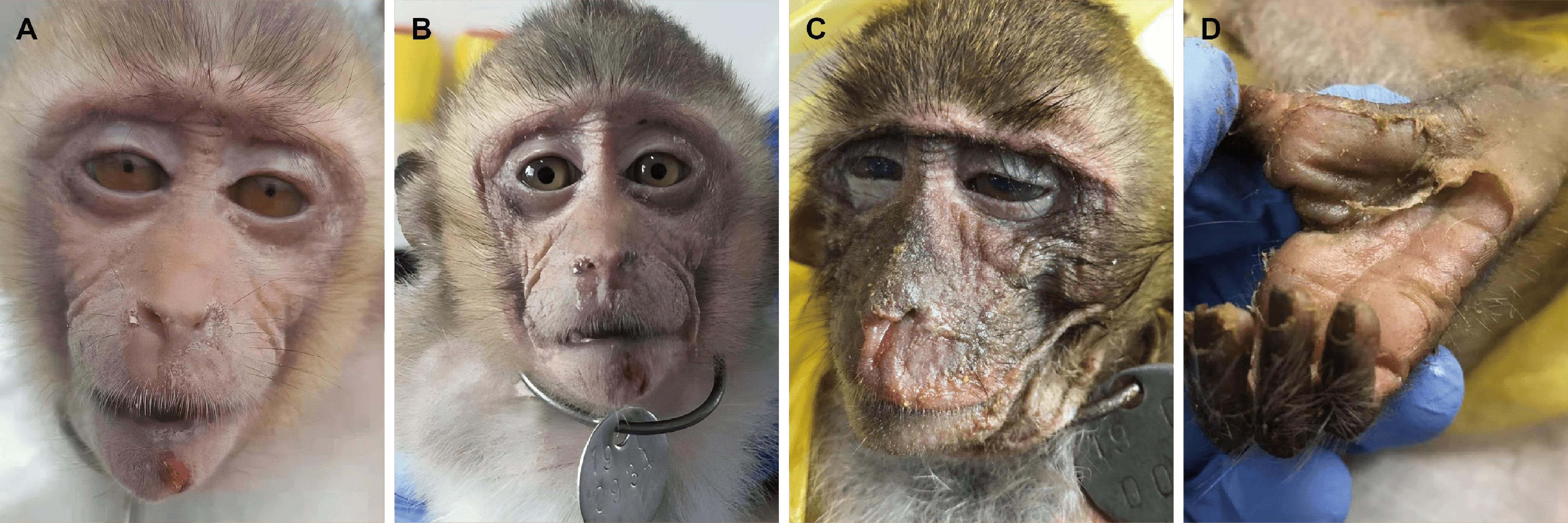
Figure 1 Clinical symptoms of the infected cynomolgus monkeys
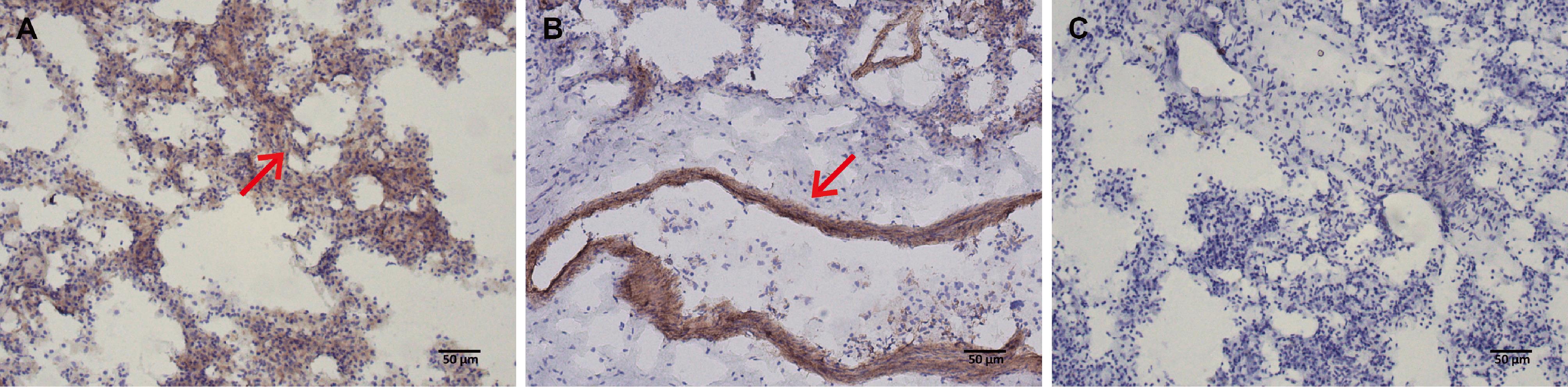
Figure 2 Detection of viral protein expression in lung tissue sections of the deceased monkey by immunohistochemical staining (DAB staining, ×100)
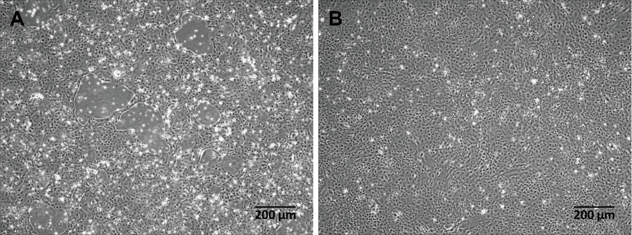
Figure 3 Observation of cytopathic effect after inoculation of MDCK cells with skin tissue filtrate from the deceased monkey
病毒株 Strains | CDV/CYN/SUZ/2020 | CDV/dog/HCM/33/140816 | ZC1310M | CYN07-dV | BJ-01 | MKY-KM08 | CDV1_TH/2014 | MCL-18-Li-1/1 | 007Lm | CDV3 |
|---|---|---|---|---|---|---|---|---|---|---|
| CDV/CYN/SUZ/2020 | 100.00 | - | - | - | - | - | - | - | - | - |
| CDV/dog/HCM/33/140816 | 98.86 | 100.00 | - | - | - | - | - | - | - | - |
| ZC1310M | 98.44 | 98.73 | 100.00 | - | - | - | - | - | - | - |
| CYN07-dV | 97.05 | 97.31 | 97.70 | 100.00 | - | - | - | - | - | - |
| BJ-01 | 97.05 | 97.30 | 97.69 | 99.92 | 100.00 | - | - | - | - | - |
| MKY-KM08 | 96.93 | 97.18 | 97.56 | 99.64 | 99.64 | 100.00 | - | - | - | - |
| CDV1_TH/2014 | 94.28 | 94.54 | 94.84 | 94.62 | 94.61 | 94.57 | 100.00 | - | - | - |
| MCL-18-Li-1/1 | 93.54 | 93.78 | 94.11 | 93.87 | 93.87 | 93.82 | 94.06 | 100.00 | - | - |
| 007Lm | 93.93 | 94.25 | 94.57 | 94.53 | 94.51 | 94.46 | 94.57 | 93.95 | 100.00 | - |
| CDV3 | 92.14 | 92.37 | 92.68 | 92.43 | 92.42 | 92.43 | 92.34 | 91.96 | 92.76 | 100.00 |
Table 1 Sequence identity (similarity) comparison between the full-length open reading frame nucleotide sequence of the CDV strain isolated from the skin tissue of the deceased monkey and prevalent Asian CDV strains
病毒株 Strains | CDV/CYN/SUZ/2020 | CDV/dog/HCM/33/140816 | ZC1310M | CYN07-dV | BJ-01 | MKY-KM08 | CDV1_TH/2014 | MCL-18-Li-1/1 | 007Lm | CDV3 |
|---|---|---|---|---|---|---|---|---|---|---|
| CDV/CYN/SUZ/2020 | 100.00 | - | - | - | - | - | - | - | - | - |
| CDV/dog/HCM/33/140816 | 98.86 | 100.00 | - | - | - | - | - | - | - | - |
| ZC1310M | 98.44 | 98.73 | 100.00 | - | - | - | - | - | - | - |
| CYN07-dV | 97.05 | 97.31 | 97.70 | 100.00 | - | - | - | - | - | - |
| BJ-01 | 97.05 | 97.30 | 97.69 | 99.92 | 100.00 | - | - | - | - | - |
| MKY-KM08 | 96.93 | 97.18 | 97.56 | 99.64 | 99.64 | 100.00 | - | - | - | - |
| CDV1_TH/2014 | 94.28 | 94.54 | 94.84 | 94.62 | 94.61 | 94.57 | 100.00 | - | - | - |
| MCL-18-Li-1/1 | 93.54 | 93.78 | 94.11 | 93.87 | 93.87 | 93.82 | 94.06 | 100.00 | - | - |
| 007Lm | 93.93 | 94.25 | 94.57 | 94.53 | 94.51 | 94.46 | 94.57 | 93.95 | 100.00 | - |
| CDV3 | 92.14 | 92.37 | 92.68 | 92.43 | 92.42 | 92.43 | 92.34 | 91.96 | 92.76 | 100.00 |
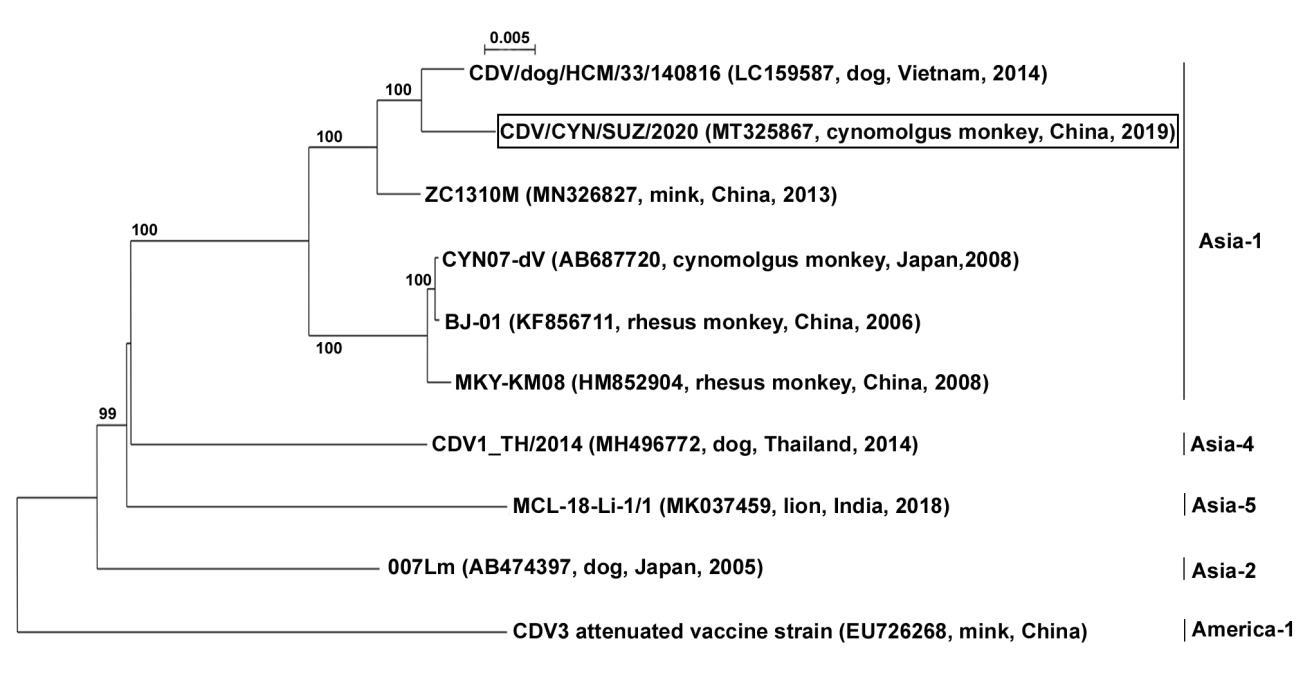
Figure 4 Phylogenetic analysis based on full-length open reading frame sequences of different CDV strains (the CDV strain newly isolated from the skin tissue of the deceased monkey in this study is marked with a black box)
| [1] | TROGU T, CASTELLI A, CANZIANI S, et al. Detection and molecular characterization of canine distemper virus in wildlife from northern Italy[J]. Pathogens, 2022, 11(12):1557. DOI:10.3390/pathogens11121557 . |
| [2] | SUN Z Z, LI A X, YE H H, et al. Natural infection with canine distemper virus in hand-feeding Rhesus monkeys in China[J]. Vet Microbiol, 2010, 141(3-4):374-378. DOI:10.1016/j.vetmic. 2009.09.024 . |
| [3] | DUQUE-VALENCIA J, SARUTE N, OLARTE-CASTILLO X A, et al. Evolution and interspecies transmission of canine distemper virus-an outlook of the diverse evolutionary landscapes of a multi-host virus[J]. Viruses, 2019, 11(7):582. DOI:10.3390/v11070582 . |
| [4] | OLEAGA Á, VÁZQUEZ C B, ROYO L J, et al. Canine distemper virus in wildlife in south-western Europe[J]. Transbound Emerg Dis, 2022, 69(4): e473-e485. DOI:10.1111/tbed.14323 . |
| [5] | LIANG J, WANG T T, WANG Q, et al. Prevalence of canine distemper in minks, foxes and raccoon dogs from 1983 to 2023 in Asia, North America, South America and Europe[J]. Front Vet Sci, 2024, 11:1394631. DOI:10.3389/fvets. 2024.1394631 . |
| [6] | CHENG Y N, WANG J K, ZHANG M, et al. Isolation and sequence analysis of a canine distemper virus from a raccoon dog in Jilin Province, China[J]. Virus Genes, 2015, 51(2):298-301. DOI:10.1007/s11262-015-1236-3 . |
| [7] | WILKES R P. Canine distemper virus in endangered species: species jump, clinical variations, and vaccination[J]. Pathogens, 2022, 12(1):57. DOI:10.3390/pathogens12010057 . |
| [8] | DI FRANCESCO C E, SMOGLICA C, DI PIRRO V, et al. Molecular detection and phylogenetic analysis of canine distemper virus in marsican brown bear (Ursus arctos marsicanus)[J]. Animals, 2022, 12(14):1826. DOI:10.3390/ani12141826 . |
| [9] | FRANZO G, DE VILLIERS L, COETZEE L M, et al. Unveiling the molecular epidemiology of canine distemper virus in Namibia: an expected pathogen showing an unexpected origin[J]. Heliyon, 2024, 10(15): e34805. DOI:10.1016/j.heliyon. 2024.e34805 . |
| [10] | SAKAI K J, NAGATA N, AMI Y, et al. Lethal canine distemper virus outbreak in cynomolgus monkeys in Japan in 2008[J]. J Virol, 2013, 87(2):1105-1114. DOI:10.1128/JVI.02419-12 . |
| [11] | QIU W, ZHENG Y, ZHANG S F, et al. Canine distemper outbreak in rhesus monkeys, China[J]. Emerg Infect Dis, 2011, 17(8):1541-1543. DOI:10.3201/eid1708.101153 . |
| [12] | 陆承平. 兽医微生物学[M]. 3版. 北京: 中国农业出版社, 2001: 525-526. |
| LU C P. Veterinary microbiology[M]. 3rd ed. Beijing: China Agricultural Press, 2001:525-526. | |
| [13] | YOSHIKAWA Y, OCHIKUBO F, MATSUBARA Y, et al. Natural infection with canine distemper virus in a Japanese monkey (Macaca fuscata)[J]. Vet Microbiol, 1989, 20(3):193-205. DOI:10.1016/0378-1135(89)90043-6 . |
| [14] | 麦博, 王弘义, 盘宝进, 等. 灵长类动物感染犬瘟热[J]. 中国比较医学杂志, 2012, 22(6):72-76. DOI: 10.3969/j.issn.1671-7856.2012.06.016 . |
| MAI B, WANG H Y, PAN B J, et al. Natural infection with canine distemper virus in Primates[J]. Chin J Comp Med, 2012, 22(6):72-76. DOI: 10.3969/j.issn.1671-7856.2012.06.016 . | |
| [15] | 李慧敏. 犬瘟热病毒的分离鉴定及其抗原蛋白的研究[D]. 成都: 四川农业大学, 2006. |
| LI H M. Isolation and identification of canine distemper virus and study on its antigenic protein[D]. Chengdu: Sichuan Agricultural University, 2006. | |
| [16] | 黄国君, 岳华, 杨发龙, 等. SYBR GreenⅠ荧光定量RT-PCR检测犬瘟热病毒方法的建立及应用[J]. 中国预防兽医学报, 2008, 30(6):450-454. DOI: 10.3724/SP.J.1011.2008.00534 . |
| HUANG G J, YUE H, YANG F L, et al. Development of a SYBR GreenⅠ real-time RT-PCR assay for detection of canine distemper virus[J]. Chin J Prev Vet Med, 2008, 30(6):450-454. DOI: 10.3724/SP.J.1011.2008.00534 . | |
| [17] | GOUY M, GUINDON S, GASCUEL O. SeaView version 4: a multiplatform graphical user interface for sequence alignment and phylogenetic tree building[J]. Mol Biol Evol, 2010, 27(2):221-224. DOI:10.1093/molbev/msp259 . |
| [18] | GUINDON S, GASCUEL O. A simple, fast, and accurate algorithm to estimate large phylogenies by maximum likelihood[J]. Syst Biol, 2003, 52(5):696-704. DOI:10.1080/10635150390235520 . |
| [19] | NGUYEN D V, SUZUKI J, MINAMI S, et al. Isolation and phylogenetic analysis of canine distemper virus among domestic dogs in Vietnam[J]. J Vet Med Sci, 2017, 79(1):123-127. DOI:10.1292/jvms.16-0394 . |
| [20] | QUINTERO-GIL C, RENDON-MARIN S, MARTINEZ-GUTIERREZ M, et al. Origin of canine distemper virus: consolidating evidence to understand potential zoonoses[J]. Front Microbiol, 2019, 10:1982. DOI:10.3389/fmicb.2019.01982 . |
| [21] | RIVERA-MARTÍNEZ A, RODRÍGUEZ-ALARCÓN C A, ADAME-GALLEGOS J R, et al. Canine distemper virus: origins, mutations, diagnosis, and epidemiology in Mexico[J]. Life, 2024, 14(8):1002. DOI:10.3390/life14081002 . |
| [22] | SHESHBERADARAN H, NORRBY E, MCCULLOUGH K C, et al. The antigenic relationship between measles, canine distemper and rinderpest viruses studied with monoclonal antibodies[J]. J Gen Virol, 1986, 67 ( Pt 7):1381-1392. DOI:10.1099/0022-1317-67-7-1381 . |
| [23] | ROUXEL R N, SVITEK N, VON MESSLING V. A chimeric measles virus with canine distemper envelope protects ferrets from lethal distemper challenge[J]. Vaccine, 2009, 27(36):4961-4966. DOI:10.1016/j.vaccine.2009.05.096 . |
| [24] | OTSUKI N, SEKIZUKA T, SEKI F, et al. Canine distemper virus with the intact C protein has the potential to replicate in human epithelial cells by using human nectin4 as a receptor[J]. Virology, 2013, 435(2):485-492. DOI:10.1016/j.virol.2012.10.033 . |
| [25] | RENDON-MARIN S, RUÍZ-SAENZ J. Universal peptide-based potential vaccine design against canine distemper virus (CDV) using a vaccinomic approach[J]. Sci Rep, 2024, 14(1):16605. DOI:10.1038/s41598-024-67781-5 . |
| [26] | SWATI, DEKA D, UPPAL S K, et al. Isolation and phylogenetic characterization of Canine distemper virus from India[J]. Virusdisease, 2015, 26(3):133-140. DOI:10.1007/s13337-015-0256-x . |
| [27] | GUERCIO A, MIRA F, DI BELLA S, et al. Biomolecular analysis of canine distemper virus strains in two domestic ferrets (Mustela Putorius furo)[J]. Vet Sci, 2023, 10(6):375. DOI:10.3390/vetsci10060375 . |
| [28] | MOCHIZUKI M, HASHIMOTO M, HAGIWARA S, et al. Genotypes of canine distemper virus determined by analysis of the hemagglutinin genes of recent isolates from dogs in Japan[J]. J Clin Microbiol, 1999, 37(9):2936-2942. DOI:10.1128/JCM.37.9.2936-2942.1999 . |
| [29] | SHI N, ZHANG L, YU X H, et al. Insight into an outbreak of canine distemper virus infection in masked palm civets in China[J]. Front Vet Sci, 2021, 8:728238. DOI:10.3389/fvets.2021.728238 . |
| [1] | WEI Yanye, SHEN Guo, ZHANG Pengfei, SHI Songping, HU Jiahao, ZHANG Xuzhe, HUA Huiyuan, HUA Guanyang, LU Hongzheng, ZENG Yong, JI Feng, WEI Zhumei. Dynamic Monitoring and Correlation Analysis of General Body Indicators, Blood Glucose, and Blood Lipid in Obese Cynomolgus Monkeys [J]. Laboratory Animal and Comparative Medicine, 2025, 45(1): 30-36. |
| [2] | Ping YANG, Li CUI, Cheng YU, Zhiyue WEN. Effects of Bevacizumab Injection on the Skin Wound Healing in Cynomolgus Monkeys [J]. Laboratory Animal and Comparative Medicine, 2023, 43(1): 21-29. |
| [3] | Zhumei WEI, Guo SHEN, Zhenming LI, Yong ZENG, Feng JI, Jihong YANG. Measurement and Analysis of Bone Mineral Content and Bone Mineral Density in Healthy Cynomolgus Monkeys at Different Ages [J]. Laboratory Animal and Comparative Medicine, 2022, 42(5): 409-415. |
| [4] | GAO Shiping, LI Feng, ZHA Sifan. High-fat Diet Induced Cynomolgus Monkey Model of Non-alcoholic Fatty Liver Disease [J]. Laboratory Animal and Comparative Medicine, 2020, 40(2): 123-127. |
| [5] | LI Liang-shun, PAN Shi-you, ZHI Yuan-fang, BAI shuai-zhou, SUN Jian-hua, GONG Li-kun, REN Jin. Diagnosis and Treatment for Intussusceptions in One Case of Cynomolgus Monkey [J]. Laboratory Animal and Comparative Medicine, 2019, 39(3): 236-238. |
| [6] | WEI Zhu-mei, YANG Bo, LI Zhen-ming, HUAN Yin-hua, SU Ke-long, YANG Ji-hong. Continuous Monitoring on Blood Glucose and Insulin Levels in Obesity and Diabetes Cynomolgus Monkeys [J]. Laboratory Animal and Comparative Medicine, 2015, 35(5): 394-397. |
| [7] | YANG Bo, WEI Zhu-mei, DENG Xin-ning, HE Nai-zhi, YIN Feng-ri, LI Zhen-ming, YANG Ji-hong. Established Method of Measurement for Gastrocnemius Volume in Cynomolgus Monkeys by MRI Detection [J]. Laboratory Animal and Comparative Medicine, 2014, 34(4): 314-318. |
| [8] | SANG Chuan-lan, DUO Hai-gang, ZHANG Yong-Bin, CHEN Jia, Rao Jun-hua. Preliminary Probe of Detection of Simian Varicella Virus Antibodies in Cynomolgus Monkey [J]. Laboratory Animal and Comparative Medicine, 2012, 32(5): 446-448. |
| [9] | SHI Qiao-juan, LI wei, SA Xiao-ying. Application of Cynomolgus Monkey in Medical Science [J]. Laboratory Animal and Comparative Medicine, 2012, 32(4): 358-365. |
| [10] | ZENG Xian-cheng, JI Long-feng, ZHANG Ling-li, YAN Dong-ming, ZHOU Chao, JIANG Zi-rui, PAN Xue-ying, CHANG Yan, MA Jing. Determination of Cortisol in Plasma of Cynomolgus Monkeys by LC-MS/MS and Stress Monitoring [J]. Laboratory Animal and Comparative Medicine, 2011, 31(6): 441-446. |
| [11] | MA Nan-hua1,LI Zong-qiang2,QIN Jian1,ZHENG Li-ping1,ZHU Xu1,HUANG Sheng-xian1,XIE Li-ping3. Changes of Serum Levels of Estrogen and Progestogen during Menstrual Cycle in Normal Female Cynomolgus Monkeys [J]. Laboratory Animal and Comparative Medicine, 2008, 28(2): 106-109. |
| [12] | SUN Xiao-mei,YE You-song,TONG Pin-fen,DAI Jie-jie. Discussion on Genetic Control of Captive Nonhuman Primate Colonies [J]. Laboratory Animal and Comparative Medicine, 2007, 27(1): 67-70. |
| [13] | CHENG Shu-jun1,2,LI Hai-biao1. Progress on Embryonic Stem Cells in Nonhuman Primates [J]. Laboratory Animal and Comparative Medicine, 2004, 24(2): 114-118. |
| Viewed | ||||||
|
Full text |
|
|||||
|
Abstract |
|
|||||