
Laboratory Animal and Comparative Medicine ›› 2025, Vol. 45 ›› Issue (3): 368-375.DOI: 10.12300/j.issn.1674-5817.2024.166
• Quality Control of Laboratory Animals • Previous Articles Next Articles
KONG Zhihao1, WEI Xiaofeng1( )(
)( ), YU Lingzhi1()(), FENG Liping1, ZHU Qi1, SHI Guojun2, WANG Chen2
Received:2024-11-12
Revised:2025-02-05
Online:2025-06-25
Published:2025-06-25
Contact:
WEI Xiaofeng, YU Lingzhi
CLC Number:
KONG Zhihao,WEI Xiaofeng,YU Lingzhi,et al. Isolation and Identification of Staphylococcus xylosus in Nude Mice with Squamous Skin Scurfs[J]. Laboratory Animal and Comparative Medicine, 2025, 45(3): 368-375. DOI: 10.12300/j.issn.1674-5817.2024.166.
Add to citation manager EndNote|Ris|BibTeX
URL: https://www.slarc.org.cn/dwyx/EN/10.12300/j.issn.1674-5817.2024.166
Figure 1 Detection of Corynebacterium bovis was negative by real-time fluorescence PCR
Figure 2 Colony morphology on mannitol salt agar plate (A) and blood agar plate (B) and Gram staining microscopic examination (C) of the isolated strain
生化项目 Biochemical items | 鉴定结果 Results |
|---|---|
| D-苦杏仁甙 D-amygdalin | - |
磷脂酰肌醇磷脂酶C Phosphatidylinositol phospholipase C | - |
| D-木糖 D-xylose | + |
| 精氨酸双水解酶1 Arginine dihydrolase 1 | + |
| β-半乳糖苷酶 β-galactosidase | + |
| α-葡糖苷酶 α-glucosidase | + |
丙氨酸-苯丙氨酸-脯氨酸芳胺酶 Ala-Phe-Pro-arylamidase | - |
| 环糊精 Cyclodextrin | - |
| L-天冬氨酸芳胺酶 L-aspartate arylamidase | - |
| β-半乳糖吡喃糖苷酶 β-galactopyranosidase | - |
| α-甘露糖苷酶 α-mannosidase | - |
| 磷酸酶 Phosphatase | + |
| 亮氨酸芳胺酶 Leucine arylamidase | - |
| L-脯氨酸芳胺酶 L-proline arylamidase | - |
| β-葡萄糖醛酸酶 β-glucuronidase | + |
| α-半乳糖苷酶 α-galactosidase | - |
| L-吡咯烷酮芳胺酶 L-pyrrolidonyl-arylamidase | + |
| β-葡萄糖醛酸酶 β-glucuronidase | + |
| 丙氨酸芳胺酶 Alanine arylamidase | - |
| 酪氨酸芳胺酶 Tyrosine arylamidase | - |
| D-山梨醇 D-sorbitol | - |
| 尿素酶 Urease | + |
| 多黏菌素B Polymyxin B | - |
| D-半乳糖 D-galactose | - |
| D-核糖 D-ribose | + |
| L-乳酸盐碱化 L-lactate alkalinization | + |
| 乳糖 Lactose | + |
| N-乙酰-D-氨基葡萄糖 N-acetyl-D-glucosamine | + |
| D-麦芽糖 D-maltose | + |
| 杆菌肽耐药 Bacitracin resistance | + |
| 新生霉素耐药 Novobiocin resistance | + |
| 6.5%NaCl中生长 Growth in 6.5% NaCl | + |
| D-甘露醇 D-mannitol | + |
| D-甘露糖 D-mannose | + |
| 甲基-β-D-吡喃葡萄糖苷 Methyl-β-D-glucopyranoside | + |
| 支链淀粉 Pullulan | - |
| D-棉子糖 D-raffinose | - |
| O/129耐药 O/129 resistance | + |
| 水杨素 Salicin | - |
| 蔗糖 Saccharose/Sucrose | + |
| D-海藻糖 D-trehalose | + |
| 精氨酸双水解酶2 Arginine dihydrolase 2 | - |
| 奥普托欣耐药 Optochin resistance | + |
Table 1 Biochemical identification results indicating that the isolated strain was Staphylococcus xylosus
生化项目 Biochemical items | 鉴定结果 Results |
|---|---|
| D-苦杏仁甙 D-amygdalin | - |
磷脂酰肌醇磷脂酶C Phosphatidylinositol phospholipase C | - |
| D-木糖 D-xylose | + |
| 精氨酸双水解酶1 Arginine dihydrolase 1 | + |
| β-半乳糖苷酶 β-galactosidase | + |
| α-葡糖苷酶 α-glucosidase | + |
丙氨酸-苯丙氨酸-脯氨酸芳胺酶 Ala-Phe-Pro-arylamidase | - |
| 环糊精 Cyclodextrin | - |
| L-天冬氨酸芳胺酶 L-aspartate arylamidase | - |
| β-半乳糖吡喃糖苷酶 β-galactopyranosidase | - |
| α-甘露糖苷酶 α-mannosidase | - |
| 磷酸酶 Phosphatase | + |
| 亮氨酸芳胺酶 Leucine arylamidase | - |
| L-脯氨酸芳胺酶 L-proline arylamidase | - |
| β-葡萄糖醛酸酶 β-glucuronidase | + |
| α-半乳糖苷酶 α-galactosidase | - |
| L-吡咯烷酮芳胺酶 L-pyrrolidonyl-arylamidase | + |
| β-葡萄糖醛酸酶 β-glucuronidase | + |
| 丙氨酸芳胺酶 Alanine arylamidase | - |
| 酪氨酸芳胺酶 Tyrosine arylamidase | - |
| D-山梨醇 D-sorbitol | - |
| 尿素酶 Urease | + |
| 多黏菌素B Polymyxin B | - |
| D-半乳糖 D-galactose | - |
| D-核糖 D-ribose | + |
| L-乳酸盐碱化 L-lactate alkalinization | + |
| 乳糖 Lactose | + |
| N-乙酰-D-氨基葡萄糖 N-acetyl-D-glucosamine | + |
| D-麦芽糖 D-maltose | + |
| 杆菌肽耐药 Bacitracin resistance | + |
| 新生霉素耐药 Novobiocin resistance | + |
| 6.5%NaCl中生长 Growth in 6.5% NaCl | + |
| D-甘露醇 D-mannitol | + |
| D-甘露糖 D-mannose | + |
| 甲基-β-D-吡喃葡萄糖苷 Methyl-β-D-glucopyranoside | + |
| 支链淀粉 Pullulan | - |
| D-棉子糖 D-raffinose | - |
| O/129耐药 O/129 resistance | + |
| 水杨素 Salicin | - |
| 蔗糖 Saccharose/Sucrose | + |
| D-海藻糖 D-trehalose | + |
| 精氨酸双水解酶2 Arginine dihydrolase 2 | - |
| 奥普托欣耐药 Optochin resistance | + |
Figure 3 Phylogenetic tree analysis based on single-copy orthologue of the isolated strain
Figure 4 Clinical manifestations of scaling in different groups of nude mice infected with isolated bacterial suspension
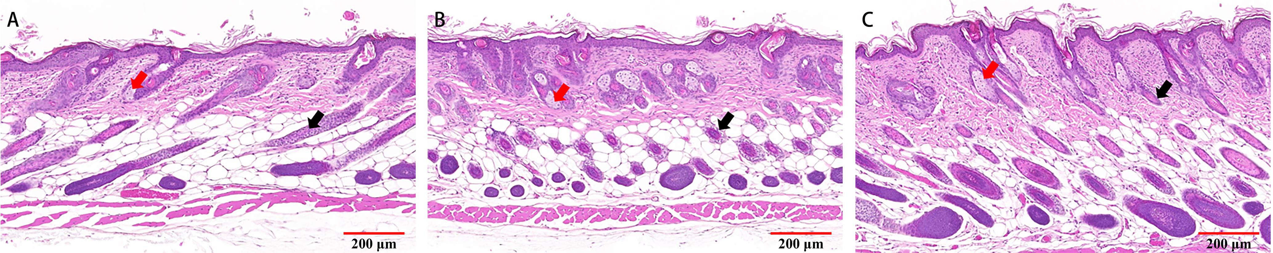
Figure 5 Pathological changes in skin tissues of nude mice with squamous skin scurfs after infection with isolated bacteria (HE staining, ×100)
| [1] | SCANZIANI E, GOBBI A, CRIPPA L, et al. Hyperkeratosis-associated coryneform infection in severe combined immunodeficient mice[J]. Lab Anim, 1998, 32(3):330-336. DOI:10.1258/002367798780559239 . |
| [2] | KIM T H, KIM D S, HAN J H, et al. Detection of Corynebacterium bovis infection in athymic nude mice from a research animal facility in Korea[J]. J Vet Sci, 2014, 15(4):583-586. DOI:10.4142/jvs.2014.15.4.583 . |
| [3] | MANUEL C A, PUGAZHENTHI U, LESZCZYNSKI J K. Surveillance of a ventilated rack system for Corynebacterium bovis by sampling exhaust-air manifolds[J]. J Am Assoc Lab Anim Sci, 2016, 55(1):58-65. |
| [4] | RUSSO M, INVERNIZZI A, GOBBI A, et al. Diffuse scaling dermatitis in an athymic nude mouse[J]. Vet Pathol, 2013, 50(4):722-726. DOI:10.1177/0300985812463408 . |
| [5] | TAVAKKOL Z, SAMUELSON D, DELANCEY PULCINI E, et al. Resident bacterial flora in the skin of C57BL/6 mice housed under SPF conditions[J]. J Am Assoc Lab Anim Sci, 2010, 49(5):588-591. |
| [6] | GOZALO A S, HOFFMANN V J, BRINSTER L R, et al. Spontaneous Staphylococcus xylosus infection in mice deficient in NADPH oxidase and comparison with other laboratory mouse strains[J]. J Am Assoc Lab Anim Sci, 2010, 49(4):480-486. |
| [7] | JACKSON S H, GALLIN J I, HOLLAND S M. The p47phox mouse knock-out model of chronic granulomatous disease[J]. J Exp Med, 1995, 182(3):751-758. DOI:10.1084/jem.182.3.751 . |
| [8] | PIZZOLLA A, HULTQVIST M, NILSON B, et al. Reactive oxygen species produced by the NADPH oxidase 2 complex in monocytes protect mice from bacterial infections[J]. J Immunol, 2012, 188(10):5003-5011. DOI:10.4049/jimmunol. 1103430 . |
| [9] | LAUBACH V E, SHESELY E G, SMITHIES O, et al. Mice lacking inducible nitric oxide synthase are not resistant to lipopolysaccharide-induced death[J]. Proc Natl Acad Sci USA, 1995, 92(23):10688-10692. DOI:10.1073/pnas.92.23.10688 . |
| [10] | WON Y S, KWON H J, OH G T, et al. Identification of Staphylococcus xylosus isolated from C57BL/6J-Nos2tm1Lau mice with dermatitis[J]. Microbiol Immunol, 2002, 46(9): 629-632. DOI: 10.1111/j.1348-04212002.tb02744.x . |
| [11] | 中国实验动物学会. 实验动物 牛棒状杆菌检测方法: T/CALAS 20—2017 [S]. 北京: 科学出版社, 2017. |
| Chinese Association for Laboratory Animal Sciences. Laboratory animal–Method for examination ofCoryne-bacterium bovis: T/CALAS 20—2017[S]. Beijing:Science Press, 2017. | |
| [12] | 中国实验动物学会. 实验动物 木糖葡萄球菌检测方法: T/CALAS 92—2020[S/OL]. [2024-12-30]. https://www.ttbz.org.cn/Pdfs/Index/?ftype=st&pms=74557. |
| Chinese Association for Laboratory Animal Sciences. Laboratory animal–Method for the detection ofStaphyl-ococcus xylosus:T/CALAS 92—2020[S/OL]. [2024-12-30]. https://www.ttbz.org.cn/Pdfs/Index/?ftype=st&pms=74557. | |
| [13] | NATSIS N E, COHEN P R. Coagulase-negative Staphylococcus skin and soft tissue infections[J]. Am J Clin Dermatol, 2018, 19(5):671-677. DOI:10.1007/s40257-018-0362-9 . |
| [14] | BRADFIELD J F, WAGNER J E, BOIVIN G P, et al. Epizootic fatal dermatitis in athymic nude mice due to Staphylococcus xylosus [J]. Lab Anim Sci, 1993, 43(1):111-113. |
| [15] | 于灵芝, 冯丽萍, 孔志豪, 等. 木糖葡萄球菌实时荧光定量PCR检测方法的建立及其应用[J]. 中国实验动物学报, 2024, 32(1):73-79. DOI: 10.3969/j.issn.1005-4847.2024.01.010 . |
| YU L Z, FENG L P, KONG Z H, et al. Establishment of qPCR method to detect Staphylococcus xylosus and its application[J]. Acta Lab Anim Sci Sin, 2024, 32(1):73-79. DOI: 10.3969/j.issn.1005-4847.2024.01.010 . | |
| [16] | BATTAGLIA M, GARRETT-SINHA L A. Staphylococcus xylosus and Staphylococcus aureus as commensals and pathogens on murine skin[J]. Lab Anim Res, 2023, 39(1):18. DOI: 10.1186/s42826-023-00169-0 . |
| [17] | RESHAMWALA K, CHEUNG G Y C, HSIEH R C, et al. Identification and characterization of the pathogenic potential of phenol-soluble modulin toxins in the mouse commensal Staphylococcus xylosus [J]. Front Immunol, 2022, 13:999201. DOI:10.3389/fimmu.2022.999201 . |
| [18] | ACUFF N V, LAGATTA M, NAGY T, et al. Severe dermatitis associated with spontaneous Staphylococcus xylosus infection in Rag -/- Tpl2-/- mice[J]. Comp Med, 2017, 67(4):344-349. |
| [19] | LI Z Q, DONG J X, WANG M, et al. Resveratrol ameliorates liver fibrosis induced by nonpathogenic Staphylococcus in BALB/c mice through inhibiting its growth[J]. Mol Med, 2022, 28(1):52. DOI:10.1186/s10020-022-00463-y . |
| [20] | QU Q W, CUI W Q, XING X X, et al. Rutin, a natural inhibitor of IGPD protein, partially inhibits biofilm formation in Staphylococcus xylosus ATCC700404 in vitro and in vivo [J]. Front Pharmacol, 2021, 12:728354. DOI:10.3389/fphar.2021. 728354 . |
| [21] | EYERICH K, PENNINO D, SCARPONI C, et al. IL-17 in atopic eczema: linking allergen-specific adaptive and microbial-triggered innate immune response[J]. J Allergy Clin Immunol, 2009, 123(1):59-66.e4. DOI:10.1016/j.jaci.2008.10.031 . |
| [22] | NIEBUHR M, GATHMANN M, SCHARONOW H, et al. Staphylococcal alpha-toxin is a strong inducer of interleukin-17 in humans[J]. Infect Immun, 2011, 79(4):1615-1622. DOI:10.1128/IAI.00958-10 . |
| [23] | KIM Y, LEE Y S, YANG J Y, et al. The resident pathobiont Staphylococcus xylosus in Nfkbiz-deficient skin accelerates spontaneous skin inflammation[J]. Sci Rep, 2017, 7(1):6348. DOI:10.1038/s41598-017-05740-z . |
| [1] | MENG Yu, LIANG Dongli, ZHENG Linlin, ZHOU Yuanyuan, WANG Zhaoxia. Optimization and Evaluation of Conditions for Orthotopic Nude Mouse Models of Human Liver Tumor Cells [J]. Laboratory Animal and Comparative Medicine, 2024, 44(5): 511-522. |
| [2] | Shanshan ZHAI, Liang LIANG, Yingying CAO, Zhuxin LI, Qing WANG, Junyu TAO, Chenxia YUN, Jing LENG, Haibo TANG. Diagnosis of Trichoepithelioma in a Tree Shrew and Observation of Cell Biological Characteristics [J]. Laboratory Animal and Comparative Medicine, 2023, 43(4): 440-445. |
| [3] | JIA Huan-huan, ZENG Ye-wen, LUO Ting, GONG Bao-yong, MAI Dong-mei, PAN Ying-chun, ZHAO Wei-bo. Effects of Different Concentrations of Chromium-containing Bedding Materials on Toxity of Blood and Organs in BALB/c Nude Mice and KM Mice [J]. Laboratory Animal and Comparative Medicine, 2019, 39(6): 454-461. |
| [4] | TU Jue, ZHOU Wei-min, Cai Yue-qin, Ling Yun, Chen Min-li. Expression of Phospholipase A2 in Different Xenografted Tumor Models of Nude Mice [J]. Laboratory Animal and Comparative Medicine, 2019, 39(4): 298-304. |
| [5] | SONG Deng-peng, RAO Hong, HAN An-yan, WU Fu-yun, CHEN De-sen. Effects of Icariin on Human BALB/c-nu Prostate Cancer Model in Nude Mice [J]. Laboratory Animal and Comparative Medicine, 2018, 38(6): 428-433. |
| [6] | YANG Liu, YANG Hua, LIU Yun, CHEN Jia, GONG Ming-fu, SHU Tong-sheng. Feasibility Study on Intravenous Administration of Nanoparticles via Retrobulbar Vein in Nude Mice [J]. Laboratory Animal and Comparative Medicine, 2016, 36(2): 117-120. |
| [7] | ZHANG Yan, FAN Wen-xi, XU Yi-mei, SHI Sheng, GUI You-jun, YAN Shun-sheng. Effect of β-carboline derivative on Subcutaneous Transplant Hepatocellular Carcinoma Growth in BALB/c Nude Mice [J]. Laboratory Animal and Comparative Medicine, 2013, 33(4): 290-295. |
| [8] | YIN Bei-pei, LU Hong-qi, SHEN Long-hai, LI Bing-sheng. The Inhibiting Effect of Coix Seed Oil Injection on Xenograft Model of Human Liver Cancer Cell SMMC-7721 [J]. Laboratory Animal and Comparative Medicine, 2012, 32(2): 133-136. |
| [9] | CHEN Juan, YU Ying-hao. Inhibitory Effects of Sporoderm-broken Ganoderma Lucidum Spores on T-cell lymphoma [J]. Laboratory Animal and Comparative Medicine, 2011, 31(6): 426-431. |
| [10] | GAO Ai-xia1,LUO Ju-dong2,WU Qing-ting3,LI Hou-da3,XUE Zhi-mou1. Antitumor Effect of Anthocyanidin on Lung Cancer NCI-H460 Cells in vitro and in vivo [J]. Laboratory Animal and Comparative Medicine, 2008, 28(2): 85-89. |
| [11] | LI Zhi-Ling-1, 2 , LIU Xiang-Yun-12, ZHANG Xiao-Fang-1, WANG Lei-1, 2 , XIE Shu-Wu-1, 2 , SUN Yu-1, XIE Chen-Jing-1, WU Jian-Hui-1, CAO Lin-1, SUN Zu-Yue-1. Establishing Human Prostate Cancer Models from Different Cells and Its Metastatic Character [J]. Laboratory Animal and Comparative Medicine, 2006, 26(4): 207-212. |
| [12] | YAN Ming-Xia-1, 2 , YAO Ming-2, LIU Qian-3, ZHOU Guang-Xing-1, YANG Ping-1, YANG Fei-1, KONG Han-Wei-2, ZHU Ping-Mei-1, XU Lan-Wen-1. Liver Metastaic human Gastric Carcinoma Established in Nude Mice Using Orthotopic Organ Selection of Metastasis [J]. Laboratory Animal and Comparative Medicine, 2005, 25(2): 72-76. |
| [13] | YAN Ming-Xia-1, 2 , XU Lan-Wen-1, YAO Ming-2, ZHOU Guang-Xing-1, FAN Shi-Ming-2, KONG Han-Wei-2, QI Jin-Fang-2, ZHU Ping-Mei-1. Biological Properties of Orthotopic Implant Models for Human Gastric Carcinoma in Nude and SCID Mice [J]. Laboratory Animal and Comparative Medicine, 2005, 25(1): 8-12. |
| [14] | CHEN Ling-ji, DING Jian. The Criteria of New Anticancer Agents Evaluation in Human Tumor Nude Mice Model——The preclinical criteria of NCI and EORTC investigation [J]. Laboratory Animal and Comparative Medicine, 2001, 21(4): 247-247. |
| [15] | CHENG Feng, QIAN Guan-xiang, CHEN Shi-shu. Improving Tansplantation of Human Gastric Adenocarcinoma Tissue in Nude Mice [J]. Laboratory Animal and Comparative Medicine, 2001, 21(1): 29-32. |
| Viewed | ||||||
|
Full text |
|
|||||
|
Abstract |
|
|||||