
Laboratory Animal and Comparative Medicine ›› 2025, Vol. 45 ›› Issue (3): 269-278.DOI: 10.12300/j.issn.1674-5817.2025.030
• Animal Models of Human Diseases • Previous Articles Next Articles
XIAO Linlin1,2, YANG Yixuan1,2, LI Shanshan1,2, LUO Lanshiyu1,2, YIN Siwei1,2, SUN Juming1, SHI Wei1, OUYANG Yiqiang1( )(
)( ), LI Xiyi3()()
Received:2025-03-01
Revised:2025-04-14
Online:2025-06-25
Published:2025-06-25
Contact:
OUYANG Yiqiang, LI Xiyi
CLC Number:
XIAO Linlin,YANG Yixuan,LI Shanshan,et al. Establishment of a Rat Model of Alzheimer's Disease by Introducing Human Triple Mutant APP Gene into Hippocampus via Brain Stereotactic Technology[J]. Laboratory Animal and Comparative Medicine, 2025, 45(3): 269-278. DOI: 10.12300/j.issn.1674-5817.2025.030.
Add to citation manager EndNote|Ris|BibTeX
URL: https://www.slarc.org.cn/dwyx/EN/10.12300/j.issn.1674-5817.2025.030
Figure 1 Schematic diagram of recombinant adeno-associated viral vector structure
Figure 2 In vivo imaging of rats two weeks and six months after virus injection
Figure 3 Novel object recognition test to assess recognition memory of rats
Figure 4 APP gene expression and sequencing results in rat hippocampus
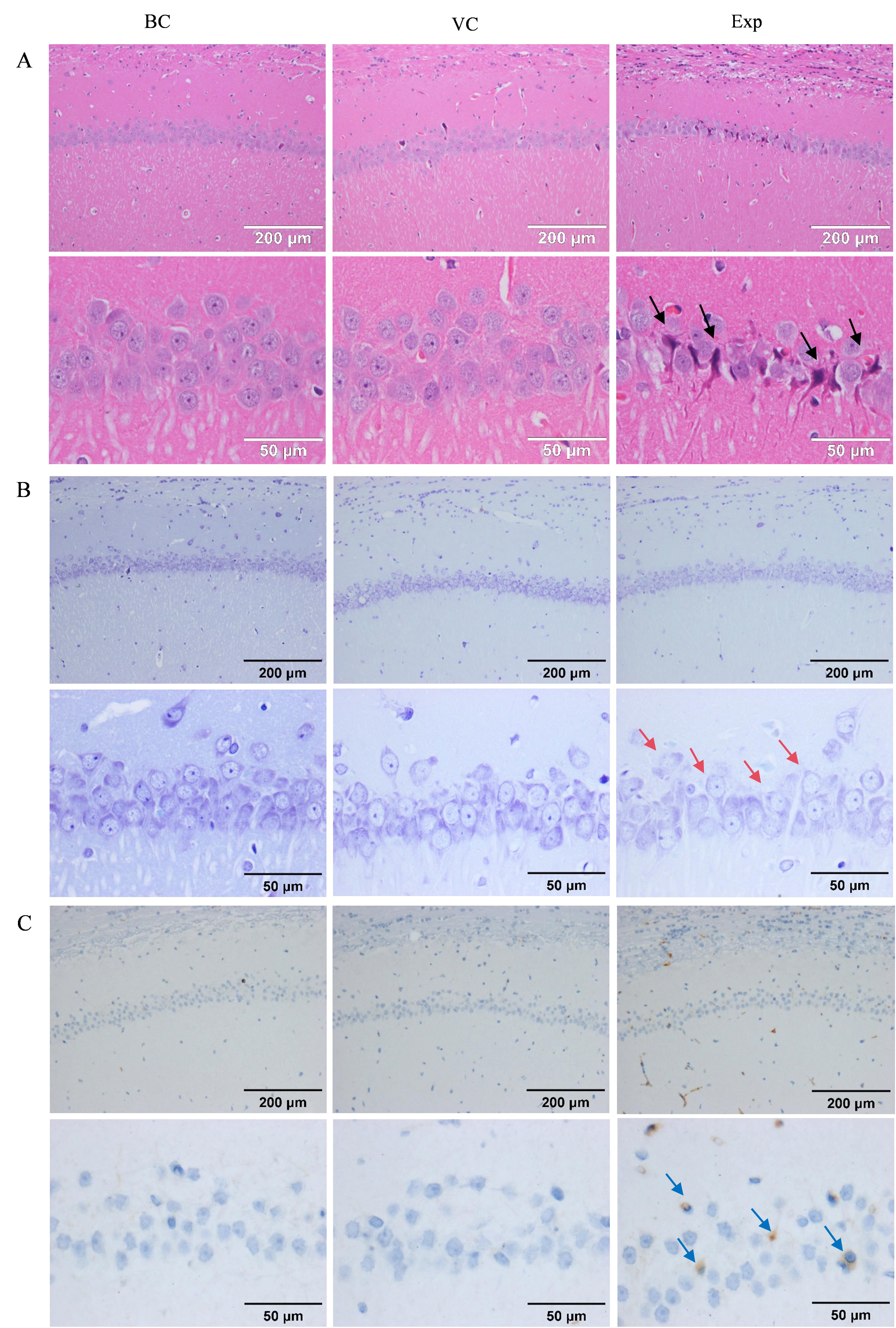
Figure 5 HE staining (A), Nissl staining (B), and immunohistochemical staining (C) results of rat hippocampal region
| [1] | 王刚, 齐金蕾, 刘馨雅, 等. 中国阿尔茨海默病报告2024[J]. 诊断学理论与实践, 2024, 23(3):219-256. DOI: 10.16150/j.1671-2870.2024.03.001 . |
| WANG G, QI J L, LIU X Y, et al. China Alzheimer report 2024[J]. J Diagn Concepts Pract, 2024, 23(3):219-256. DOI: 10.16150/j.1671-2870.2024.03.001 . | |
| [2] | SCHELTENS P, DE STROOPER B, KIVIPELTO M, et al. Alzheimer's disease[J]. Lancet, 2021, 397(10284):1577-1590. DOI:10.1016/s0140-6736(20)32205-4 . |
| [3] | SCHELTENS P, BLENNOW K, BRETELER M M, et al. Alzheimer's disease[J]. Lancet, 2016, 388(10043):505-517. DOI:10.1016/s0140-6736(15)01124-1 . |
| [4] | BREIJYEH Z, KARAMAN R. Comprehensive review on Alzheimer's disease: causes and treatment[J]. Molecules, 2020, 25(24):5789. DOI:10.3390/molecules25245789 . |
| [5] | JUCKER M, WALKER L C. Alzheimer's disease: From immunotherapy to immunoprevention[J]. Cell, 2023, 186(20):4260-4270. DOI:10.1016/j.cell.2023.08.021 . |
| [6] | ZHOU B, LU J G, SIDDU A, et al. Synaptogenic effect of APP-Swedish mutation in familial Alzheimer's disease[J]. Sci Transl Med, 2022, 14(667): eabn9380. DOI:10.1126/scitranslmed.abn9380 . |
| [7] | KIM M, BEZPROZVANNY I. Analysis of non-amyloidogenic mutations in APP supports loss of function hypothesis of Alzheimer's disease[J]. Int J Mol Sci, 2023, 24(3):2092. DOI:10.3390/ijms24032092 . |
| [8] | ARMBRUST F, BICKENBACH K, MARENGO L, et al. The Swedish dilemma - the almost exclusive use of APPswe-based mouse models impedes adequate evaluation of alternative β-secretases[J]. Biochim Biophys Acta Mol Cell Res, 2022, 1869(3):119164. DOI:10.1016/j.bbamcr.2021.119164 . |
| [9] | SCHILLING S, PRADHAN A, HEESCH A, et al. Differential effects of familial Alzheimer's disease-causing mutations on amyloid precursor protein (APP) trafficking, proteolytic conversion, and synaptogenic activity[J]. Acta Neuropathol Commun, 2023, 11(1):87. DOI:10.1186/s40478-023-01577-y . |
| [10] | JAWORSKI T, DEWACHTER I, LECHAT B, et al. AAV-tau mediates pyramidal neurodegeneration by cell-cycle re-entry without neurofibrillary tangle formation in wild-type mice[J]. PLoS One, 2009, 4(10): e7280. DOI:10.1371/journal.pone. 0007280 . |
| [11] | XIA D, LIANOGLOU S, SANDMANN T, et al. Novel App knock-in mouse model shows key features of amyloid pathology and reveals profound metabolic dysregulation of microglia[J]. Mol Neurodegener, 2022, 17(1):41. DOI:10.1186/s13024-022-00547-7 . |
| [12] | CLAYTON K, DELPECH J C, HERRON S, et al. Plaque associated microglia hyper-secrete extracellular vesicles and accelerate tau propagation in a humanized APP mouse model[J]. Mol Neurodegener, 2021, 16(1):18. DOI:10.1186/s13024-021-00440-9 . |
| [13] | TESSON L, COZZI J, MÉNORET S, et al. Transgenic modifications of the rat genome[J]. Transgenic Res, 2005, 14(5):531-546. DOI:10.1007/s11248-005-5077-z . |
| [14] | PANG K L, JIANG R C, ZHANG W, et al. An App knock-in rat model for Alzheimer's disease exhibiting Aβ and tau pathologies, neuronal death and cognitive impairments[J]. Cell Res, 2022, 32(2):157-175. DOI:10.1038/s41422-021-00582-x . |
| [15] | PUPO A, FERNÁNDEZ A, LOW S H, et al. AAV vectors: The Rubik's cube of human gene therapy[J]. Mol Ther, 2022, 30(12):3515-3541. DOI:10.1016/j.ymthe.2022.09.015 . |
| [16] | ASCHAUER D F, KREUZ S, RUMPEL S. Analysis of transduction efficiency, tropism and axonal transport of AAV serotypes 1, 2, 5, 6, 8 and 9 in the mouse brain[J]. PLoS One, 2013, 8(9): e76310. DOI:10.1371/journal.pone.0076310 . |
| [17] | ROSTAGNO A A. Pathogenesis of Alzheimer's disease[J]. Int J Mol Sci, 2022, 24(1):107. DOI:10.3390/ijms24010107 . |
| [18] | KAYED R, LASAGNA-REEVES C A. Molecular mechanisms of amyloid oligomers toxicity[J]. J Alzheimers Dis, 2013, 33(): S67-S78. DOI:10.3233/JAD-2012-129001 . |
| [19] | FOLKESSON R, MALKIEWICZ K, KLOSKOWSKA E, et al. A transgenic rat expressing human APP with the Swedish Alzheimer's disease mutation[J]. Biochem Biophys Res Commun, 2007, 358(3):777-782. DOI:10.1016/j.bbrc.2007.04.195 . |
| [20] | KLOSKOWSKA E, PHAM T M, NILSSON T, et al. Cognitive impairment in the Tg6590 transgenic rat model of Alzheimer's disease[J]. J Cell Mol Med, 2010, 14(6B):1816-1823. DOI:10.1111/j.1582-4934.2009.00809.x . |
| [21] | FENG W X, ZHANG Y L, WANG Z, et al. Microglia prevent beta-amyloid plaque formation in the early stage of an Alzheimer's disease mouse model with suppression of glymphatic clearance[J]. Alzheimers Res Ther, 2020, 12(1):125. DOI:10.1186/s13195-020-00688-1 . |
| [22] | LI Z Y, ZHANG Y, MENG X B, et al. A novel DPP-4 inhibitor Gramcyclin A attenuates cognitive deficits in APP/PS1/tau triple transgenic mice via enhancing brain GLP-1-dependent glucose uptake[J]. Phytother Res, 2022, 36(3):1297-1309. DOI:10.1002/ptr.7387 . |
| [23] | HAMPTON D W, WEBBER D J, BILICAN B, et al. Cell-mediated neuroprotection in a mouse model of human tauopathy[J]. J Neurosci, 2010, 30(30):9973-9983. DOI:10.1523/JNEUROSCI. 0834-10.2010 . |
| [24] | KIM H Y, LEE D K, CHUNG B R, et al. Intracerebroventricular injection of amyloid-β peptides in normal mice to acutely induce Alzheimer-like cognitive deficits[J]. J Vis Exp, 2016(109):53308. DOI:10.3791/53308 . |
| [25] | SU Y C, WALKER J R, PARK Y, et al. Novel NanoLuc substrates enable bright two-population bioluminescence imaging in animals[J]. Nat Methods, 2020, 17(8):852-860. DOI:10.1038/s41592-020-0889-6 . |
| [26] | TIAN X D, ZHANG Y Y, LI X Y, et al. A luciferase prosubstrate and a red bioluminescent calcium indicator for imaging neuronal activity in mice[J]. Nat Commun, 2022, 13(1):3967. DOI:10.1038/s41467-022-31673-x . |
| [1] | Liya YANG, Tao SONG, Jialin HE, Yiming GUO, Mingkang QI, Hanbi WANG, Huiping WANG. Establishment of a Vaginal Atrophy Rat Model and its Application in Pharmacodynamic Evaluation [J]. Laboratory Animal and Comparative Medicine, 2022, 42(6): 531-540. |
| [2] | Xinpeng LU, Rong LIU, Wenbo Huang, Jin ZHAO, Hongtao LI. A Comparative Study of Chronic Obstructive Pulmonary Disease Rat Models Established by Different Methods [J]. Laboratory Animal and Comparative Medicine, 2022, 42(3): 201-206. |
| [3] | AN Xin, LIU Lanjing, BU Jianping. Changes of Retinal Morphology and Expressions of RhoA and ROCK-2 in Ocular Ischemic Syndrome Rats [J]. Laboratory Animal and Comparative Medicine, 2020, 40(5): 397-. |
| [4] | XIAO Kunlin, ZHANG Rui, Sun Hong, XIAO Kuntai, MA Jianbing. Evaluation of Monoiodoacetic Acid-induced Knee Osteoarthritis SD Rats with Diseasse Progression at Different Time Points [J]. Laboratory Animal and Comparative Medicine, 2020, 40(1): 47-52. |
| [5] | LI Min, CHANG Xue-hui, ZHANG Liang-zhi, LEI Zhen, LUO Shen. Comparative Analysis on Rat Models of Parkinson's Disease Established by Injecting 6- hydroxy Dopamine with Different Sites [J]. Laboratory Animal and Comparative Medicine, 2018, 38(4): 261-266. |
| [6] | GAO Feng, ZHANG Shi-geng, ZHANG Nan, ZHANG Bu-yi, DAI Yu-liang, LI Shao-jiang. Establishment of Over Active Bladder Model Associated with Chronic Cystitis in Rat [J]. Laboratory Animal and Comparative Medicine, 2016, 36(5): 340-344. |
| [7] | FENG Jia, XIANG Yang, XIA Yan, CHEN An-ping, YANG Nian-an, GONG Shu-shi, YUAN Lin. Comparative Study on Three Kinds of Osteoporosis Model in Rats [J]. Laboratory Animal and Comparative Medicine, 2015, 35(1): 52-55. |
| [8] | QIAO ming, LIU Lan-ying, WANG He-sheng. Establishment and Evaluation of Chronic Allergic Asthma Models in Rat [J]. Laboratory Animal and Comparative Medicine, 2014, 34(1): 79-82. |
| [9] | SHI Zhi-chong, YANG Jun, TAN Yan-bin, SUN Yu-li, YU Xiao-qi, PAN Jie, XIAO Pin. Effect of a Kind of Strong-bone Capsule on Increasing Bone Mineral Density in Castrated Female Rat [J]. Laboratory Animal and Comparative Medicine, 2011, 31(2): 111-114. |
| [10] | ZHOU Wen-jiang1,2,TAO Lin-lin3. Advances in Application of Animal Model Infected with Cryptococcus neoformans [J]. Laboratory Animal and Comparative Medicine, 2010, 30(3): 220-224. |
| [11] | LI Dong-mei1,2,HU Wen-juan1,2,LIU Xiang-yun1,2,ZHANG Xiao-fang2,YAN Han2,XIE Shu-wu2,WU Jian-hui2,SUN Zu-yue2. Study on Experimental Autoimmune Prostatitis in Rats [J]. Laboratory Animal and Comparative Medicine, 2008, 28(3): 151-154. |
| [12] | LIU Na-xin1,GUAN Min-qiang2,CHEN Tong-ke2,ZHAO Hui-ling2,CHEN Xi-wen2. Modification and Comparison of Two Methods for Creating Rats Models of Acute Necrotizing Pancreatitis [J]. Laboratory Animal and Comparative Medicine, 2008, 28(2): 110-113. |
| [13] | ZHANG Da-wei1,YANG Yong-mei1 JIN Xing2,ZHANG Jing-yong2,MENG Hong3. Establishment of Thromboangitis Obliterans Model in Rat [J]. Laboratory Animal and Comparative Medicine, 2003, 23(4): 203-206. |
| [14] | RONG Zhao-hui, QU Jie-ming, HE Li-xian, LI Xi-ying. Study on Inflammatory Reaction and Lung Injury in Pneumocystis Carinii Pneumonia Rats [J]. Laboratory Animal and Comparative Medicine, 2001, 21(4): 198-204. |
| [15] | HE Li-qun, NIE Yong-hong, ZHOU Shi-Ling. A New Rat Model of Uric Acid Nephropathy [J]. Laboratory Animal and Comparative Medicine, 2001, 21(1): 22-25. |
| Viewed | ||||||
|
Full text |
|
|||||
|
Abstract |
|
|||||