Laboratory Animal and Comparative Medicine ›› 2020, Vol. 40 ›› Issue (5): 397-.DOI: 10.3969/j.issn.1674-5817.2020.05.006
Previous Articles Next Articles
AN Xin, LIU Lanjing, BU Jianping
Received:
Online:
Published:
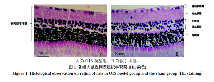
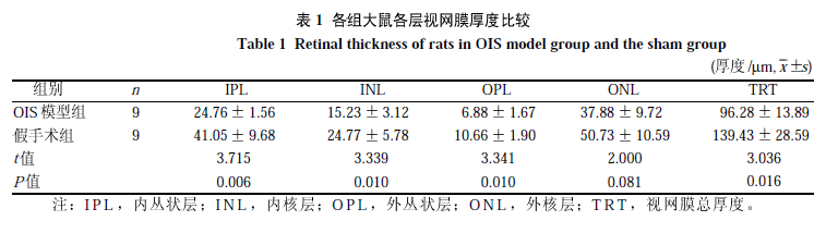
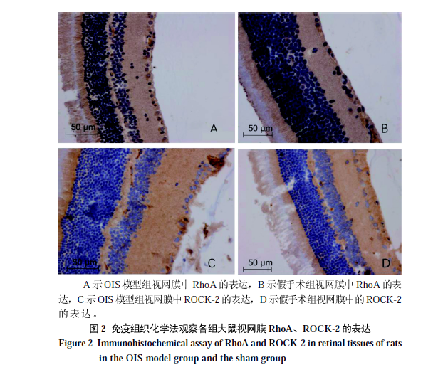
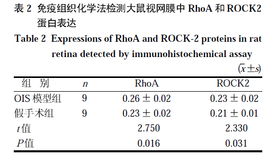
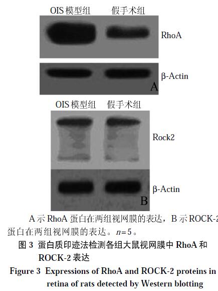
Abstract: Objective To set up an ideal rat model for ocular ischemic syndrome (OIS), and to investigate the differences in the retinal morphology and the expressions of Ras-related monomer guanosine triphosphate kinase A (RhoA) and Rho-associated coiled coil forming protein kinase-2 (ROCK-2) in rat retina. Methods Twenty BN rats were randomly divided into two groups: the sham group and OIS model group (by bilateral carotid artery occlusion, BCCAO). Overall retinal thickness and retinal cell density were evaluated by histological analysis three months after modeling, respectively. The protein expressions of RhoA and ROCK-2 were also examined by immunohistochemical staining and Western blotting. Results Compared with the sham group, the number of retinal ganglion cells (RGC) was diminished in the model group (P<0.01). The significant decrease of thickness was found in the total retina, inner plexiform layer (IPL), inner nuclear layer (INL) and outer plexiform layer (OPL)(P<0.05); however, no change was evident in the outer nuclear layers (ONL) (P>0.05). The expressions of RhoA and ROCK-2 in the model group were significantly increased as compared with the sham group by immunohistochemical staining and Western blotting analysis (the former P<0.01, the latter P<0.05). Conclusion RhoA and ROCK-2 are significantly expressed in the OIS model group, which maybe provide new ideas for the treatment of this disease.
Key words: Ocular ischemic syndrome, Rat models, RhoA, ROCK-2
CLC Number:
R73-3
Q95-33
AN Xin, LIU Lanjing, BU Jianping. Changes of Retinal Morphology and Expressions of RhoA and ROCK-2 in Ocular Ischemic Syndrome Rats[J]. Laboratory Animal and Comparative Medicine, 2020, 40(5): 397-.
0 / / Recommend
Add to citation manager EndNote|Ris|BibTeX
URL: https://www.slarc.org.cn/dwyx/EN/10.3969/j.issn.1674-5817.2020.05.006
https://www.slarc.org.cn/dwyx/EN/Y2020/V40/I5/397