
Laboratory Animal and Comparative Medicine ›› 2026, Vol. 46 ›› Issue (4): 487-497.DOI: 10.12300/j.issn.1674-5817.2025.180
• Animal Models of Human Diseases • Previous Articles
LEI Jianjun, SUO Chenhao, ZHANG He( )
)
Received:2025-11-05
Revised:2026-01-23
Online:2026-08-25
Published:2026-08-22
Correspondence to:
ZHANG He
CLC Number:
LEI Jianjun,SUO Chenhao,ZHANG He. Resveratrol Alleviates Blood‑Brain Barrier Damage in Cerebral Ischemia‑Reperfusion Rat Models through Modulation of the CCL3/CCR5 Signaling Pathway[J]. Laboratory Animal and Comparative Medicine, 2026, 46(4): 487-497. DOI: 10.12300/j.issn.1674-5817.2025.180.
Add to citation manager EndNote|Ris|BibTeX
URL: https://www.slarc.org.cn/dwyx/EN/10.12300/j.issn.1674-5817.2025.180
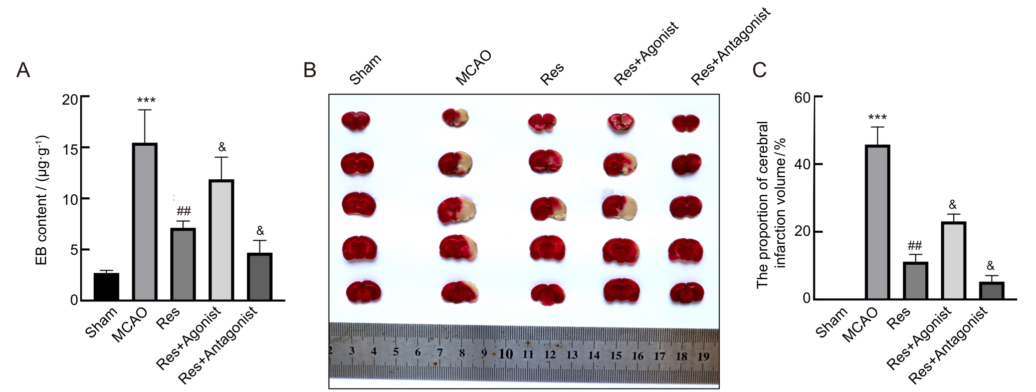
Figure 1 Effects of resveratrol on brain injury in middle cerebral artery occlusion rats
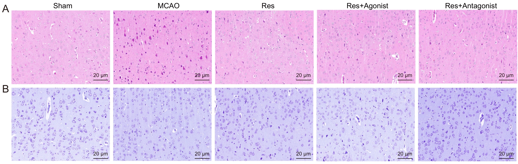
Figure 2 Effects of resveratrol on neuronal damage in the brain tissue of middle cerebral artery occlusion rats
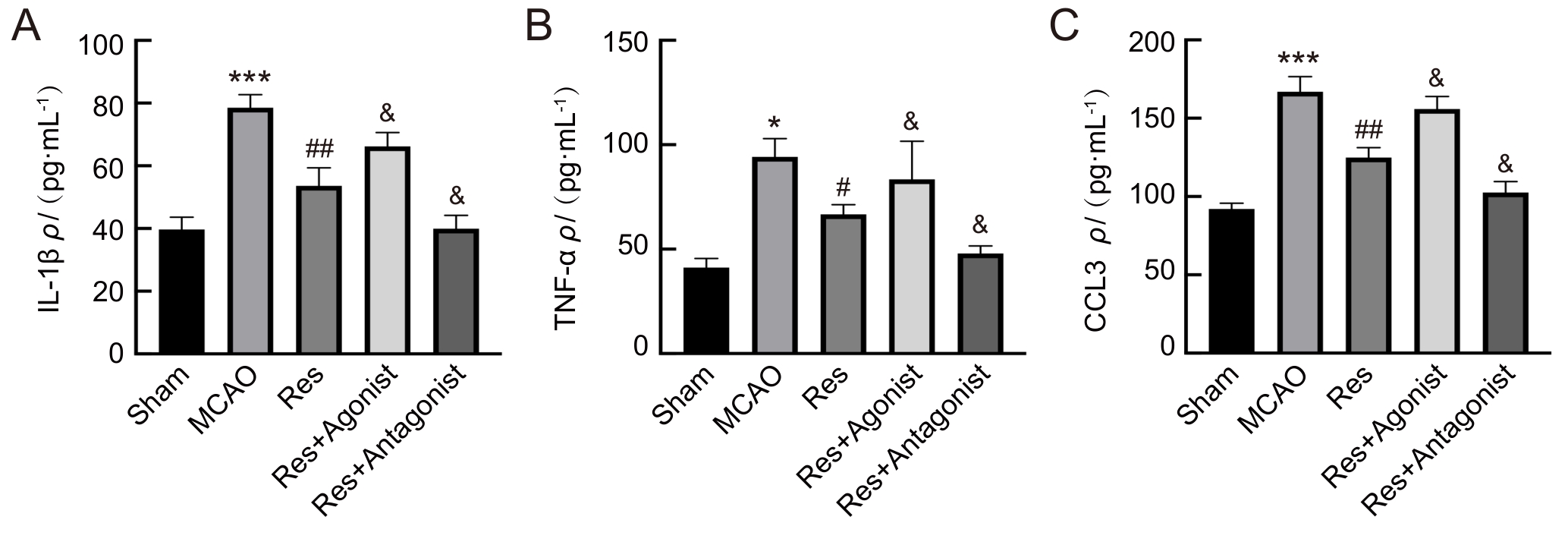
Figure 3 Effects of resveratrol on pro-inflammatory cytokines in peripheral blood of rats with middle cerebral artery occlusion
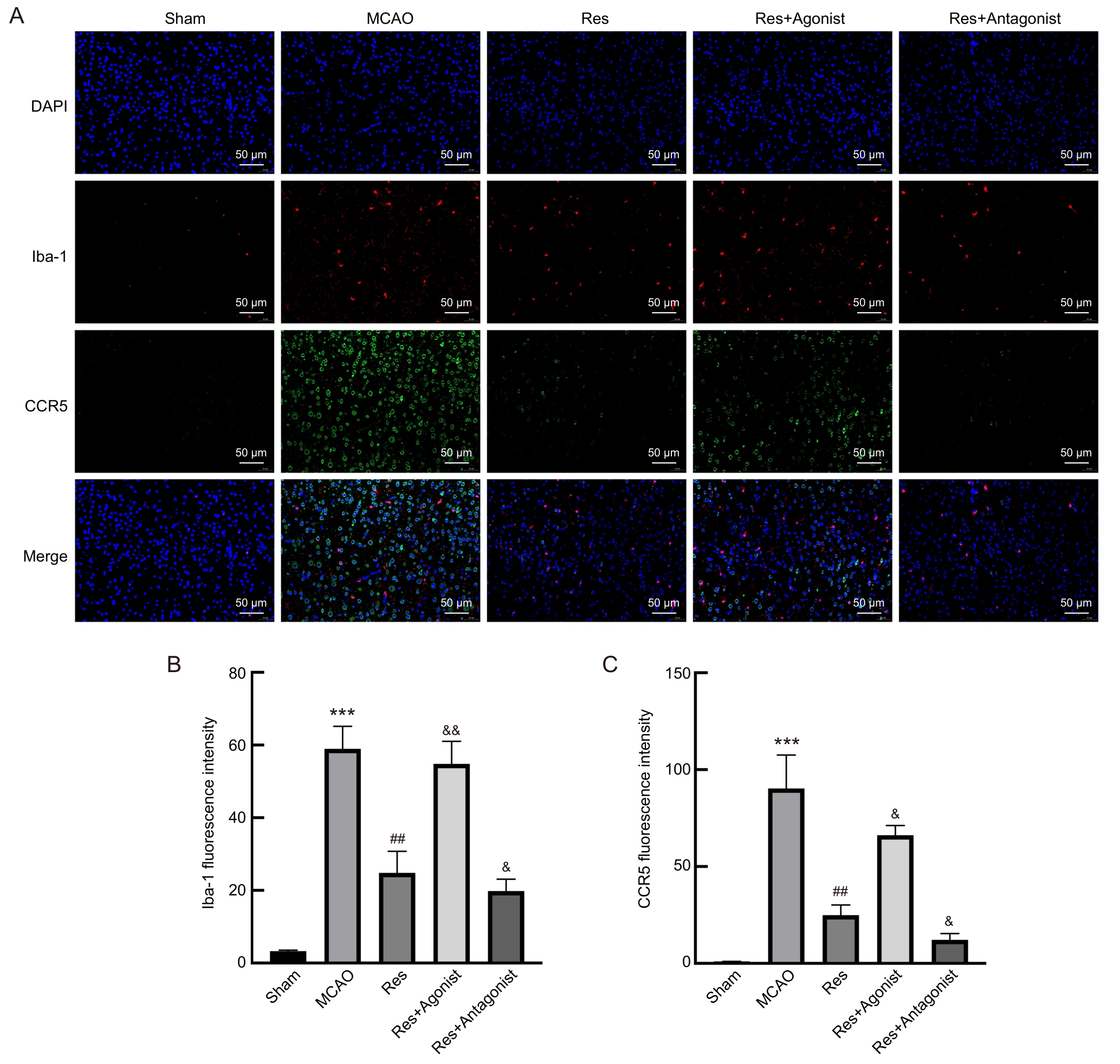
Figure 4 Effects of resveratrol on microglial activation in rats with middle cerebral artery occlusion
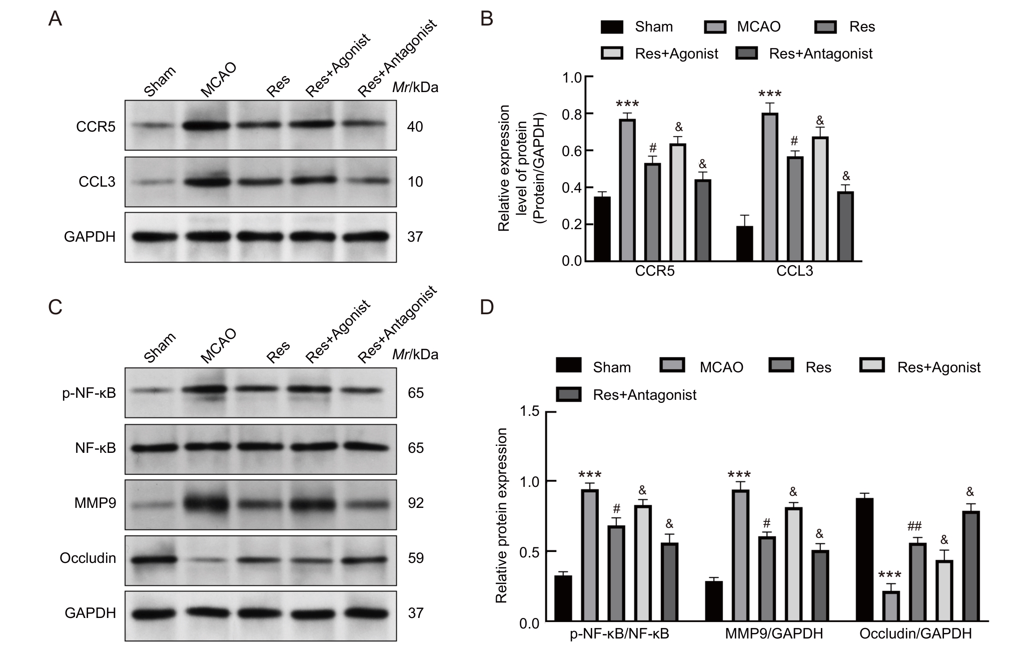
Figure 5 Effects of resveratrol treatment on CCL3/CCR5 signaling pathway and blood-brain barrier-related proteins in brain tissues of rats with middle cerebral artery occlusion
| [1] | 刘佳, 叶岩荣, 沈赟, 等. 银杏内酯B通过调控脑内T细胞特性及与胶质细胞间相互作用促进缺血性脑卒中小鼠的神经功能恢复[J]. 实验动物与比较医学, 2024, 44(2): 139-148. DOI: 10.12300/j.issn.1674-5817.2023.121 . |
| LIU J, YE Y R, SHEN Y, et al. Ginkgolide B promotes neural function recovery of ischemic stroke mice by regulating characteristics of brain T cells and their interactions with glial cells[J]. Lab Anim Comp Med, 2024, 44(2): 139-148. DOI: 10.12300/j.issn.1674-5817.2023.121 . | |
| [2] | RAFFAELE S, CLAUSEN B H, MANNELLA F C, et al. Characterisation of GPR17-expressing oligodendrocyte precursors in human ischaemic lesions and correlation with reactive glial responses[J]. J Pathol, 2025, 265(2): 226-243. DOI: 10.1002/path.6381 . |
| [3] | ALIDADI M, OMIDI N, ABDI M, et al. Melatonin ameliorates astrogliosis and microgliosis in a cuprizone demyelinating mouse model[J]. Biochem Biophys Rep, 2025, 41: 101929. DOI: 10.1016/j.bbrep.2025.101929 . |
| [4] | LIU X, TAO X W, XIONG Z, et al. Mechanism of HDAC2 regulating Nrf2 acetylation level in neuronal ferroptosis of neonatal rats with hypoxic-ischemic brain injury[J]. Brain Inj, 2025, 39(8): 635-645. DOI: 10.1080/02699052.2025.2468309 . |
| [5] | SHI K B, LI H D, CHANG T, et al. Bone marrow hematopoiesis drives multiple sclerosis progression[J]. Cell, 2022, 185(13): 2234-2247.e17. DOI: 10.1016/j.cell.2022.05.020 . |
| [6] | KNOX E G, ABURTO M R, CLARKE G, et al. The blood-brain barrier in aging and neurodegeneration[J]. Mol Psychiatry, 2022, 27(6): 2659-2673. DOI: 10.1038/s41380-022-01511-z . |
| [7] | AKINLAJA Y O, NISHIYAMA A. Glial modulation of synapse development and plasticity: oligodendrocyte precursor cells as a new player in the synaptic quintet[J]. Front Cell Dev Biol, 2024, 12: 1418100. DOI: 10.3389/fcell.2024.1418100 . |
| [8] | HUNTEMER-SILVEIRA A, PATIL N, BRICKNER M A, et al. Strategies for oligodendrocyte and myelin repair in traumatic CNS injury[J]. Front Cell Neurosci, 2021, 14: 619707. DOI: 10.3389/fncel.2020.619707 . |
| [9] | WANG F, YE Z H, YIN X J, et al. Electroacupuncture at 5/100 Hz alleviates neuropathic pain in rats by inhibiting the CCL3/CCR5 axis in the spinal cord[J]. Acupunct Med, 2025, 43(2): 95-103. DOI: 10.1177/09645284251327197 . |
| [10] | JAFARI M, DAMANI R. Blood pressure variability and outcome after acute intracerebral hemorrhage[J]. J Neurol Sci, 2020, 413: 116766. DOI: 10.1016/j.jns.2020.116766 . |
| [11] | BAI M L, SUN R Z, CAO B, et al. Monocyte-related cytokines/chemokines in cerebral ischemic stroke[J]. CNS Neurosci Ther, 2023, 29(12): 3693-3712. DOI: 10.1111/cns.14368 . |
| [12] | ESCOBAR-PESO A, MARTÍNEZ-ALONSO E, MASJUAN J, et al. Development of pharmacological strategies with therapeutic potential in ischemic stroke[J]. Antioxidants, 2023, 12(12): 2102. DOI: 10.3390/antiox12122102 . |
| [13] | 牟凌梅, 李雪梅, 文君. 白藜芦醇调节TREM-1表达减轻大鼠脑缺血再灌注损伤后细胞焦亡[J]. 四川医学, 2025, 46(6): 628-634. DOI: 10.16252/j.cnki.issn1004-0501-2025.06.006 . |
| MOU L M, LI X M, WEN J. Resveratrol regulates TREM-1 expression to alleviate cell pyroptosis after cerebral ischemia-reperfusion injury in rats[J]. Sichuan Med J, 2025, 46(6): 628-634. DOI: 10.16252/j.cnki.issn1004-0501-2025.06.006 . | |
| [14] | 张小良, 高赛红, 杨迎春, 等. 白藜芦醇激活自噬抑制焦亡减轻脑缺血再灌注损伤[J]. 中国临床药理学与治疗学, 2024, 29(11): 1240-1248. DOI: 10.12092/j.issn.1009-2501.2024.11.005 . |
| ZHANG X L, GAO S H, YANG Y C, et al. Resveratrol activates autophagy and inhibits pyroptosis to alleviate cerebral ischemia-reperfusion injury[J]. Chin J Clin Pharmacol Ther, 2024, 29(11): 1240-1248. DOI: 10.12092/j.issn.1009-2501.2024.11.005 . | |
| [15] | AN Y, SU G, CHEN W, et al. Research progress on the mechanisms of microglial extracellular vesicles affecting the prognosis of ischemic stroke[J]. Neurochem Int, 2025, 185: 105949. DOI: 10.1016/j.neuint.2025.105949 . |
| [16] | LÓPEZ-MORALES M A, CASTELLÓ-RUIZ M, BURGUETE M C, et al. Effect and mechanisms of resveratrol in animal models of ischemic stroke: a systematic review and Bayesian meta-analysis[J]. J Cereb Blood Flow Metab, 2023, 43(12): 2013-2028. DOI: 10.1177/0271678x231206236 . |
| [17] | WANG C X, WANG J Y, ZHU Z C, et al. Spotlight on pro-inflammatory chemokines: regulators of cellular communication in cognitive impairment[J]. Front Immunol, 2024, 15: 1421076. DOI: 10.3389/fimmu.2024.1421076 . |
| [18] | GUO Q X, DU A N, WANG J Y, et al. Integrated bioinformatic analyses reveal immune molecular markers and regulatory networks for cerebral ischemia-reperfusion[J]. Front Biosci (Landmark Ed), 2023, 28(8): 179. DOI: 10.31083/j.fbl2808179 . |
| [19] | DASDELEN M F, CAGLAYAN A B, ER S, et al. Social isolation initiated post-weaning augments ischemic brain injury by promoting pro-inflammatory responses[J]. Exp Neurol, 2024, 375: 114729. DOI: 10.1016/j.expneurol.2024.114729 . |
| [20] | FESTA B P, SIDDIQI F H, JIMENEZ-SANCHEZ M, et al. Microglial-to-neuronal CCR5 signaling regulates autophagy in neurodegeneration[J]. Neuron, 2023, 111(13): 2021-2037.e12. DOI: 10.1016/j.neuron.2023.04.006 . |
| [21] | WANG L F, LI W J, ZHANG X Y, et al. Resveratrol prevents age-related heart impairment through inhibiting the Notch/NF-κB pathway[J]. Food Sci Nutr, 2024, 12(2): 1035-1045. DOI: 10.1002/fsn3.3817 . |
| [22] | BASHEER N, MUHAMMADI M K, FREITES C L, et al. TLR4-mediated chronic neuroinflammation has no effect on tangle pathology in a tauopathy mouse model[J]. Front Aging Neurosci, 2024, 16: 1468602. DOI: 10.3389/fnagi.2024.1468602 . |
| [23] | LI W B, ZHANG Y D, YANG F, et al. Molecular mechanism by which TRPC6 regulates calcium signaling and neuroinflammation in the onset and development of ischemic stroke: a review[J]. Altern Ther Health Med, 2024, 30(8): 170-175. |
| [24] | DENG L, ZHANG J Y, CHEN S, et al. miR-671-5p upregulation attenuates blood–brain barrier disruption in the ischemia stroke model via the NF-кB/MMP-9 signaling pathway[J]. Mol Neurobiol, 2023, 60(7): 3824-3838. DOI: 10.1007/s12035-023-03318-7 . |
| [25] | DENG H J, XUE P F, ZHOU X G, et al. CCL4/CCR5 regulates chondrocyte biology and OA progression[J]. Cytokine, 2024, 183: 156746. DOI: 10.1016/j.cyto.2024.156746 . |
| [1] | TANG Jianping, ZHAO Liya, ZHAO Ying. Screening and Analysis of Microsatellite Genetic Markers in Commonly Used Inbred Rat Strains [J]. Laboratory Animal and Comparative Medicine, 2026, 46(3): 388-396. |
| [2] | LI Jiafei, ZHANG Zhenhao, WANG Shuo, TIAN Ge, WEN Shuang, YAN Yuxue, CUI Ran, YE Zhen, CUI Yongchun. Effects of Autonomic Neuromodulators on Atrial Electrical Remodeling and Histopathological Changes in a Rat Model of Atrial Fibrillation [J]. Laboratory Animal and Comparative Medicine, 2026, 46(3): 321-331. |
| [3] | AI Xiufeng, ZHANG Lizong, FANG Mingsun, LÜ Dongying, CHEN Chu, CAI Zhaowei, WANG Dejun. Analysis of Differences in the Intestinal Flora of Rats and Mice after Drinking Chlorinated Water Based on 16S rRNA Sequencing [J]. Laboratory Animal and Comparative Medicine, 2026, 46(3): 437-445. |
| [4] | TANG Xiaohang, GU Yingmin, LÜ Yangyang, HUANG Mingshu, TIAN Xuesong. Evaluation of the Histological Staining Performance of Rat Eyeball Sections Prepared Using a Self-Developed Fixative [J]. Laboratory Animal and Comparative Medicine, 2026, 46(2): 261-270. |
| [5] | SONG Jing, YANG Zongtong, LI Xiaojing, LI Zifa, SU Fengyun, XU Dongchuan, SUI Zaiyun. Effects of Xiebai San on the Morphological Structures of Lung and Intestinal Tissues and Expression Levels of PI3K and Akt in Rats with Allergic Asthma [J]. Laboratory Animal and Comparative Medicine, 2026, 46(2): 191-204. |
| [6] | JIANG Haitao, YUAN Hantao, HUANG Wenting, YANG Rongrong, CHEN Xiaochun, YU Baoqing, LI Sibo. Regulation of Rat Intervertebral Disc Annulus Fibrosus Cell Proliferation and Apoptosis by Yaoshu Zhuyu Fang via miR-17-5P/MDM2/p53 Pathway [J]. Laboratory Animal and Comparative Medicine, 2026, 46(1): 55-65. |
| [7] | XU Yingtao, WANG Mengmeng, LIN Ping, CHI Haitao, WANG Yi, BAI Ying. Exosomes Treat Ischemic Stroke by Regulation of Ferroptosis Through the NRF2/SLC7A11/GPX4 Pathway in Mice [J]. Laboratory Animal and Comparative Medicine, 2026, 46(1): 20-31. |
| [8] | YANG Lin, JIN Meng, WU Hanqing, LI Shun, ZHOU Xiaohui. Establishment and Preliminary Analysis of an AG6 Mouse Encephalopathy Model Induced by Vaccinia Virus Tiantan Strain Infection [J]. Laboratory Animal and Comparative Medicine, 2026, 46(1): 3-10. |
| [9] | GAO Chaoqi, ZHU Zhibo, SUN Xiandong. Application Progress and Classification Analysis of Rat Vascular Remodeling Models [J]. Laboratory Animal and Comparative Medicine, 2025, 45(5): 542-550. |
| [10] | LUO Yifan, ZHANG Zhenwei, MEI Lu, SHI Yeping, XING Yitong, ZHANG Zeqi, LI Chuxin, HAN Chunxia, YANG Pingshun, CHEN Qiusheng. Telocytes-Mediated Effects and Mechanisms of Anointing and Massage Therapy Using Oligopeptide-Herbal Medicine Composite Against Obesity in Rats [J]. Laboratory Animal and Comparative Medicine, 2025, 45(5): 551-560. |
| [11] | QIN Chao, LI Shuangxing, ZHAO Tingting, JIANG Chenchen, ZHAO Jing, YANG Yanwei, LIN Zhi, WANG Sanlong, WEN Hairuo. Study on the 90-day Feeding Experimental Background Data of SD Rats for Drug Safety Evaluation [J]. Laboratory Animal and Comparative Medicine, 2025, 45(4): 439-448. |
| [12] | LIU Liyu, JI Bo, LIU Xiaoxuan, FANG Yang, ZHANG Ling, GUO Tingting, QUAN Ye, LI Hewen, LIU Yitian. Exploration of Rat Fetal Lung Tissue Fixation Methods [J]. Laboratory Animal and Comparative Medicine, 2025, 45(4): 432-438. |
| [13] | PAN Yicong, JIANG Wenhong, HU Ming, QIN Xiao. Optimization of Surgical Procedure and Efficacy Evaluation of Aortic Calcification Model in Rats with Chronic Kidney Disease [J]. Laboratory Animal and Comparative Medicine, 2025, 45(3): 279-289. |
| [14] | LIU Zhiwei, YANG Ran, LIAN Hao, ZHANG Yu, JIN Lilun. Cartilage Protection and Anti-Inflammatory Effects of Fraxetin on Monosodium Iodoacetate-Induced Rat Model of Osteoarthritis [J]. Laboratory Animal and Comparative Medicine, 2025, 45(3): 259-268. |
| [15] | JIANG Meng, HAO Shulan, TONG Liguo, ZHONG Qiming, GAO Zhenfei, WANG Yonghui, WANG Xixing, JI Haijie. Dynamic Evaluation of Vinorelbine-Induced Phlebitis of Dorsalis Pedis Vein in a Rat Model [J]. Laboratory Animal and Comparative Medicine, 2025, 45(3): 251-258. |
| Viewed | ||||||
|
Full text |
|
|||||
|
Abstract |
|
|||||
