



Laboratory Animal and Comparative Medicine
• XXXX XXXX •
LI Limei1,2( ), HUANG Xueping1,2, CHEN Jianqing1,2, HE Xiukui2,3(
 )()
)()
Online:2026-07-28
Correspondence to:
HE Xiukui
CLC Number:
LI Limei,HUANG Xueping,CHEN Jianqing,et al. Comparison of Five Methods for Induction of Model Rat with Diminished Ovarian Reserve[J]. Laboratory Animal and Comparative Medicine. DOI: 10.12300/j.issn.1674-5817.2025.205.
Add to citation manager EndNote|Ris|BibTeX
URL: https://www.slarc.org.cn/dwyx/EN/10.12300/j.issn.1674-5817.2025.205
组别 Group | 动情周期 Estrous cycle | χ2 值 χ2 value | P值 P value | ||
|---|---|---|---|---|---|
正常 Regular | 紊乱 Dsorder | 延长 Delayed | |||
| 对照组 Control group | 6(100%) | 0(0%) | 0(0%) | ||
| CTX单次组 Single-dose CTX group | 2(33.3%) | 3(50.0%) | 1(16.7%) | 6.00 | 0.05 |
| CTX两次组 Double-dose CTX group | 2(33.3%) | 3(50.0%) | 1(16.7%) | 6.00 | 0.05 |
| CTX多次组 Multiple-dose CTX group | 1(16.7%) | 5(83.3%) | 0(0%) | 8.57 | 0.003* |
| 雷公藤组 Tripterygium wilfordii group | 1(16.7%) | 4(66.7%) | 1(16.7%) | 8.57 | 0.014* |
| 制动+LTS组 Restraint stress +LTS group | 2(33.3%) | 3(50.0%) | 1(16.7%) | 6.00 | 0.05 |
| 围绝经期组 Perimenopausal group | 1(16.7%) | 3(50.0%) | 2(33.3%) | 8.57 | 0.014* |
Table 1 Changes of estrous cycle of rats in each group
组别 Group | 动情周期 Estrous cycle | χ2 值 χ2 value | P值 P value | ||
|---|---|---|---|---|---|
正常 Regular | 紊乱 Dsorder | 延长 Delayed | |||
| 对照组 Control group | 6(100%) | 0(0%) | 0(0%) | ||
| CTX单次组 Single-dose CTX group | 2(33.3%) | 3(50.0%) | 1(16.7%) | 6.00 | 0.05 |
| CTX两次组 Double-dose CTX group | 2(33.3%) | 3(50.0%) | 1(16.7%) | 6.00 | 0.05 |
| CTX多次组 Multiple-dose CTX group | 1(16.7%) | 5(83.3%) | 0(0%) | 8.57 | 0.003* |
| 雷公藤组 Tripterygium wilfordii group | 1(16.7%) | 4(66.7%) | 1(16.7%) | 8.57 | 0.014* |
| 制动+LTS组 Restraint stress +LTS group | 2(33.3%) | 3(50.0%) | 1(16.7%) | 6.00 | 0.05 |
| 围绝经期组 Perimenopausal group | 1(16.7%) | 3(50.0%) | 2(33.3%) | 8.57 | 0.014* |
组别 Group | 体重增长率/% Growth rate of body mass/% | ||
|---|---|---|---|
第1周 Week 1 | 第2周 Week 2 | 第2周-第1周 Week 2-week 1 | |
| 对照组 Control group | 9.58±2.67#●○♥ | 19.49±5.79#●○♥♣ | 9.92±7.62#●○♥♣ |
| CTX单次组 Single-dose CTX group | 8.07±3.22#●○♥ | 17.85±1.85#○♥♣ | 9.78±3.84#●○♥♣ |
| CTX两次组 Double-dose CTX group | 14.34±4.31*#♠ | 9.10±3.50*#♠♣ | -5.24±6.69*♠ |
| CTX多次组 Multiple-dose CTX group | 10.24±4.45#●○ | 1.44±6.72*●○♠♥ | -8.80±5.05*#♠ |
| 雷公藤组 Tripterygium wilfordii group | 16.86±3.60*#♠♣ | 9.00±6.65*#♠♣ | -7.86±6.97*♠ |
| 制动+LTS组 Restraint stress +LTS group | 17.71±5.02*#♠♣ | 12.91±2.88*#♣ | -4.80±7.11*♠ |
| 围绝经期组 Perimenopausal group | 1.76±2.23*●○♠♥♣ | 0.12±0.58*●○♠♥ | -1.64±2.18*♠♣ |
| F值 F value | 13.207 | 15.820 | 10.655 |
| P值 P value | 0.000 | 0.000 | 0.000 |
Table 2 Comparison of weekly body weight growth rate in each group
组别 Group | 体重增长率/% Growth rate of body mass/% | ||
|---|---|---|---|
第1周 Week 1 | 第2周 Week 2 | 第2周-第1周 Week 2-week 1 | |
| 对照组 Control group | 9.58±2.67#●○♥ | 19.49±5.79#●○♥♣ | 9.92±7.62#●○♥♣ |
| CTX单次组 Single-dose CTX group | 8.07±3.22#●○♥ | 17.85±1.85#○♥♣ | 9.78±3.84#●○♥♣ |
| CTX两次组 Double-dose CTX group | 14.34±4.31*#♠ | 9.10±3.50*#♠♣ | -5.24±6.69*♠ |
| CTX多次组 Multiple-dose CTX group | 10.24±4.45#●○ | 1.44±6.72*●○♠♥ | -8.80±5.05*#♠ |
| 雷公藤组 Tripterygium wilfordii group | 16.86±3.60*#♠♣ | 9.00±6.65*#♠♣ | -7.86±6.97*♠ |
| 制动+LTS组 Restraint stress +LTS group | 17.71±5.02*#♠♣ | 12.91±2.88*#♣ | -4.80±7.11*♠ |
| 围绝经期组 Perimenopausal group | 1.76±2.23*●○♠♥♣ | 0.12±0.58*●○♠♥ | -1.64±2.18*♠♣ |
| F值 F value | 13.207 | 15.820 | 10.655 |
| P值 P value | 0.000 | 0.000 | 0.000 |
组别 Group | 卵巢重量/mg Ovarian weight/mg | 子宫重量/mg Uterine weight/mg | 卵巢指数/%(mg/g) Ovarian index/%(mg/g) | 子宫指数/%(mg/g) Uterine index/%(mg/g) |
|---|---|---|---|---|
| 对照组Control group | 122.68±5.09 | 412.87±50.75 | 56.71±2.48# | 190.01±13.32 |
| CTX单次组Single-dose CTX group | 124.35±13.07 | 408.87±75.22# | 59.19±5.95# | 195.13±39.13 |
| CTX两次组Double-dose CTX group | 125.35±10.53 | 422.52±110.21 | 61.85±5.18# | 208.29±53.33 |
| CTX多次组Multiple-dose CTX group | 112.47±14.05 | 399.1±114.50# | 62.16±6.05# | 219.57±55.00 |
| 雷公藤组Tripterygium wilfordii group | 118.67±15.37 | 358.83±36.25# | 57.30±7.99# | 173.65±22.36 |
| 制动+LTS组Restraint stress +LTS group | 132.48±15.34 | 387.55±58.68# | 60.59±6.00# | 177.01±22.03 |
| 围绝经期组Perimenopausal group | 123.27±29.16 | 703.03±150.86 | 27.58±6.46*●○♠♥♣ | 157.04±30.99 |
| F值F value | 0.869 | 9.430 | 25.748 | 2.018 |
| P值P value | 0.527 | 0.000 | 0.000 | 0.089 |
Table 3 Comparison of ovary and uterus weight,ovarian and uterine index in each group
组别 Group | 卵巢重量/mg Ovarian weight/mg | 子宫重量/mg Uterine weight/mg | 卵巢指数/%(mg/g) Ovarian index/%(mg/g) | 子宫指数/%(mg/g) Uterine index/%(mg/g) |
|---|---|---|---|---|
| 对照组Control group | 122.68±5.09 | 412.87±50.75 | 56.71±2.48# | 190.01±13.32 |
| CTX单次组Single-dose CTX group | 124.35±13.07 | 408.87±75.22# | 59.19±5.95# | 195.13±39.13 |
| CTX两次组Double-dose CTX group | 125.35±10.53 | 422.52±110.21 | 61.85±5.18# | 208.29±53.33 |
| CTX多次组Multiple-dose CTX group | 112.47±14.05 | 399.1±114.50# | 62.16±6.05# | 219.57±55.00 |
| 雷公藤组Tripterygium wilfordii group | 118.67±15.37 | 358.83±36.25# | 57.30±7.99# | 173.65±22.36 |
| 制动+LTS组Restraint stress +LTS group | 132.48±15.34 | 387.55±58.68# | 60.59±6.00# | 177.01±22.03 |
| 围绝经期组Perimenopausal group | 123.27±29.16 | 703.03±150.86 | 27.58±6.46*●○♠♥♣ | 157.04±30.99 |
| F值F value | 0.869 | 9.430 | 25.748 | 2.018 |
| P值P value | 0.527 | 0.000 | 0.000 | 0.089 |
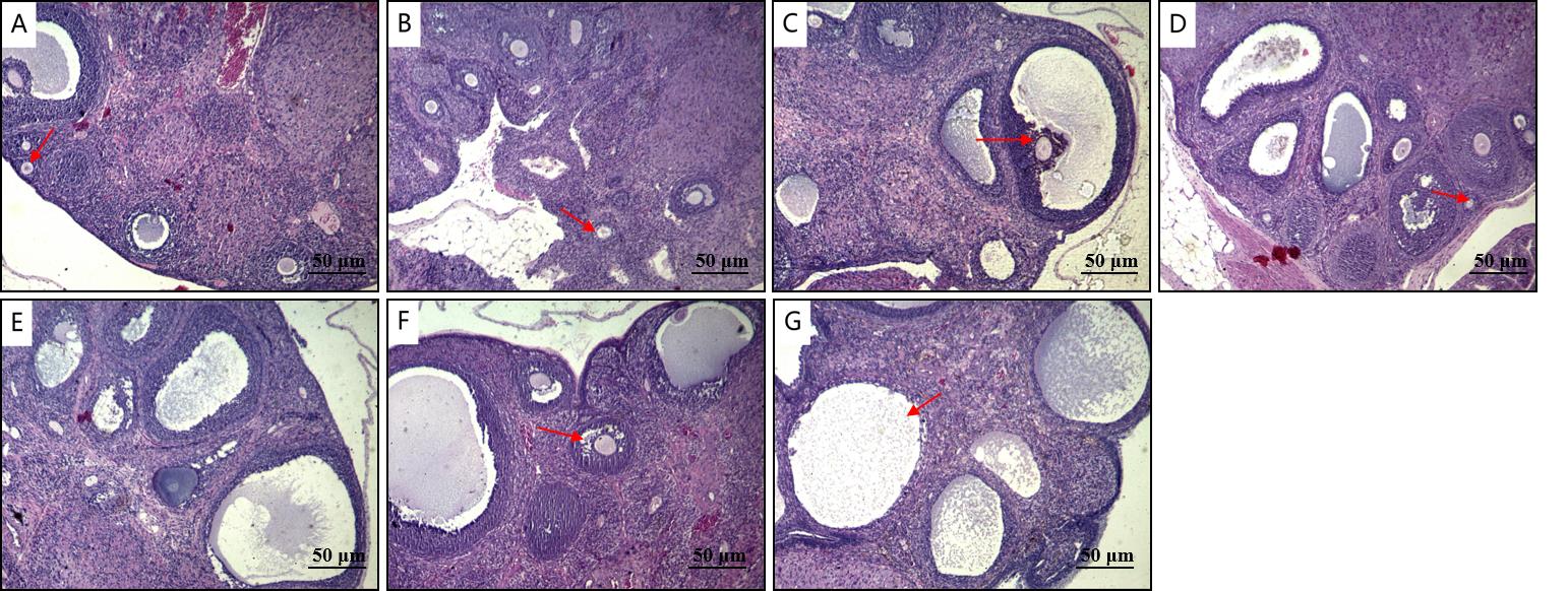
Figure 1 HE staining of rat ovary (×100) : A. control group B. single-dose CTX group C. double-dose CTX group D. multiple-dose CTX group E. tripterygium wilfordii group F. restraint stress +LTS group G. perimenopausal group
组别 group | 原始卵泡数 Number of primordial follicles | 初级卵泡数 Number of primary follicles | 次级卵泡数 Number of secondary follicle | 成熟卵泡数 Number of mature follicles | 闭锁卵泡数 Number of atretic follicles |
|---|---|---|---|---|---|
对照组 Control group | 10.67±0.58#○♠♥♣ | 8.33±0.58a | 8.33±1.16abceff | 1.67±1.16 | 3.67±1.16#●○♠♥♣ |
CTX单次组 Single-dose CTX group | 5.67±1.53*# | 11.33±0.58n | 13.67±2.31*# | 0.33±0.58 | 7.67±1.16*●○♣ |
CTX两次组 Double-dose CTX group | 4.67±0.58* | 11.33±3.22 | 14.00±4.36*# | 0.33±0.58 | 9.67±1.16*●○ |
CTX多次组 Multiple-dose CTX group | 6.33±0.58*# | 10.33±1.16 | 14.00±3.61*# | 0.67±1.16 | 12.00±1.73*#♠ |
雷公藤组 Tripterygium wilfordii group | 6.33±0.58*# | 13.33±5.03 | 13.00±1.73# | 2.00±0.00 | 13.00±1.73*#♠♥ |
制动+LTS组 Restraint stress +LTS group | 6.33±1.16*# | 11.00±1.73 | 14.67±2.31*# | 0.33±0.58 | 13.33±0.58*#♠♥ |
围绝经期组 Perimenopausal group | 3.00±1.73*●○♠♣ | 6.33±4.93 | 1.00±1.73*●○♠♥♣ | 0.00±0.00 | 8.67±1.53*●○♣ |
F值 F value | 14.375 | 1.692 | 10.449 | 3.333 | 19.614 |
P值 P value | 0.000 | 0.195 | 0.000 | 0.030 | 0.000 |
Table 4 Comparison of ovarian follicle count at all levels in each group
组别 group | 原始卵泡数 Number of primordial follicles | 初级卵泡数 Number of primary follicles | 次级卵泡数 Number of secondary follicle | 成熟卵泡数 Number of mature follicles | 闭锁卵泡数 Number of atretic follicles |
|---|---|---|---|---|---|
对照组 Control group | 10.67±0.58#○♠♥♣ | 8.33±0.58a | 8.33±1.16abceff | 1.67±1.16 | 3.67±1.16#●○♠♥♣ |
CTX单次组 Single-dose CTX group | 5.67±1.53*# | 11.33±0.58n | 13.67±2.31*# | 0.33±0.58 | 7.67±1.16*●○♣ |
CTX两次组 Double-dose CTX group | 4.67±0.58* | 11.33±3.22 | 14.00±4.36*# | 0.33±0.58 | 9.67±1.16*●○ |
CTX多次组 Multiple-dose CTX group | 6.33±0.58*# | 10.33±1.16 | 14.00±3.61*# | 0.67±1.16 | 12.00±1.73*#♠ |
雷公藤组 Tripterygium wilfordii group | 6.33±0.58*# | 13.33±5.03 | 13.00±1.73# | 2.00±0.00 | 13.00±1.73*#♠♥ |
制动+LTS组 Restraint stress +LTS group | 6.33±1.16*# | 11.00±1.73 | 14.67±2.31*# | 0.33±0.58 | 13.33±0.58*#♠♥ |
围绝经期组 Perimenopausal group | 3.00±1.73*●○♠♣ | 6.33±4.93 | 1.00±1.73*●○♠♥♣ | 0.00±0.00 | 8.67±1.53*●○♣ |
F值 F value | 14.375 | 1.692 | 10.449 | 3.333 | 19.614 |
P值 P value | 0.000 | 0.195 | 0.000 | 0.030 | 0.000 |
组别 Group | 雌二醇ρ/(pg·mL-1) E2ρ/(pg·mL-1) | 黄体生成素ρ/(pg·mL-1) LHρ/(pg·mL-1) | 促卵泡激素ρ/(pg·mL-1) FSHρ/(pg·mL-1) | 抗苗勒氏管激素ρ/(ng·mL-1) AMHρ/(ng·mL-1) |
|---|---|---|---|---|
| 对照组 Control group | 310.02±36.80# | 353.33±64.79#●♣ | 10.11±0.83○♥ | 1.16±0.32#●♠ |
| CTX单次组 Single-dose CTX group | 304.40±28.63# | 473.64±101.95#● | 11.35±4.80 | 1.65±0.58*○♥♣ |
| CTX两次组 Double-dose CTX group | 321.28±36.98# | 448.60±103.37#● | 7.08±0.97* | 0.97±0.35#●♠ |
| CTX多次组 Multiple-dose CTX group | 325.62±25.30# | 508.77±130.94* | 8.18±1.19 | 0.90±0.47#●♠ |
| 雷公藤组 Tripterygium wilfordii group | 310.43±36.89# | 499.03±122.67 | 8.24±0.83* | 1.13±0.31#●♠ |
| 制动+LTS组 Restraint stress +LTS group | 271.99±42.61♥♣ | 631.27±200.25*♠♥ | 8.78±1.62 | 1.92±0.43* |
| 围绝经期组 Perimenopausal group | 250.93±30.33*○♠♥♣ | 634.50±149.51*♠♥ | 8.73±1.93 | 1.81±0.34* |
| F值 F value | 3.827 | 2.484 | 3.497 | 6.232 |
| P值 P value | 0.005 | 0.042 | 0.008 | 0.000 |
Table 5 Comparison of serum FSH, LH, E2 and AMH in each group
组别 Group | 雌二醇ρ/(pg·mL-1) E2ρ/(pg·mL-1) | 黄体生成素ρ/(pg·mL-1) LHρ/(pg·mL-1) | 促卵泡激素ρ/(pg·mL-1) FSHρ/(pg·mL-1) | 抗苗勒氏管激素ρ/(ng·mL-1) AMHρ/(ng·mL-1) |
|---|---|---|---|---|
| 对照组 Control group | 310.02±36.80# | 353.33±64.79#●♣ | 10.11±0.83○♥ | 1.16±0.32#●♠ |
| CTX单次组 Single-dose CTX group | 304.40±28.63# | 473.64±101.95#● | 11.35±4.80 | 1.65±0.58*○♥♣ |
| CTX两次组 Double-dose CTX group | 321.28±36.98# | 448.60±103.37#● | 7.08±0.97* | 0.97±0.35#●♠ |
| CTX多次组 Multiple-dose CTX group | 325.62±25.30# | 508.77±130.94* | 8.18±1.19 | 0.90±0.47#●♠ |
| 雷公藤组 Tripterygium wilfordii group | 310.43±36.89# | 499.03±122.67 | 8.24±0.83* | 1.13±0.31#●♠ |
| 制动+LTS组 Restraint stress +LTS group | 271.99±42.61♥♣ | 631.27±200.25*♠♥ | 8.78±1.62 | 1.92±0.43* |
| 围绝经期组 Perimenopausal group | 250.93±30.33*○♠♥♣ | 634.50±149.51*♠♥ | 8.73±1.93 | 1.81±0.34* |
| F值 F value | 3.827 | 2.484 | 3.497 | 6.232 |
| P值 P value | 0.005 | 0.042 | 0.008 | 0.000 |
| [1] | 卵巢储备功能减退临床诊治专家共识专家组, 中华预防医学会生育力保护分会生殖内分泌生育保护学组. 卵巢储备功能减退临床诊治专家共识[J]. 生殖医学杂志, 2022, 31(4): 425-434. |
| Expert Group of Consensus on Clinical Diagnosis & Management of Diminished Ovarian Reserve, Reproductive Endocrinology & Fertility Preservation Section of Chinese Society on Fertility Preservation under Chinese Preventive Medicine Association. Consensus on clinical diagnosis and management of diminished ovarian reserve[J]. J Reprod Med, 2022, 31(4): 425-434. | |
| [2] | 黄慧, 邓亚胜, 梁天薇, 等. 卵巢储备功能减退动物模型的造模方法评价与分析[J]. 实验动物与比较医学, 2023, 43(4): 422-428. DOI: 10.12300/j.issn.1674-5817.2023.032 . |
| HUANG H, DENG Y S, LIANG T W, et al. Evaluation and analysis of modeling methods for animal models with diminished ovarian reserve[J]. Lab Anim Comp Med, 2023, 43(4): 422-428. DOI: 10.12300/j.issn.1674-5817.2023.032 . | |
| [3] | 李丽美, 陶莉莉, 和秀魁, 等. 环磷酰胺建立SD大鼠卵巢功能减退模型的研究[J]. 重庆医学, 2019, 48(13): 2180-2183, 2188. DOI: 10.3969/j.issn.1671-8348.2019.13.005 . |
| LI L M, TAO L L, HE X K, et al. Study on cyclophosphamide for constructing SD rat diminished ovarian reserve model[J]. Chongqing Med, 2019, 48(13): 2180-2183, 2188. DOI: 10.3969/j.issn.1671-8348.2019.13.005 . | |
| [4] | 黄雪君, 孙冬梅, 陈玉兴, 等. 护卵颗粒对重复制动应激致卵巢功能减退模型大鼠的影响[J]. 世界中医药, 2016, 11(3): 502-506. DOI: 10.3969/j.issn.1673-7202.2016.03.035 . |
| HUANG X J, SUN D M, CHEN Y X, et al. Effect of hu Luan ke Li on diminished ovarian reserve rats with repeated immobilization stress[J]. World Chin Med, 2016, 11(3): 502-506. DOI: 10.3969/j.issn.1673-7202.2016.03.035 . | |
| [5] | 张子怡, 陈宝军, 张英杰, 等. 肾阳虚、肾阴虚证大鼠模型的建立与稳定性观察[J]. 福建中医药, 2015, 46(1): 51-54. DOI: 10.13260/j.cnki.jfjtcm.010856 . |
| ZHANG Z Y, CHEN B J, ZHANG Y J, et al. Establishment and stability observation of rat model with kidney-Yang deficiency and kidney-Yin deficiency syndrome[J]. Fujian J Tradit Chin Med, 2015, 46(1): 51-54. DOI: 10.13260/j.cnki.jfjtcm.010856 . | |
| [6] | 陈思充, 陈子豪, 高青华, 等. 不同阴道涂片方法的比较及对大鼠动情周期的判断[J]. 解剖科学进展, 2023, 29(1): 93-95, 102. DOI: 10.16695/j.cnki.1006-2947.2023.01.025 . |
| CHEN S C, CHEN Z H, GAO Q H, et al. Comparison of different vaginal smears and judgment of estrous cycle in rats[J]. Prog Anat Sci, 2023, 29(1): 93-95, 102. DOI: 10.16695/j.cnki.1006-2947.2023.01.025 . | |
| [7] | 范雅祺, 郑燕鹏, 周子婷, 等. 雷公藤建立卵巢早衰大鼠模型的实验研究[J]. 中国实验动物学报, 2023, 31(3): 355-364. DOI: 10.3969/j.issn.1005-4847.2023.03.010 . |
| FAN Y Q, ZHENG Y P, ZHOU Z T, et al. Establishment of a rat model of premature ovarian failure induced by Tripterygium wilfordii [J]. Acta Lab Anim Sci Sin, 2023, 31(3): 355-364. DOI: 10.3969/j.issn.1005-4847.2023.03.010 . | |
| [8] | TOURAINE P, CHABBERT-BUFFET N, PLU-BUREAU G, et al. Premature ovarian insufficiency[J]. Nat Rev Dis Primers, 2024, 10: 63. DOI: 10.1038/s41572-024-00547-5 . |
| [9] | 陈佳芳, 雷磊. 早发性卵巢功能不全动物模型评价思路[J]. 中华中医药杂志, 2023, 38(9): 4255-4258. |
| CHEN J F, LEI L. Evaluation thought of premature ovarian insufficiency animal models[J]. China J Tradit Chin Med Pharm, 2023, 38(9): 4255-4258. | |
| [10] | LE D C, NGO M T, KUO Y C, et al. Secretome from estrogen-responding human placenta-derived mesenchymal stem cells rescues ovarian function and circadian rhythm in mice with cyclophosphamide-induced primary ovarian insufficiency[J]. J Biomed Sci, 2024, 31(1): 95. DOI: 10.1186/s12929-024-01085-8 . |
| [11] | SENGUPTA P. The laboratory rat: relating its age with human's[J]. Int J Prev Med, 2013, 4(6): 624-630. |
| [12] | 陈燕霞, 袁苑, 马堃, 等. 定坤丹对雷公藤多苷诱导卵巢储备功能低下小鼠性激素和卵泡计数的影响[J]. 中国实验方剂学杂志, 2020, 26(14): 78-84. DOI: 10.13422/j.cnki.syfjx.20201339 . |
| CHEN Y X, YUAN Y, MA K, et al. Effect of dingkundan on sex hormone and follicle count in mice of Tripterygium wilfordii polyglycosides induced diminished ovarian reserve[J]. Chin J Exp Tradit Med Formulae, 2020, 26(14): 78-84. DOI: 10.13422/j.cnki.syfjx.20201339 . | |
| [13] | 苏德慧, 苏会娜, 谈诚, 等. 不同剂量环磷酰胺诱发卵巢早衰小鼠模型对衰老和凋亡的影响及机制探讨[J]. 中国妇产科临床杂志, 2025, 26(2): 129-133. DOI: 10.13390/j.issn.1672-1861.2025.02.009 . |
| SU D H, SU H N, TAN C, et al. Effects and mechanisms of cyclophosphamide-induced premature ovarian failure in mouse models: the impact of different doses on senescence and apoptosis[J]. Chin J Clin Obstet Gynecol, 2025, 26(2): 129-133. DOI: 10.13390/j.issn.1672-1861.2025.02.009 . | |
| [14] | 林华丹, 王旭, 陈春宏, 等. 三种卵巢储备功能减退小鼠模型的效果比较[J]. 中国比较医学杂志, 2025, 35(5): 51-59. DOI: 10.3969/j.issn.1671-7856.2025.05.005 . |
| LIN H D, WANG X, CHEN C H, et al. Comparison of three methods for induction of model mice with diminished ovarian reserve[J]. Chin J Comp Med, 2025, 35(5): 51-59. DOI: 10.3969/j.issn.1671-7856.2025.05.005 . | |
| [15] | GONFLONI S, JODICE C, GUSTAVINO B, et al. DNA damage stress response and follicle activation: signaling routes of mammalian ovarian reserve[J]. Int J Mol Sci, 2022, 23(22): 14379. DOI: 10.3390/ijms232214379 . |
| [16] | 贡磊磊, 王晓霞, 封学伟, 等. 不同浓度环磷酰胺诱导早发性卵巢功能不全小鼠模型及作用机制研究[J]. 实验动物与比较医学, 2025, 45(4): 403-410. DOI: 10.12300/j.issn.1674-5817.2024.194 . |
| GONG L L, WANG X X, FENG X W, et al. A mouse model and mechanism study of premature ovarian insufficiency induced by different concentrations of cyclophosphamide[J]. Lab Anim Comp Med, 2025, 45(4): 403-410. DOI: 10.12300/j.issn.1674-5817.2024.194 . | |
| [17] | STRINGER J M, ALESI L R, WINSHIP A L, et al. Beyond apoptosis: evidence of other regulated cell death pathways in the ovary throughout development and life[J]. Hum Reprod Update, 2023, 29(4): 434-456. DOI: 10.1093/humupd/dmad005 . |
| [18] | 王凤, 张会永, 李芹, 等. 肝郁证动物模型造模方法与模型评价研究进展[J]. 中华中医药杂志, 2021, 36(2): 934-939. |
| WANG F, ZHANG H Y, LI Q, et al. Research progress on animal modeling methods and model evaluation of liver depression syndrome[J]. China J Tradit Chin Med Pharm, 2021, 36(2): 934-939. | |
| [19] | 韦柳彤, 赖东梅. 慢性应激与卵巢功能减退的相关性研究进展[J]. 上海交通大学学报(医学版), 2024, 44(12): 1601-1606. DOI: 10.3969/j.issn.1674-8115.2024.12.014 . |
| WEI L T, LAI D M. Research progress in the correlation between chronic stress and ovarian dysfunction[J]. J Shanghai Jiaotong Univ Med Sci, 2024, 44(12): 1601-1606. DOI: 10.3969/j.issn.1674-8115.2024.12.014 . | |
| [20] | 丁杨, 向菁菁, 吴慧华, 等. 基础FSH/LH比值及年龄因素在高龄女性患者体外受精-胚胎移植治疗中的预测价值[J]. 实用医学杂志, 2016, 32(10): 1669-1672. DOI: 10.3969/j.issn.1006-5725.2016.10.035 . |
| DING Y, XIANG J J, WU H H, et al. Predictive value of basic FSH/LH ratio and age factors in in vitro fertilization-embryo transfer treatment for elderly female patients[J]. J Pract Med, 2016, 32(10): 1669-1672. DOI: 10.3969/j.issn.1006-5725.2016.10.035 . | |
| [21] | 尚丹丹, 刘萍, 刘利贞, 等. 卵巢抵抗综合征和早发性卵巢功能不全诊断与个体化管理进展[J]. 实用医学杂志, 2025, 41(1): 146-152. DOI: 10.3969/j.issn.1006-5725.2025.01.024 . |
| SHANG D D, LIU P, LIU L Z, et al. Diagnosis and advances in individualized management of resistant ovary syndrome and premature ovarian insufficiency[J]. J Pract Med, 2025, 41(1): 146-152. DOI: 10.3969/j.issn.1006-5725.2025.01.024 . | |
| [22] | 宗国霞, 王威, 夏宇跃, 等. 早发性卵巢功能不全患者外周血CD3+ HLA-DR细胞水平与卵巢储备功能的相关性[J]. 实用医学杂志, 2019, 35(13): 2108-2111. DOI: 10.3969/j.issn.1006-5725.2019.13.017 . |
| ZONG G X, WANG W, XIA Y Y, et al. Correlation between peripheral blood CD3+ HLA-DR cell levels and ovarian reserve function in patients with premature ovarian insufficiency[J]. J Pract Med, 2019, 35(13): 2108-2111. DOI: 10.3969/j.issn.1006-5725.2019.13.017 . |
| [1] | LI Hongman, CHEN Yutong, SHI Yingpei, WANG Yijing, PAN Yan, XU Tong, ZHOU Yi, DENG Qiyue, LIU Xue. A New Perspective on Mental Health Assessment in Laboratory Animals: Stress Response Monitoring Based on Hair Characteristics [J]. Laboratory Animal and Comparative Medicine, 2026, 46(4): 553-563. |
| [2] | MA Wenjing, LIU Yanping, LI Yifan, GUO Huan, XIE Lihui, DU Longji, MA Jiali, WANG Hongping, LI Jiamin. Practice in Validity Period Management of Disinfectants, Feed, and Bedding in Laboratory Animal Facilities under Good Laboratory Practice Management System [J]. Laboratory Animal and Comparative Medicine, 2026, 46(4): 593-603. |
| [3] | LI Zifa, LIU Shan, SUN Wei, ZHAO Feng, WEI Sheng, LI Qin, XU Kaiyong. Reflections on Building a Science-Education Integration System for Laboratory Animal Science: A Case Study of Shandong University of Traditional Chinese Medicine [J]. Laboratory Animal and Comparative Medicine, 2026, 46(4): 604-610. |
| [4] | QIANG Sujing, LI Dongting, XU Jinhua, CAI Li, WEI Siyu, ZOU Jialei, DING Yuqiang. Identification and Analysis of the Klebsiella pneumoniae Species Complex in Laboratory Rats and Mice [J]. Laboratory Animal and Comparative Medicine, 2026, 46(4): 564-572. |
| [5] | ZHAO Yulei, HAN Bingbing, WANG Xingxin, YANG Guangjian, WANG Yajie, TANG Xiaoyong. Progress in Disease-Syndrome-Integrated Mouse Models of Lung Cancer [J]. Laboratory Animal and Comparative Medicine, 2026, 46(4): 531-540. |
| [6] | LEI Jianjun, SUO Chenhao, ZHANG He. Resveratrol Alleviates Blood‑Brain Barrier Damage in Cerebral Ischemia‑Reperfusion Rat Models through Modulation of the CCL3/CCR5 Signaling Pathway [J]. Laboratory Animal and Comparative Medicine, 2026, 46(4): 487-497. |
| [7] | WANG Lu, WANG Qingjie, YANG Chunhong, ZHANG Yijian. Application of New Approach Methodologies in Biomedical Field and Their Implementation of "3R" Principle [J]. Laboratory Animal and Comparative Medicine, 2026, 46(4): 573-582. |
| [8] | PAN Linqin, DENG Xiangliang, LUO Yunxia. Advances in Integrative Translational Research on Animal Models of Ischemic Stroke in Traditional Chinese and Western Medicine [J]. Laboratory Animal and Comparative Medicine, 2026, 46(3): 344-356. |
| [9] | WANG Juan, XU Jiahui, TIAN Yunyuan, ZHANG Mengmeng, LI Min, WANG Siwang, LI Yao. Comparison and Behavioral Observation of Two Female Mice Models of Ulcerative Colitis [J]. Laboratory Animal and Comparative Medicine, 2026, 46(3): 332-343. |
| [10] | CHEN Bing, XIE Xiaojie, TAO Tifu, WANG Jingdong, ZOU Yixing. Research on the Current Situation and Countermeasures for Training and Education of Laboratory Animal Practitioners in Sichuan Province [J]. Laboratory Animal and Comparative Medicine, 2026, 46(3): 456-463. |
| [11] | TANG Jianping, ZHAO Liya, ZHAO Ying. Screening and Analysis of Microsatellite Genetic Markers in Commonly Used Inbred Rat Strains [J]. Laboratory Animal and Comparative Medicine, 2026, 46(3): 388-396. |
| [12] | BU Yu, HOU Jinting, LI Yuanyuan, SHA Jingtao, XIE Chenlu, WANG Wengang, SUN Xingwei. A Review and Evaluation of Integrated Disease and Syndrome Animal Models for Hemorrhoids in Traditional Chinese and Western Medicine [J]. Laboratory Animal and Comparative Medicine, 2026, 46(3): 357-366. |
| [13] | WANG Xiuran, LI Hao, CHEN Zhengtao, YU Yang, ZHANG Suying, TAO Ru, WANG Kezhou. Innovation and Practice in the Construction of "Three-in-one" Talent Training Systems for Laboratory Animal Professionals in Medical Colleges [J]. Laboratory Animal and Comparative Medicine, 2026, 46(3): 446-455. |
| [14] | LI Jiafei, ZHANG Zhenhao, WANG Shuo, TIAN Ge, WEN Shuang, YAN Yuxue, CUI Ran, YE Zhen, CUI Yongchun. Effects of Autonomic Neuromodulators on Atrial Electrical Remodeling and Histopathological Changes in a Rat Model of Atrial Fibrillation [J]. Laboratory Animal and Comparative Medicine, 2026, 46(3): 321-331. |
| [15] | CHEN Lin, ZHANG Housen, AI Man, QI Chongyang. Research on the High-Quality Development Path of Laboratory Animal Management in Jiangsu Province Empowered by Digital Transformation [J]. Laboratory Animal and Comparative Medicine, 2026, 46(3): 426-436. |
| Viewed | ||||||
|
Full text |
|
|||||
|
Abstract |
|
|||||
