




Laboratory Animal and Comparative Medicine ›› 2023, Vol. 43 ›› Issue (6): 647-653.DOI: 10.12300/j.issn.1674-5817.2023.058
• Case Reports • Previous Articles Next Articles
Zhuxin LI1, Liang LIANG1( ), Yingying CAO1, Shanshan ZHAI1, Yinhan DAI1, Xia HE1, Junyu TAO1,2, Jing LENG1,2,3, Haibo TANG1,2(
 )()
)()
Received:2023-05-06
Revised:2023-08-08
Online:2023-12-25
Published:2023-12-25
Correspondence to:
Haibo TANG
CLC Number:
Zhuxin LI,Liang LIANG,Yingying CAO,et al. Diagnosis of a Primary Pleomorphic Liposarcoma in a Tree Shrew(Tupaia belangeri subsp. yaoshanensis)[J]. Laboratory Animal and Comparative Medicine, 2023, 43(6): 647-653. DOI: 10.12300/j.issn.1674-5817.2023.058.
Add to citation manager EndNote|Ris|BibTeX
URL: https://www.slarc.org.cn/dwyx/EN/10.12300/j.issn.1674-5817.2023.058
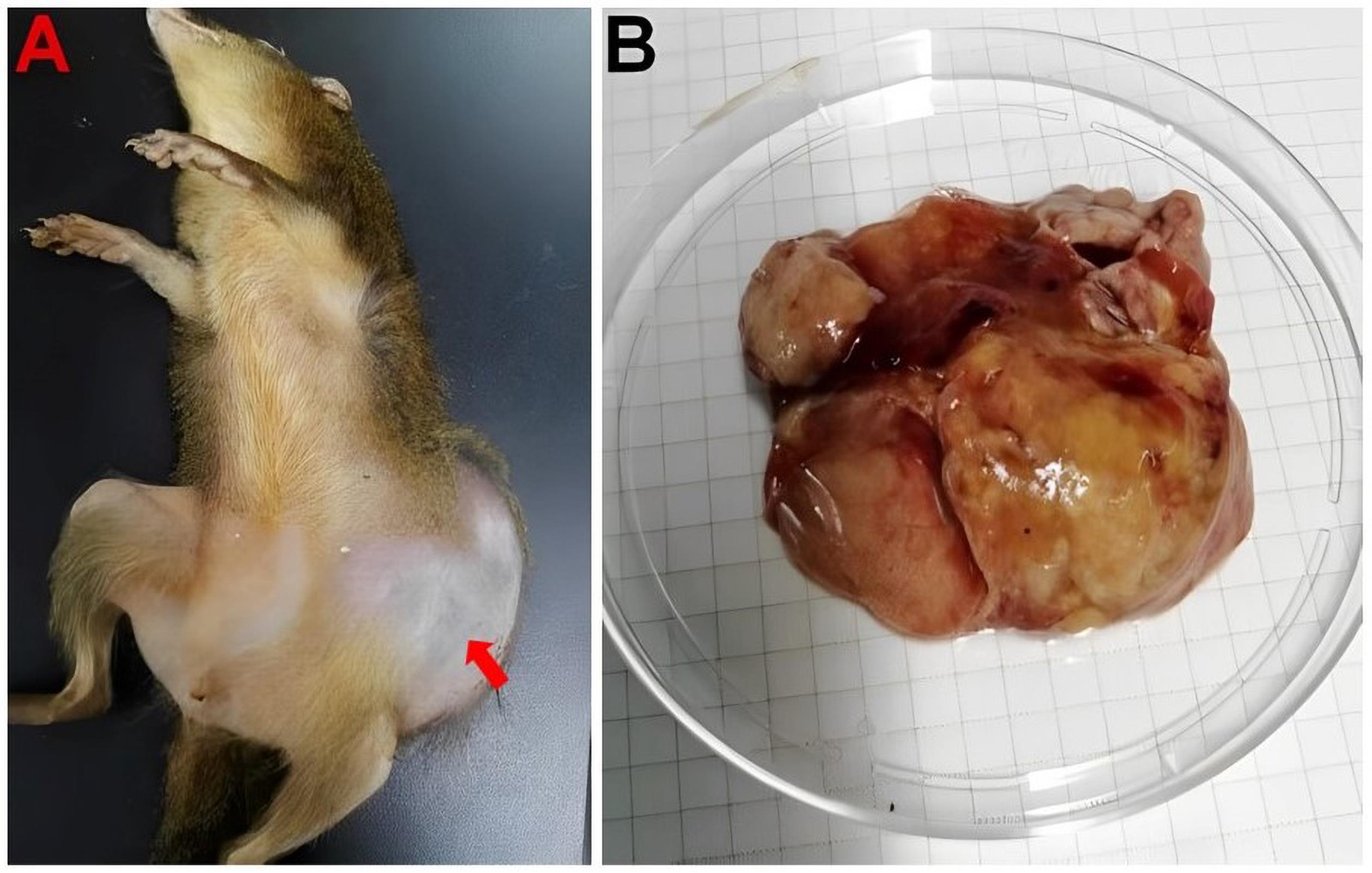
Figure 1 The overall morphology and autopsy of Tupaia belangeri subsp.yaoshanensis
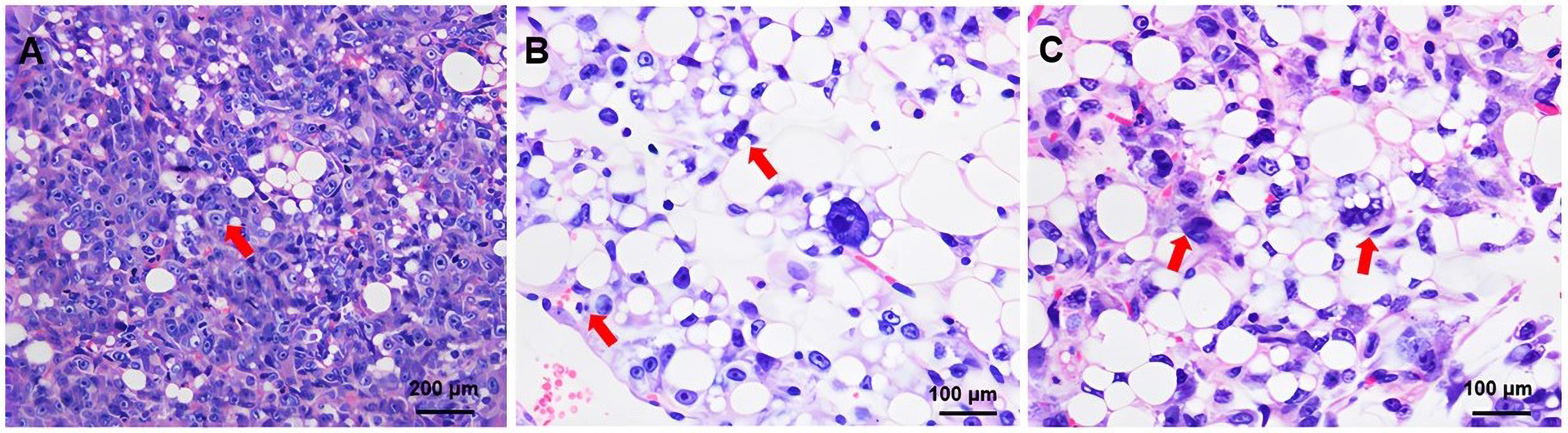
Figure 2 Histopathological changes of primary tumor in Tupaia belangeri subsp. yaoshanensis (HE staining)
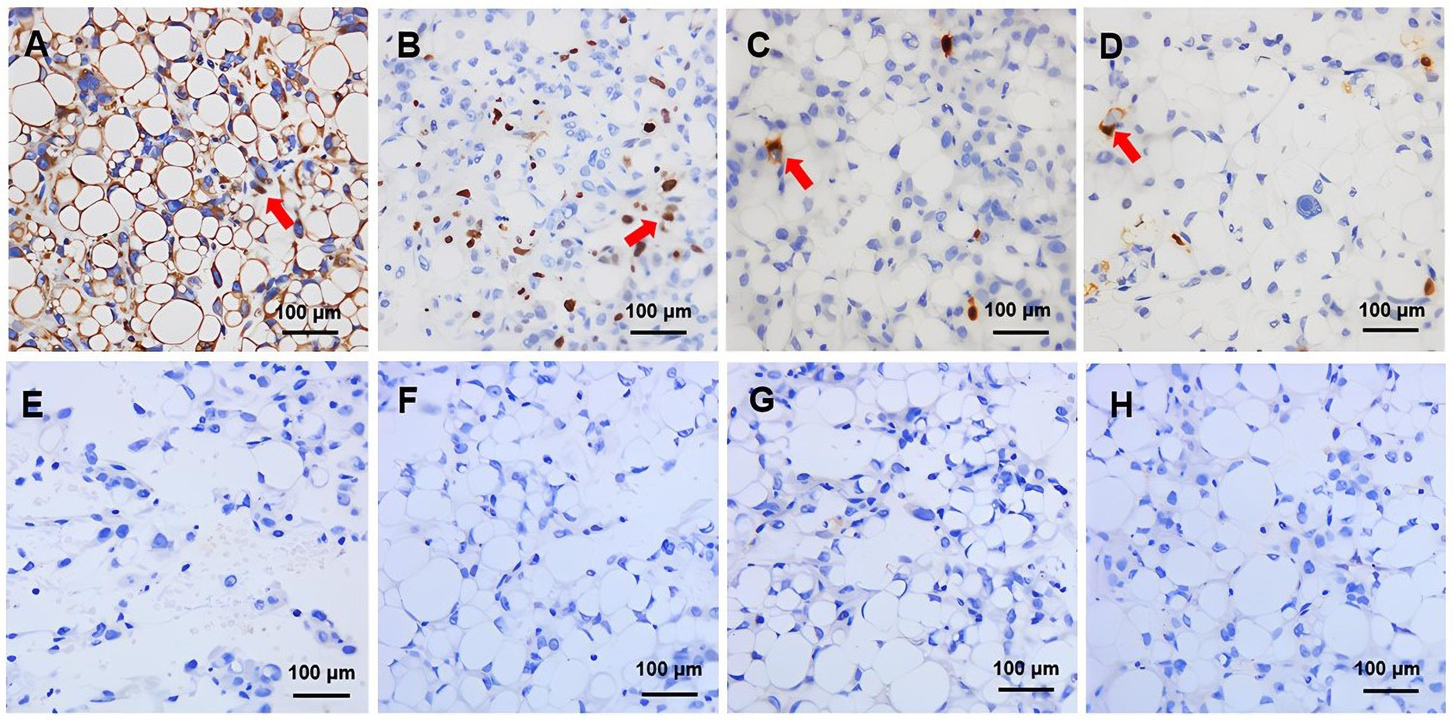
Figure 3 Immunohistochemical detection of primary tumors suggestive of pleomorphic liposarcoma in the Tupaia belangeri subsp.yaoshanensis (DAB stain)
| 1 | DEI TOS A P. Classification of pleomorphic sarcomas: where are we now?[J]. Histopathology, 2006, 48(1):51-62. DOI: 10.1111/j.1365-2559.2005.02289.x . |
| 2 | WANG L, REN W M, ZHOU X Y, et al. Pleomorphic liposarcoma: a clinicopathological, immunohistochemical and molecular cytogenetic study of 32 additional cases[J]. Pathol Int, 2013, 63(11):523-531. DOI: 10.1111/pin.12104 . |
| 3 | WANG L W, LUO R L, XIONG Z M, et al. Pleomorphic liposarcoma[J]. Medicine, 2018, 97(8): e9986. DOI: 10.1097/md.0000000000009986 . |
| 4 | 宋亚宁, 曹永宽. 胆囊三角区巨大脂肪肉瘤一例[J]. 中华临床医师杂志(电子版), 2012, 6(5):1368. |
| SONG Y N, CAO Y K. A case of giant liposarcoma in gallbladder triangle[J]. Chin J Clin Electron Ed, 2012, 6(5):1368. | |
| 5 | 季旸, 秦杰, 李霞, 等. 肢体不同病理亚型脂肪肉瘤超声学表现[J]. 医学影像学杂志, 2022, 32(7): 1215-1218. DOI: 10.3969/j.issn.1001-7399.2010.01.024 . |
| JI Y, QIN J, LI X, et al. The ultrasound performance of liposarcoma in different subtypes of pathology[J]. J Med Imaging, 2022, 32(7): 1215-1218. DOI: 10.3969/j.issn.1001-7399.2010.01.024 . | |
| 6 | 李元歌, 陈武标, 郁成, 等. 脂肪肉瘤CT及MRI的影像特征与病理分型的对照分析[J]. 医学影像学杂志, 2020, 30(2): 303-307. DOI: 10.13315/j.cnki.cjcep.2018.06.024 . |
| LI Y G, CHEN W B, YU C, et al. Comparative analysis of CT and MRI imaging features and pathological classification of liposarcoma[J]. J Med Imaging, 2020, 30(2): 303-307. DOI: 10.13315/j.cnki.cjcep.2018.06.024 . | |
| 7 | ANDERSON W J, JO V Y. Pleomorphic liposarcoma: Updates and current differential diagnosis[J]. Semin Diagn Pathol, 2019, 36(2):122-128. DOI: 10.1053/j.semdp.2019.02.007 . |
| 8 | APOSTOLOU G, BITELI M, CHATZIPANTELIS P. Cytopathological diagnosis of metastatic pleomorphic liposarcoma in the lung: a report of a case correlated with the histopathology of the primary tumour[J]. Diagn Cytopathol, 2009, 37(9):667-670. DOI: 10.1002/dc.21085 . |
| 9 | VON MEHREN M, KANE J M, BUI M M, et al. NCCN guidelines insights: soft tissue sarcoma, version 1.2021[J]. J Natl Compr Canc Netw, 2020, 18(12):1604-1612. DOI: 10.6004/jnccn.2020.0058 . |
| 10 | EWING E. Soft tissue and visceral sarcomas: ESMO Clinical Practice Guidelines for diagnosis, treatment and follow-up[J]. Annals of Oncology, 2012, 23(): vii92–vii99.doi:10.1093/annonc/mds253 . |
| 11 | 中华医学会肿瘤学分会, 中华医学会杂志社, 中国医师协会肛肠医师分会腹膜后疾病专业委员会, 等. 中国腹膜后肿瘤诊治专家共识( 2019版)[J]. 中华肿瘤杂志, 2019, 41(10):728-733. DOI:10.3760/cma.j.issn.0253-3766.2019.10.002 . |
| Chinese Medical Association, Cancer Society of Chinese Medical Association, Journal of Chinese Medical Association, et al. Expert consensus on treatment of Retroperitoneal tumors in China (Edition 2019)[J]. Chin J Oncol, 2019, 41(10):728-733. DOI: 10.3760/cma.j.issn.0253-3766.2019.10.002 . | |
| 12 | WILSON D E, REEDER D M. Mammal species of the World: a taxonomic and geographic reference[M]. 3rd ed. Maryland: Johns Hopkins University Press, 2005:104-9. |
| 13 | LI R F, ZANIN M, XIA X S, et al. The tree shrew as a model for infectious diseases research[J]. J Thorac Dis, 2018, 10(S9): S2272-S2279. DOI: 10.21037/jtd.2017.12.121 . |
| 14 | 夏巍, 赖永静, 杜龙, 等. 树鼩在人类肿瘤疾病动物模型中的应用进展[J]. 动物医学进展, 2019, 40(3): 109-113. DOI: 10.3969/j.issn.1007-5038.2019.03.022 . |
| XIA W, LAI Y J, DU L, et al. Application of tree shrew in animal models of human tumor diseases[J]. Prog Vet Med, 2019, 40(3): 109-113. DOI: 10.3969/j.issn.1007-5038.2019.03.022 . | |
| 15 | 唐海波, 梁亮, 曹颖颖, 等. 一例瑶山亚种树鼩自发隆突性皮肤纤维肉瘤的诊断[J]. 动物医学进展, 2021, 42(11):140-144. DOI: 10.3969/j.issn.1007-5038.2021.11.028 . |
| TANG H B, LIANG L, CAO Y Y, et al. Diagnosis of a spontaneous dermatofibrosarcoma protuberans from tree shrew (Tupaia belangeri subsp.yaoshanensis)[J]. Prog Vet Med, 2021, 42(11):140-144. DOI: 10.3969/j.issn.1007-5038.2021.11.028 . | |
| 16 | 张圆圆, 蒋艳玲, 文容,等. 反射式共聚焦显微镜在儿童毛发上皮瘤的诊断价值[J]. 实用皮肤病学杂志, 2021, 14(2):81-83, 87. DOI: 10.11786/sypfbxzz.1674-1293.20210205 . |
| ZHANG Y Y, JIANG Y L, WEN R, et al. The diagnostic value of reflective confocal microscopy in children with tricho-epithelioma[J]. Prac J Dermat, 2021, 14(2):81-83, 87. DOI:10.11786/sypfbxzz.1674-1293.20210205 . | |
| 17 | SBARAGLIA M, BELLAN E, DEI TOS A P. The 2020 WHO classification of soft tissue tumours: news and perspectives[J]. Pathologica, 2021, 113(2):70-84. DOI: 10.32074/1591-951X-213 . |
| 18 | LEE A T J, THWAY K, HUANG P H, et al. Clinical and molecular spectrum of liposarcoma[J]. J Clin Oncol, 2018, 36(2):151-159. DOI: 10.1200/JCO.2017.74.9598 . |
| 19 | GHADIMI M P, LIU P, PENG T S, et al. Pleomorphic liposarcoma: clinical observations and molecular variables[J]. Cancer, 2011, 117(23):5359-5369. DOI: 10.1002/cncr.26195 . |
| 20 | DOWNES K A, GOLDBLUM J R, MONTGOMERY E A, et al. Pleomorphic liposarcoma: a clinicopathologic analysis of 19 cases[J]. Mod Pathol, 2001, 14(3):179-184. DOI: 10.1038/modpathol.3880280 . |
| 21 | MIETTINEN M, ENZINGER F M. Epithelioid variant of pleomorphic liposarcoma: a study of 12 cases of a distinctive variant of high-grade liposarcoma[J]. Mod Pathol, 1999, 12(7):722-728. |
| 22 | 刘喆, 杨婕, 朱萌, 等. 多形性脂肪肉瘤4例临床病理分析[J]. 中国处方药, 2022, 20(12): 14-16. DOI: 10.3969/j.issn.1671-945X.2022.12.005 . |
| LIU Z, YANG J, ZHU M, et al. Pleomorphic liposarcoma: a clinicopathologic analysis of 4 cases[J]. J China Prescr Drug, 2022, 20(12): 14-16. DOI: 10.3969/j.issn.1671-945X.2022.12.005 . | |
| 23 | MANGHAM D. World Health Organisation classification of tumours: pathology and genetics of tumours of soft tissue and bone[M]. Lyon, France: IARC Press, 2002:427. |
| 24 | HORNICK J L, BOSENBERG M W, MENTZEL T, et al. Pleomorphic liposarcoma[J]. Am J Surg Pathol, 2004, 28(10):1257-1267. DOI: 10.1097/01.pas.0000135524.73447.4a . |
| 25 | YAN P, SUN M L, SUN Y P, et al. Effective apatinib treatment of pleomorphic liposarcoma[J]. Medicine, 2017, 96(33): e7771. DOI: 10.1097/md.0000000000007771 . |
| 26 | GEBHARD S, COINDRE J M, MICHELS J J, et al. Pleomorphic liposarcoma: clinicopathologic, immunohistochemical, and follow-up analysis of 63 cases: a study from the French Federation of Cancer Centers Sarcoma Group[J]. Am J Surg Pathol, 2002, 26(5):601-616. DOI: 10.1097/00000478-200205000-00006 . |
| 27 | NASCIMENTO A F, RAUT C P. Diagnosis and management of pleomorphic sarcomas (so-called MFH) in adults[J]. J Surg Oncol, 2008, 97(4):330-339. DOI: 10.1002/jso.20972 . |
| 28 | CRAGO A M, DICKSON M A. Liposarcoma[J]. Surg Oncol Clin N Am, 2016, 25(4):761-773. DOI: 10.1016/j.soc.2016.05.007 . |
| 29 | 施继鼎, 王占兴, 陈振声, 等. 膀胱原发多形性脂肪肉瘤1例报告[J]. 福建医药杂志, 2021, 43(3): 179-180. DOI: 10.3969/j.issn.1002-2600.2021.03.073 . |
| SHI J D, WANG Z X, CHEN Z S, et al. Primary liposarcoma pleomorphic bladder: a case report[J]. Fujian Med J, 2021, 43(3): 179-180. DOI: 10.3969/j.issn.1002-2600.2021.03.073 . | |
| 30 | CHOI J H, RO J Y. The 2020 WHO classification of tumors of soft tissue: selected changes and new entities[J]. Adv Anat Pathol, 2021, 28(1):44-58. DOI: 10.1097/PAP.0000000000000284 . |
| [1] | SUN Xiaorong, SU Dan, GUI Wenjuan, CHEN Yue. Establishment and Evaluation of a Moderate-to-Severe Knee Osteoarthritis Model in Rats Induced by Surgery [J]. Laboratory Animal and Comparative Medicine, 2024, 44(6): 597-604. |
| [2] | Minbo HOU, Tiantian CUI, Naying SU, Miaomiao ZHANG, Yongmin JIAO, Jianyan YAN, Xijie WANG, Ohira TOKO. Pathologic Diagnosis of a Pituicytoma in a Han-Wistar Rat [J]. Laboratory Animal and Comparative Medicine, 2023, 43(6): 654-658. |
| [3] | Shanshan ZHAI, Liang LIANG, Yingying CAO, Zhuxin LI, Qing WANG, Junyu TAO, Chenxia YUN, Jing LENG, Haibo TANG. Diagnosis of Trichoepithelioma in a Tree Shrew and Observation of Cell Biological Characteristics [J]. Laboratory Animal and Comparative Medicine, 2023, 43(4): 440-445. |
| [4] | Ying TAN, Wenping LIAO, Qilong GAO, Yong LI, Xinhui SHI, Jingkun WANG. Physiological Indexes and Histopathology Analysis of Sodium Iodate-Induced Retinitis Pigmentosa in Rats [J]. Laboratory Animal and Comparative Medicine, 2023, 43(2): 124-135. |
| [5] | WEN Fuli. Establishment of Eimeria stiedai Infected Rabbit Model and Nested PCR Assay [J]. Laboratory Animal and Comparative Medicine, 2020, 40(6): 477-482. |
| [6] | HUANG Jisheng, WU Shuyi, ZHAN Jinhe, NI Qingchun. Analysis of Incidence of Spontaneous Histopathological Lesions in Young SD Rats [J]. Laboratory Animal and Comparative Medicine, 2020, 40(2): 128-135. |
| [7] | XIAO Kunlin, ZHANG Rui, Sun Hong, XIAO Kuntai, MA Jianbing. Evaluation of Monoiodoacetic Acid-induced Knee Osteoarthritis SD Rats with Diseasse Progression at Different Time Points [J]. Laboratory Animal and Comparative Medicine, 2020, 40(1): 47-52. |
| [8] | ZHOU Fei, WANG Hao-an, CHEN Tao, QIU Shuang, CHEN Ke, CEN Xiao-bo. Case Report of Geniposide Induced Bilirubin Pigmentation in Sprague-Dawley Rat [J]. Laboratory Animal and Comparative Medicine, 2018, 38(4): 284-287. |
| [9] | HUANG Chao, GONG Yi-juan, TIAN Fang, WANG Yu-zhu, SUN Bing, ZHI Rui-na, IA Min-jie, DING Xun-cheng, ZHOU Xin-chu, LI Wei-hua. Establishment of Vaginal Mucosal Immuno-inflammatory Response Model in Minipig [J]. Laboratory Animal and Comparative Medicine, 2016, 36(2): 81-86. |
| [10] | LI Wei-hua1,2,WANG Hai-lan3,Gaku Ichihara4,DING Xun-cheng2,ZHOU Zhi-jun1. Comparative Study of 1-Bromopropane Induced Toxic Effects in Fisher 344/NSIc and Wistar NWN Rats [J]. Laboratory Animal and Comparative Medicine, 2010, 30(2): 105-108. |
| [11] | ZHOU Xiao-mm, BA Cai-feng, FENG Hui-quan . Experimental Study on Mouse Model Infected with Mycoplasma suis [J]. Laboratory Animal and Comparative Medicine, 2008, 28(6): 372-376. |
| [12] | XU Xiao-Ping, XU Jian-Qin, CHEN Min-Li, CHEN Fang-Ming, CHEN Liang, PENG Ding-Guo, ZHOU Wei-Min. Establishment of Rabbit Model of Allergic Rhinitis and Histological Observation of Nasal Mucosa [J]. Laboratory Animal and Comparative Medicine, 2006, 26(2): 80-82. |
| [13] | LIU Yi-1, ZHANG Chi-1, XIAO Jun-Xia-2, XI Shou-Min-1, YIN Wei-Dong-1, WANG Zong-Bao-1. Disorders of Glucose and Lipid Metabolism and Pathological Changes of Pancreas, Liver and Kidney in New Zealand White Rabbits Induced by High Sucrose,High Fat and High Cholesterol Diet [J]. Laboratory Animal and Comparative Medicine, 2005, 25(3): 151-156. |
| [14] | CHENG Shu-jun, HUANG Ren, QING Yao, TAN Wen-ya. Histopathological Observation on Spontaneous Lesion of Digestive System in Macaca mulatta [J]. Laboratory Animal and Comparative Medicine, 2003, 23(3): 131-134. |
| [15] | ZHOU Zhong-xin1,WANG Jie2, LUO Bao-ming2. Functional and Histopathological Changes of Liver in Cirrhosis Model of Mini-pigs [J]. Laboratory Animal and Comparative Medicine, 2002, 22(2): 102-105. |
| Viewed | ||||||
|
Full text |
|
|||||
|
Abstract |
|
|||||
