
Laboratory Animal and Comparative Medicine ›› 2026, Vol. 46 ›› Issue (2): 212-221.DOI: 10.12300/j.issn.1674-5817.2025.077
• Animal Models of Human Diseases • Previous Articles Next Articles
LIU Sai1, FU Bin1, LI Sidi1,2, CHEN Zhida1, ZHANG Yue1, GUO Zhongkun1, WANG Yongan1( )(
)( ), WANG Kezhou1()()
Received:2025-05-22
Revised:2025-09-11
Online:2026-04-25
Published:2026-04-18
Correspondence to:
WANG Yongan, WANG Kezhou
CLC Number:
LIU Sai,FU Bin,LI Sidi,et al. Adra2a Regulates LPS-Induced Inflammation in Hepatocytes of Lbp-/- Mice via the MAPK Signaling Pathway[J]. Laboratory Animal and Comparative Medicine, 2026, 46(2): 212-221. DOI: 10.12300/j.issn.1674-5817.2025.077.
Add to citation manager EndNote|Ris|BibTeX
URL: https://www.slarc.org.cn/dwyx/EN/10.12300/j.issn.1674-5817.2025.077
基因 Gene | 正向引物(5’-3’) Forward primer (5’-3’) | 反向引物(5’-3’) Reverse primer (5’-3’) |
|---|---|---|
| Adra2a | GTGACACTGACGCTGGTTTG | CCAGTAACCCATAACCTCGTTG |
| β-actin | GGCTGTATTCCCCTCCATCG | CCAGTTGGTAACAATGCCATGT |
| TNF-α | CCCTCACACTCAGATCATCTTCT | GCTACGACGTGGGCTACAG |
| IL-6 | TAGTCCTTCCTACCCCAATTTCC | TTGGTCCTTAGCCACTCCTTC |
| IL-1β | GCAACTGTTCCTGAACTCAACT | ATCTTTTGGGGTCCGTCAACT |
Table 1 Primer sequences
基因 Gene | 正向引物(5’-3’) Forward primer (5’-3’) | 反向引物(5’-3’) Reverse primer (5’-3’) |
|---|---|---|
| Adra2a | GTGACACTGACGCTGGTTTG | CCAGTAACCCATAACCTCGTTG |
| β-actin | GGCTGTATTCCCCTCCATCG | CCAGTTGGTAACAATGCCATGT |
| TNF-α | CCCTCACACTCAGATCATCTTCT | GCTACGACGTGGGCTACAG |
| IL-6 | TAGTCCTTCCTACCCCAATTTCC | TTGGTCCTTAGCCACTCCTTC |
| IL-1β | GCAACTGTTCCTGAACTCAACT | ATCTTTTGGGGTCCGTCAACT |
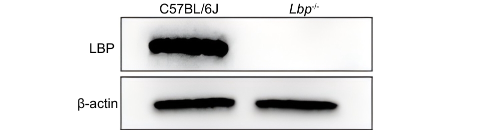
Figure 1 Expression of LBP protein in primary hepatocytes
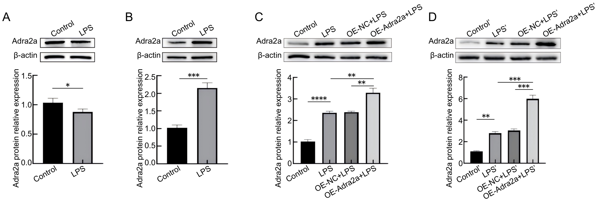
Figure 2 Changes in Adra2a protein expression after LPS and lentivirus treatment
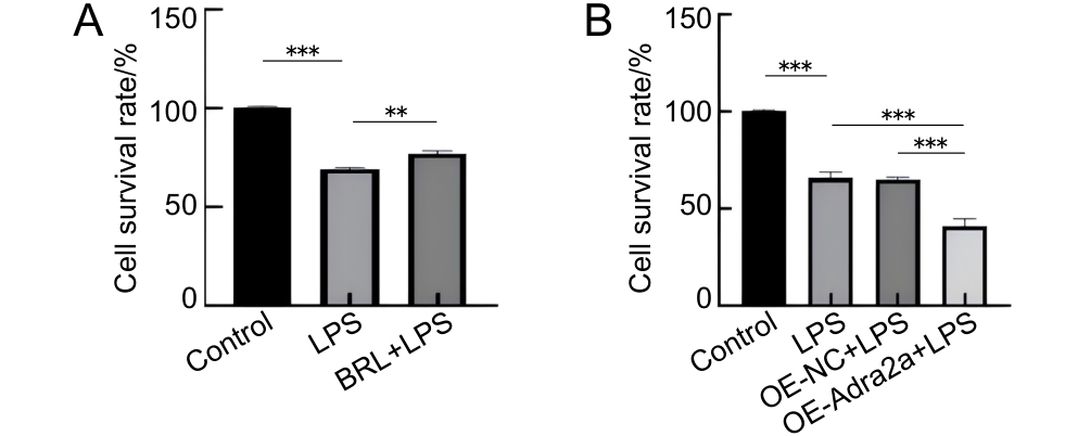
Figure 3 Effects of Adra2a inhibition and overexpression on the survival rate of primary hepatocytes from Lbp-/- mice following LPS stimulation
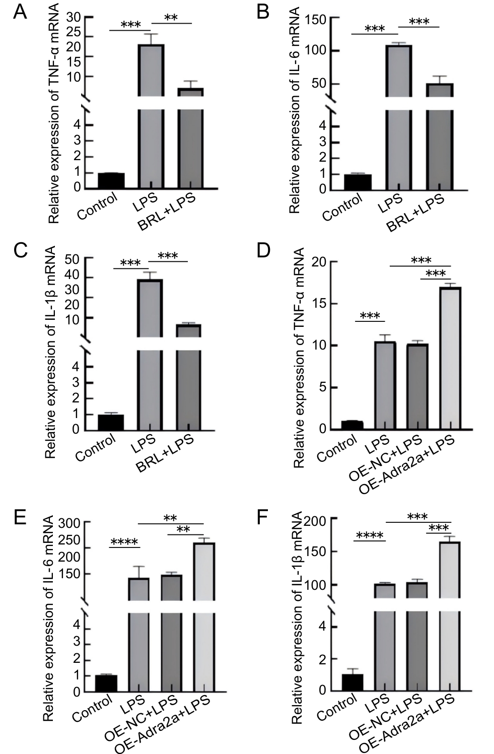
Figure 4 Effects of Adra2a inhibition (A–C) and overexpression (D–F) on the gene transcription of TNF-ɑ, IL-6, and IL-1 β in primary hepatocytes from Lbp-/- mice following LPS stimulation
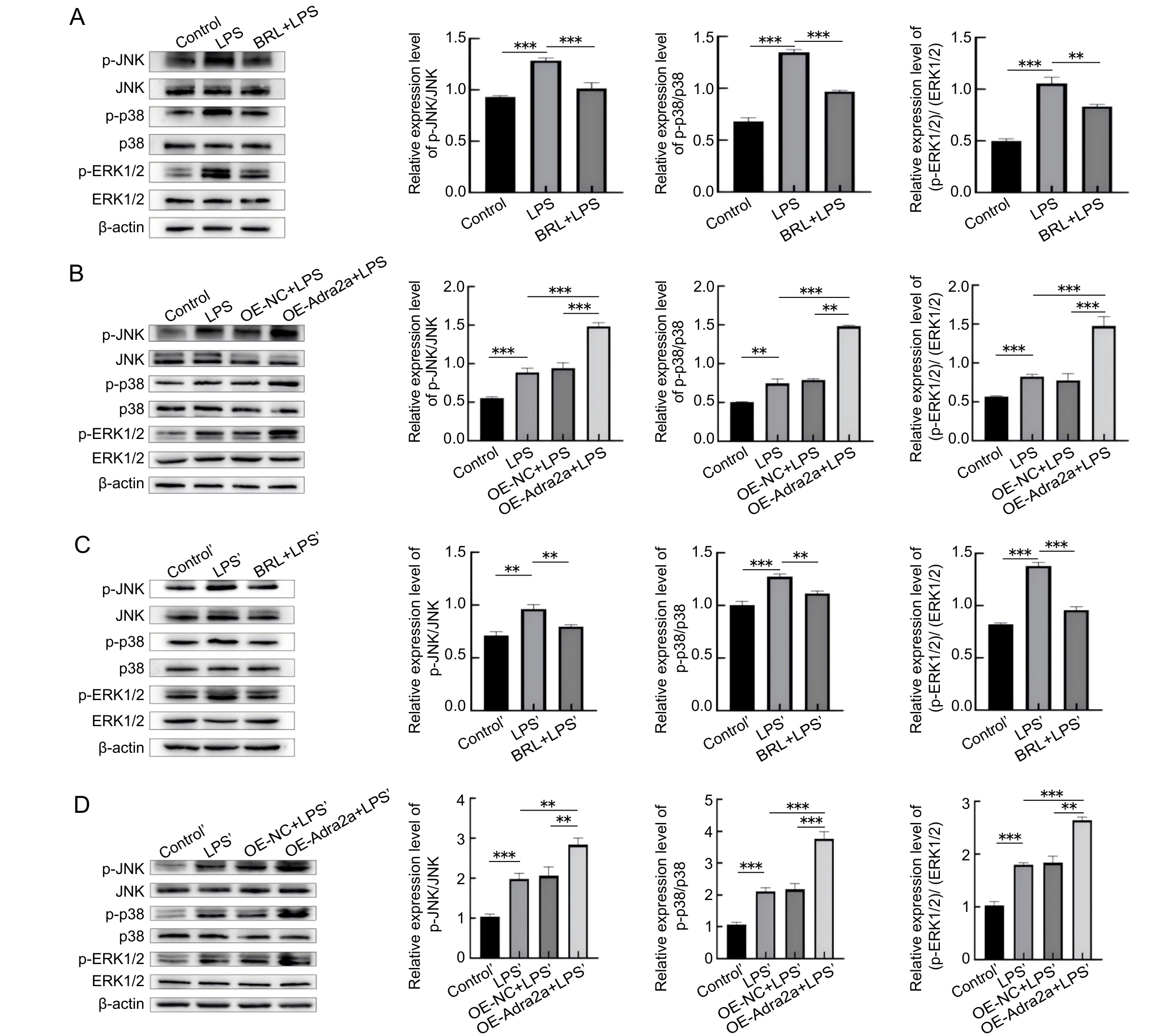
Figure 5 Effects of Adra2a inhibition and overexpression on ERK1/2, p38, and JNK protein phosphorylation inprimary hepatocytes and liver tissues of Lbp-/- mice
| [1] | DI LORENZO F, DE CASTRO C, SILIPO A, et al. Lipopolysaccharide structures of Gram-negative populations in the gut microbiota and effects on host interactions[J]. FEMS Microbiol Rev, 2019, 43(3):257-272. DOI: 10.1093/femsre/fuz002 . |
| [2] | ULEVITCH R J, TOBIAS P S. Recognition of Gram-negative bacteria and endotoxin by the innate immune system[J]. Curr Opin Immunol, 1999, 11(1):19-22. DOI: 10.1016/S0952-7915(99)80004-1 . |
| [3] | GARCIA-VELLO P, DI LORENZO F, ZUCCHETTA D, et al. Lipopolysaccharide lipid A: a promising molecule for new immunity-based therapies and antibiotics[J]. Pharmacol Ther, 2022, 230:107970. DOI: 10.1016/j.pharmthera.2021.107970 . |
| [4] | SONG Z C, MENG L L, HE Z X, et al. LBP protects hepatocyte mitochondrial function via the PPAR-CYP4A2 signaling pathway in a rat sepsis model[J]. Shock, 2021, 56(6):1066-1079. DOI: 10.1097/SHK.0000000000001808 . |
| [5] | 李思迪, 付彬, 郭中坤, 等. 利用CRISPR/Cas9技术构建脂多糖结合蛋白基因敲除小鼠[J]. 实验动物与比较医学, 2022, 42(4):294-300. DOI: 10.12300/j.issn.1674-5817.2022.002 . |
| LI S D, FU B, GUO Z K, et al. Construction of lipopolysaccaride binding protein knockout mice using CRISPR/Cas9 technology[J]. Lab Anim Comp Med, 2022, 42(4):294-300. DOI: 10.12300/j.issn.1674-5817.2022.002 . | |
| [6] | 米传靓, 付彬, 李思迪, 等. Adra1a调节LPS诱导的Lbp-/-小鼠原代肝细胞炎症反应[J]. 中国比较医学杂志, 2024, 34(5): 84-91. DOI: 10.3969/j.issn.1671-7856.2024.05.009 . |
| MI C L, FU B, LI S D, et al. Adra1a regulates LPS-induced inflammation in primary hepatocytes of Lbp-/- mice[J]. Chin J Comp Med, 2024, 34(5): 84-91. DOI: 10.3969/j.issn.1671-7856.2024.05.009 . | |
| [7] | 米传靓, 付彬, 李思迪, 等. Agtr1a调节LPS诱导的Lbp-/- 小鼠原代肝细胞炎症[J]. 中国实验动物学报, 2023, 31(8): 1021-1027. DOI: 10.3969/j.issn.1005-4847.2023.08.007 . |
| MI C L, FU B, LI S D, et al. Agtr1a regulates LPS-induced inflammation in primary hepatocytes of Lbp-/- mice[J]. Acta Lab Anim Sci Sin, 2023, 31(8): 1021-1027. DOI: 10.3969/j.issn.1005-4847.2023.08.007 . | |
| [8] | ZHOU F, CAI J, CHEN L,et al. ADRA2A contributes to airway inflammation and apoptosis in asthma through the ERK signaling in vitro and in vivo [J]. J Inflamm, 2025, 22(1): 55. DOI: 10.1186/s12950-025-00480-8 . |
| [9] | MIKSA M, DAS P, ZHOU M, et al. Pivotal role of the alpha(2A)-adrenoceptor in producing inflammation and organ injury in a rat model of sepsis[J]. PLoS One, 2009, 4(5):e5504. DOI: 10.1371/journal.pone.0005504 . |
| [10] | LIN Y, ZHU X, YAO W Z, et al. Yohimbine protects against endotoxin-induced acute lung injury by blockade of alpha 2A adrenergic receptor in rats[J]. Chin Med J, 2011, 124(7):1069-1074.DOI: 10.3760/cma.j.issn.0366-6999.2011.07.022 . |
| [11] | 任文洁, 林哲绚. 胰蛋白酶和胶原酶灌注法分离提取小鼠原代肝细胞的比较[J]. 汕头大学医学院学报, 2022, 35(4):204-209. DOI: 10.13401/j.cnki.jsumc.2022.04.003 . |
| REN W J, LIN Z X. Comparison of isolation and extraction of primary mouse hepatocytes by trypsin and collagenase perfusion[J]. J Shantou Univ Med Coll, 2022, 35(4):204-209. DOI: 10.13401/j.cnki.jsumc.2022.04.003 . | |
| [12] | RYU J K, KIM S J, RAH S H, et al. Reconstruction of LPS transfer cascade reveals structural determinants within LBP, CD14, and TLR4-MD2 for efficient LPS recognition and transfer[J]. Immunity, 2017, 46(1):38-50. DOI: 10.1016/j.immuni. 2016.11.007 . |
| [13] | MANO S S, KANEHIRA K, TANIGUCHI A. Comparison of cellular uptake and inflammatory response via toll-like receptor 4 to lipopolysaccharide and titanium dioxide nanoparticles[J]. Int J Mol Sci, 2013, 14(7):13154-13170. DOI: 10.3390/ijms140713154 . |
| [14] | LI T X, BAI J J, DU Y C, et al. Thiamine pretreatment improves endotoxemia-related liver injury and cholestatic complications by regulating galactose metabolism and inhibiting macrophage activation[J]. Int Immunopharmacol, 2022, 108:108892. DOI: 10.1016/j.intimp.2022.108892 . |
| [15] | BRASS D M, SAVOV J D, WHITEHEAD G S, et al. LPS binding protein is important in the airway response to inhaled endotoxin[J]. J Allergy Clin Immunol, 2004, 114(3):586-592. DOI: 10.1016/j.jaci.2004.04.043 . |
| [16] | MINTER R M, BI X M, BEN-JOSEF G, et al. LPS-binding protein mediates LPS-induced liver injury and mortality in the setting of biliary obstruction[J]. Am J Physiol Gastrointest Liver Physiol, 2009, 296(1): G45-G54. DOI: 10.1152/ajpgi.00041.2008 . |
| [17] | LATTIN J, ZIDAR D A, SCHRODER K, et al. G-protein-coupled receptor expression, function, and signaling in macrophages[J]. J Leukoc Biol, 2007, 82(1):16-32. DOI: 10.1189/jlb.0107051 . |
| [18] | SHARMA N, SISTLA R, ANDUGULAPATI S B. Yohimbine ameliorates liver inflammation and fibrosis by regulating oxidative stress and Wnt/β-catenin pathway[J]. Phyto-medicine, 2024, 123:155182. DOI: 10.1016/j.phymed.2023. 155182 . |
| [19] | WANG J H, LIU Y J, GUO Y S, et al. Function and inhibition of P38 MAP kinase signaling: Targeting multiple inflammation diseases[J]. Biochem Pharmacol, 2024, 220:115973. DOI: 10. 1016/j.bcp.2023.115973 . |
| [20] | AHMED T, ZULFIQAR A, ARGUELLES S, et al. Map kinase signaling as therapeutic target for neurodegeneration[J]. Pharmacol Res, 2020, 160:105090. DOI:10.1016/j.phrs. 2020. 105090 . |
| [21] | REZATABAR S, KARIMIAN A, RAMESHKNIA V, et al. RAS/MAPK signaling functions in oxidative stress, DNA damage response and cancer progression[J]. J Cell Physiol, 2019, 234(9):14951-14965. DOI: 10.1002/jcp.28334 . |
| [22] | 刘淑青. 肝细胞SOX9在肝再生中的作用和机制及肝细胞TGF-β信号通路对肝纤维化的影响和机制[D]. 上海: 中国人民解放军海军军医大学, 2023. DOI: 10.26998/d.cnki.gjuyu.2023.000324 . |
| LIU S Q. The role and underlying mechanism of hepatocellular SOX9 in liver regeneration and hepatocellular TGF-β signaling pathway in liver fibrosis[D]. Shanghai: Naval Medical University, 2023. DOI: 10.26998/d.cnki.gjuyu.2023.000324 . |
| [1] | PAN Linqin, DENG Xiangliang, LUO Yunxia. Advances in Integrative Translational Research on Animal Models of Ischemic Stroke in Traditional Chinese and Western Medicine [J]. Laboratory Animal and Comparative Medicine, 2026, 46(3): 344-356. |
| [2] | GAO Yilong, HE Xingliang, ZHOU Xiaopeng, LI Dawei, BAO Xijun, LI Laiyou. Mining Candidate Genes for Litter Size Traits in English Springer Spaniel Bitches Based on Whole Genome Resequencing [J]. Laboratory Animal and Comparative Medicine, 2026, 46(3): 378-387. |
| [3] | WANG Juan, XU Jiahui, TIAN Yunyuan, ZHANG Mengmeng, LI Min, WANG Siwang, LI Yao. Comparison and Behavioral Observation of Two Female Mice Models of Ulcerative Colitis [J]. Laboratory Animal and Comparative Medicine, 2026, 46(3): 332-343. |
| [4] | CHEN Bing, XIE Xiaojie, TAO Tifu, WANG Jingdong, ZOU Yixing. Research on the Current Situation and Countermeasures for Training and Education of Laboratory Animal Practitioners in Sichuan Province [J]. Laboratory Animal and Comparative Medicine, 2026, 46(3): 456-463. |
| [5] | TANG Jianping, ZHAO Liya, ZHAO Ying. Screening and Analysis of Microsatellite Genetic Markers in Commonly Used Inbred Rat Strains [J]. Laboratory Animal and Comparative Medicine, 2026, 46(3): 388-396. |
| [6] | BU Yu, HOU Jinting, LI Yuanyuan, SHA Jingtao, XIE Chenlu, WANG Wengang, SUN Xingwei. A Review and Evaluation of Integrated Disease and Syndrome Animal Models for Hemorrhoids in Traditional Chinese and Western Medicine [J]. Laboratory Animal and Comparative Medicine, 2026, 46(3): 357-366. |
| [7] | BU Jiwen, HUA Ye, JIN Shirong, REN Ningxin, LI Funing, DU Jiulin. Automated Management of Zebrafish Strains and Zebrafish Facilities [J]. Laboratory Animal and Comparative Medicine, 2026, 46(3): 416-425. |
| [8] | WANG Xiuran, LI Hao, CHEN Zhengtao, YU Yang, ZHANG Suying, TAO Ru, WANG Kezhou. Innovation and Practice in the Construction of "Three-in-one" Talent Training Systems for Laboratory Animal Professionals in Medical Colleges [J]. Laboratory Animal and Comparative Medicine, 2026, 46(3): 446-455. |
| [9] | LI Jiafei, ZHANG Zhenhao, WANG Shuo, TIAN Ge, WEN Shuang, YAN Yuxue, CUI Ran, YE Zhen, CUI Yongchun. Effects of Autonomic Neuromodulators on Atrial Electrical Remodeling and Histopathological Changes in a Rat Model of Atrial Fibrillation [J]. Laboratory Animal and Comparative Medicine, 2026, 46(3): 321-331. |
| [10] | CHEN Lin, ZHANG Housen, AI Man, QI Chongyang. Research on the High-Quality Development Path of Laboratory Animal Management in Jiangsu Province Empowered by Digital Transformation [J]. Laboratory Animal and Comparative Medicine, 2026, 46(3): 426-436. |
| [11] | LI Longxue, WAN Chongfan, ZHANG Qi, LEI Ruting, WANG Xiaoyue, CHENG Leyan, LAI Qi, LIU Ronghua, LIU Xuan, XU Tielong. Molecular Mechanisms of Qingfei Paidu Decoction in the Prevention and Treatment of Acute Lung Injury in Mice Based on miRNA Sequencing [J]. Laboratory Animal and Comparative Medicine, 2026, 46(3): 311-320. |
| [12] | AI Xiufeng, ZHANG Lizong, FANG Mingsun, LÜ Dongying, CHEN Chu, CAI Zhaowei, WANG Dejun. Analysis of Differences in the Intestinal Flora of Rats and Mice after Drinking Chlorinated Water Based on 16S rRNA Sequencing [J]. Laboratory Animal and Comparative Medicine, 2026, 46(3): 437-445. |
| [13] | ZHOU Yimin, ZHANG Xinyu, YANG Jianuo, LIU Mengjia, SI Cancan, YE Hailü, SUN Wenchao, LAN Tian. Development of a Triplex TaqMan Quantitative PCR Method for Simultaneous Detection of MHV, MPV, and Reo-3 [J]. Laboratory Animal and Comparative Medicine, 2026, 46(3): 408-415. |
| [14] | . [J]. Laboratory Animal and Comparative Medicine, 2026, 46(3): 464-466. |
| [15] | WANG Lu, WANG Qingjie, YANG Chunhong, ZHANG Yijian. The Application of New Approach Methodologies in the Biomedical Field and Its Implementation of "3R" Principle [J]. Laboratory Animal and Comparative Medicine, 2026, (): 1-10. |
| Viewed | ||||||
|
Full text |
|
|||||
|
Abstract |
|
|||||