



Laboratory Animal and Comparative Medicine ›› 2026, Vol. 46 ›› Issue (5): 716-726.DOI: 10.12300/j.issn.1674-5817.2025.199
• Animal Experimental Techniques and Methods • Previous Articles Next Articles
LIAN Ronggui, LIAO Naishun( )(
)( )
)
Received:2025-12-03
Revised:2026-04-20
Online:2026-10-25
Published:2026-10-16
Correspondence to:
LIAO Naishun
CLC Number:
LIAN Ronggui,LIAO Naishun. In Vivo Tracking of Adipose-Derived Mesenchymal Stem Cell Therapy for Subacute Liver Injury Using Rare-Earth Down-Conversion Nanoparticles[J]. Laboratory Animal and Comparative Medicine, 2026, 46(5): 716-726. DOI: 10.12300/j.issn.1674-5817.2025.199.
Add to citation manager EndNote|Ris|BibTeX
URL: https://www.slarc.org.cn/dwyx/EN/10.12300/j.issn.1674-5817.2025.199
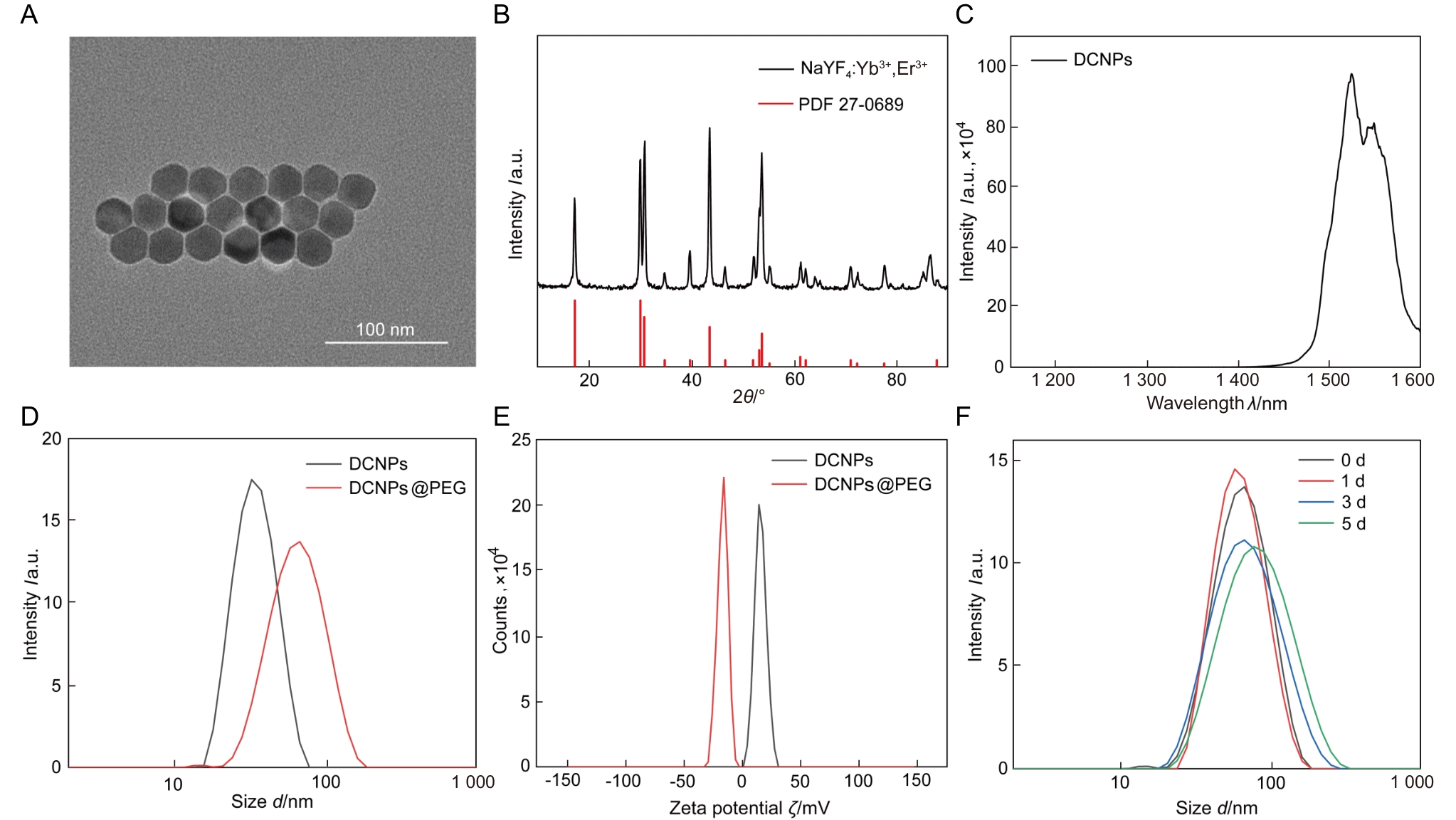
Figure 1
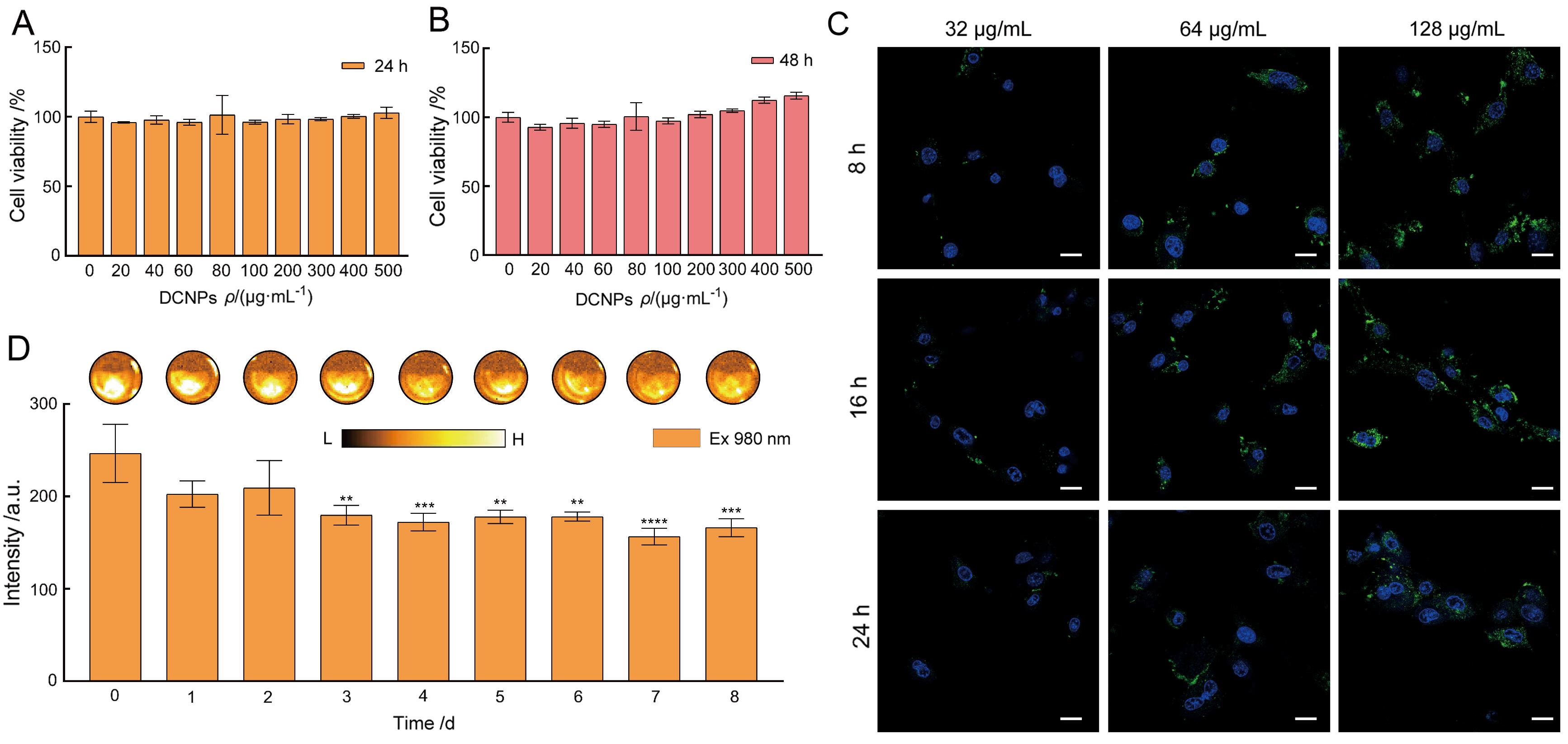
Figure 2
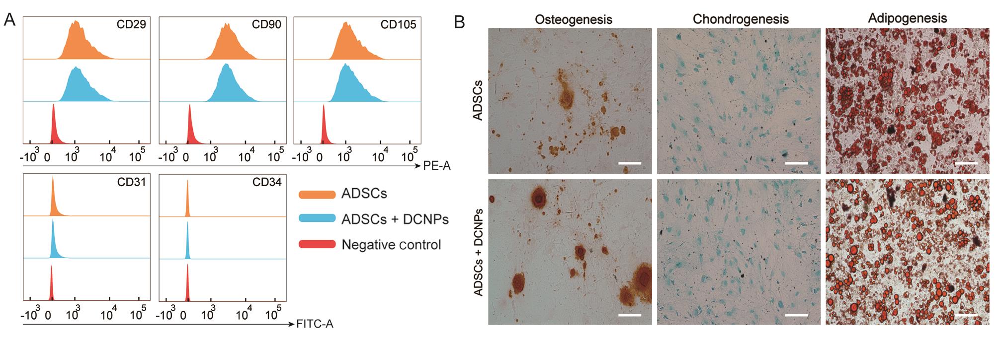
Figure 3
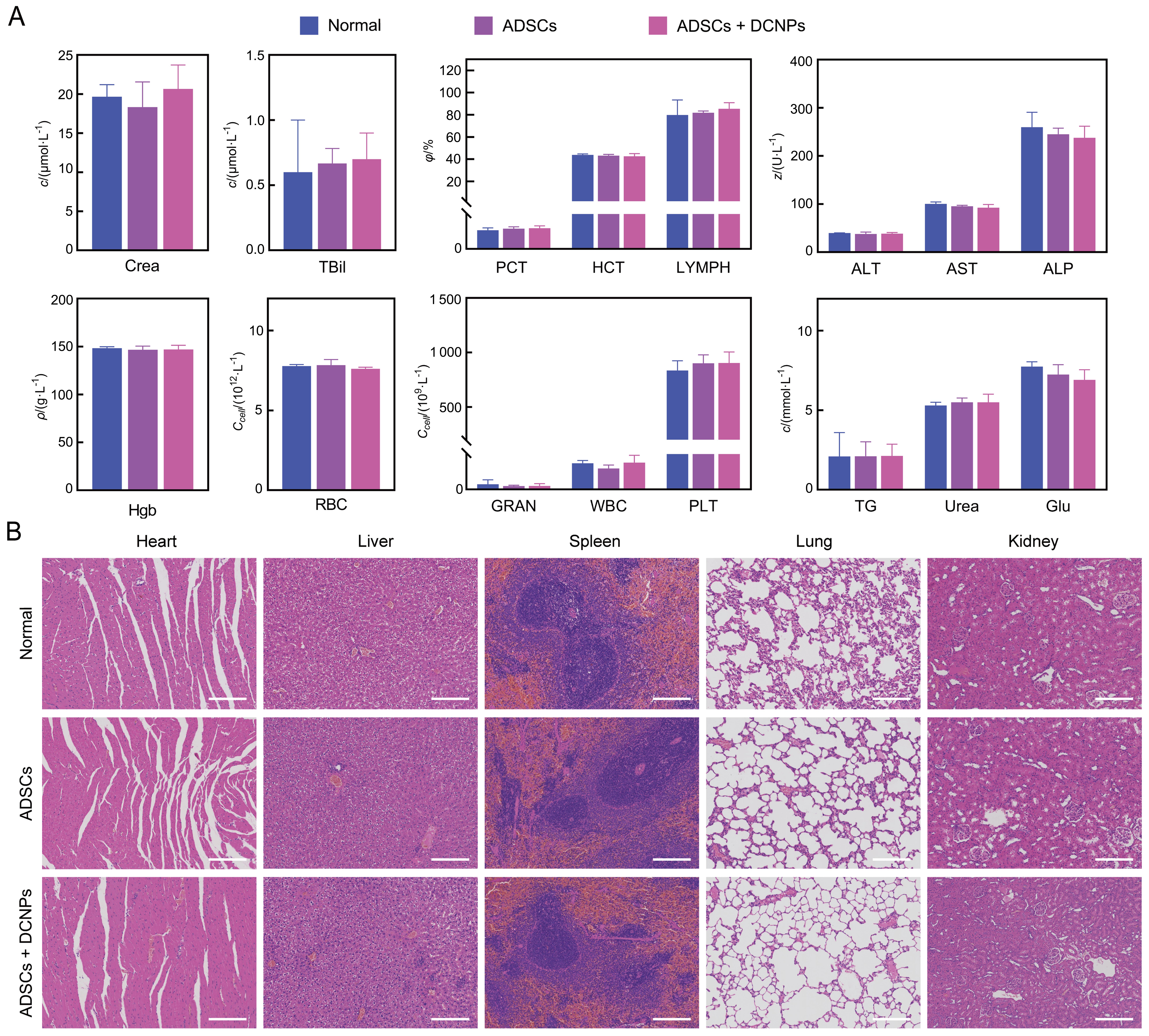
Figure 4
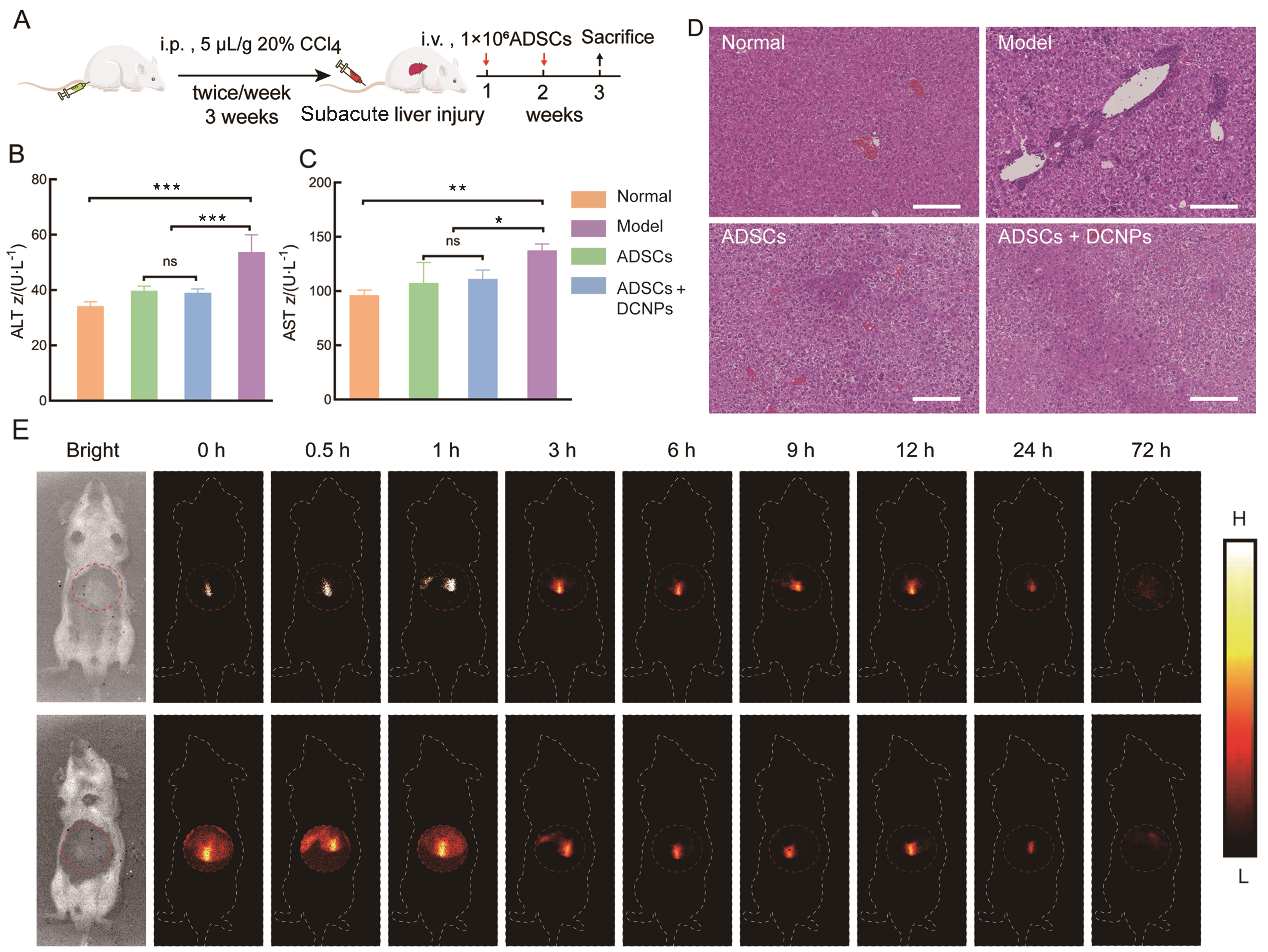
Figure 5
| [1] |
MAZINI L, ROCHETTE L, AMINE M, et al. Regenerative capacity of adipose derived stem cells (ADSCs), comparison with mesenchymal stem cells (MSCs)[J]. Int J Mol Sci, 2019, 20(10): 2523. DOI: 10.3390/ijms20102523.
|
| [2] |
MAZINI L, ROCHETTE L, ADMOU B, et al. Hopes and limits of adipose-derived stem cells (ADSCs) and mesenchymal stem cells (MSCs) in wound healing[J]. Int J Mol Sci, 2020, 21(4): 1306. DOI: 10.3390/ijms21041306.
|
| [3] |
THAWLEY V. Acute liver injury and failure[J]. Vet Clin N Am Small Anim Pract, 2017, 47(3): 617-630. DOI: 10.1016/j.cvsm. 2016.11.010.
|
| [4] |
STERODIMAS A, DE FARIA J, NICARETTA B, et al. Tissue engineering with adipose-derived stem cells (ADSCs): Current and future applications[J]. J Plast Reconstr Aesthet Surg, 2010, 63(11): 1886-1892. DOI: 10.1016/j.bjps. 2009.10.028.
|
| [5] |
NADERI N, COMBELLACK E J, GRIFFIN M, et al. The regenerative role of adipose-derived stem cells (ADSC) in plastic and reconstructive surgery[J]. Int Wound J, 2017, 14(1): 112-124. DOI: 10.1111/iwj.12569.
|
| [6] |
LEE S, CHAE D S, SONG B W, et al. ADSC-based cell therapies for musculoskeletal disorders: a review of recent clinical trials[J]. Int J Mol Sci, 2021, 22(19): 10586. DOI: 10.3390/ijms221910586.
|
| [7] |
蒋励, 陈子晗, 凡勇. 近红外二区荧光探针在活体多重成像中的研究进展[J]. 化学学报, 2024, 82(10): 1069-1085. DOI: 10.6023/A24070218.
|
|
JIANG L, CHEN Z H, FAN Y. Research progress on fluorescent probes in the second near-infrared window for in vivo multiplexed imaging[J]. Acta Chim Sin, 2024, 82(10): 1069-1085. DOI: 10.6023/A24070218.
|
|
| [8] |
XU J M, FU M, JI C Y, et al. Plasmonic-enhanced NIR-Ⅱ downconversion fluorescence beyond 1500 nm from core–shell–shell lanthanide nanoparticles[J]. Adv Opt Mater, 2023, 11(19): 2300477. DOI: 10.1002/adom.202300477.
|
| [9] |
YANG Y L, WANG S F, LU L F, et al. NIR-Ⅱ chemiluminescence molecular sensor for in vivo high-contrast inflammation imaging[J]. Angew Chem, 2020, 132(42): 18538-18543. DOI: 10.1002/ange.202007649.
|
| [10] |
WANG P Y, FAN Y, LU L F, et al. NIR-Ⅱ nanoprobes in-vivo assembly to improve image-guided surgery for metastatic ovarian cancer[J]. Nat Commun, 2018, 9: 2898. DOI: 10.1038/s41467-018-05113-8.
|
| [11] |
CHEN H J, WANG L, ZHU H, et al. NIR-Ⅱ fluorescence imaging for in vivo quantitative analysis[J]. ACS Appl Mater Interfaces, 2024, 16(22): 28011-28028. DOI: 10.1021/acsami.4c04913.
|
| [12] |
CHEN Y, PEI P, YANG Y W, et al. Noninvasive early diagnosis of allograft rejection by a granzyme B protease responsive NIR-Ⅱ bioimaging nanosensor[J]. Angew Chem Int Ed, 2023, 62(23): e202301696. DOI: 10.1002/anie.202301696.
|
| [13] |
WANG C L, LIN H X, GE X G, et al. Dye-sensitized downconversion nanoprobes with emission beyond 1500 nm for ratiometric visualization of cancer redox state[J]. Adv Funct Mater, 2021, 31(16): 2009942. DOI: 10.1002/adfm. 202009942.
|
| [14] |
DUO Y Y, ZHAO L, WANG Z G, et al. NIR-Ⅱ fluorophores: from synthesis to biological applications[J]. J Anal Test, 2023, 7(3): 245-259. DOI: 10.1007/s41664-023-00254-2.
|
| [15] |
ZHAO M Y, CHEN X Y. Recent advances in NIR-Ⅱ materials for biomedical applications[J]. Acc Mater Res, 2024, 5(5): 600-613.DOI: 10.1021/accountsmr.4c00025.
|
| [16] |
麦丽开·阿不力米提, 艾山·艾比布勒, 史深, 等. 复方卡森糖浆缓解D-氨基半乳糖引起的小鼠急性化学性肝损伤[J]. 实验动物与比较医学, 2022, 42(3): 194-200. DOI: 10.12300/j.issn.1674-5817.2021.140.
|
|
Merika·Ablimiti, Aishan·Abibule, SHI S, et al. Compound casson syrup alleviates acute chemical liver injury in mice caused by D-galactosamine[J]. Lab Anim Comp Med, 2022, 42(3): 194-200. DOI: 10.12300/j.issn.1674-5817.2021.140.
|
|
| [17] |
SI Y L, ZHAO Y L, HAO H J, et al. MSCs: Biological characteristics, clinical applications and their outstanding concerns[J]. Ageing Res Rev, 2011, 10(1): 93-103. DOI: 10.1016/j.arr.2010.08.005.
|
| [18] |
张丽丽, 万鑫, 冀菁荃, 等. 四氯化碳诱导急性肝损伤大鼠模型对心脏的影响[J]. 实验动物与比较医学, 2021, 41(6): 486-492. DOI: 10.12300/j.issn.1674-5817.2021.033.
|
|
ZHANG L L, WAN X, JI J Q, et al. Effects of carbon tetrachloride-induced acute liver injury on cardiac pathological changes in a rat model[J]. Lab Anim Comp Med, 2021, 41(6): 486-492. DOI: 10.12300/j.issn.1674-5817.2021.033.
|
|
| [19] |
ROCHETTE L, MAZINI L, MALKA G, et al. The crosstalk of adipose-derived stem cells (ADSC), oxidative stress, and inflammation in protective and adaptive responses[J]. Int J Mol Sci, 2020, 21(23): 9262. DOI: 10.3390/ijms21239262.
|
| [20] |
孟雨, 梁冬丽, 郑琳琳, 等. 人肝肿瘤细胞的裸小鼠原位癌建模条件优化及评价[J]. 实验动物与比较医学, 2024, 44(5): 511-522. DOI: 10.12300/j.issn.1674-5817.2024.048.
|
|
MENG Y, LIANG D L, ZHENG L L, et al. Optimization and evaluation of conditions for orthotopic nude mouse models of human liver tumor cells[J]. Lab Anim Comp Med, 2024, 44(5): 511-522. DOI: 10.12300/j.issn.1674-5817.2024.048.
|
|
| [21] |
PAPADOPOULOS K S, PIPERI C, KORKOLOPOULOU P. Clinical applications of adipose-derived stem cell (ADSC) exosomes in tissue regeneration[J]. Int J Mol Sci, 2024, 25(11): 5916. DOI: 10.3390/ijms25115916.
|
| [22] |
ZHONG Y, WANG J, LU K Q, et al. NIR-Ⅱ luminescent nanoparticles based-Theranostic platform for drug-induced liver injury diagnoses and ameliorates[J]. Chem Eng J, 2024, 488: 151058. DOI: 10.1016/j.cej.2024.151058.
|
| [1] | ZHANG Haitao, WEI Yalan, FAN Xiaojing, CHEN Lu, ZHUANG Xudong, WANG Xinrui. Anti-Inflammatory Effects of the Triterpenoid Saponin-Enriched Fraction from She Medicine Clematis florida and Exploration of Its Mechanisms in a Rat Model of Cervicitis [J]. Laboratory Animal and Comparative Medicine, 2026, 46(5): 616-628. |
| [2] | TIAN Yan, LAI Dong, LAI Fei, JI Changfu. Study on the Relationship Between Changes in Heparin-Binding Protein Levels and Coagulation Disorders and Vascular Endothelial Injury in a Rat Model of Sepsis [J]. Laboratory Animal and Comparative Medicine, 2026, 46(5): 700-706. |
| [3] | LI Yangjin, WU Zhenhong, HUANG Zongxiu. 28-Day Oral Toxicity Study of Ganoderma lucidum Spore Oil in Rats [J]. Laboratory Animal and Comparative Medicine, 2026, 46(5): 660-667. |
| [4] | CHEN Wanlin, DONG Qingrui, LI Mengtao, LIN Nan, ZHAO Hu, ZHANG Zaizhong, ZENG Lingjun, LI Pei, YANG Jin, WANG Yu, HUANG Aiwen. Application of Carbon Nanoparticle-Manganese Dioxide Injection in Gastrointestinal Staining-Based Marking and Localization in Rats and Pigs [J]. Laboratory Animal and Comparative Medicine, 2026, 46(5): 727-739. |
| [5] | WANG Juan, XU Jiahui, TIAN Yunyuan, ZHANG Mengmeng, LI Min, WANG Siwang, LI Yao. Comparison and Behavioral Observation of Two Female Mice Models of Ulcerative Colitis [J]. Laboratory Animal and Comparative Medicine, 2026, 46(3): 332-343. |
| [6] | AI Xiufeng, ZHANG Lizong, FANG Mingsun, LÜ Dongying, CHEN Chu, CAI Zhaowei, WANG Dejun. Analysis of Differences in the Intestinal Flora of Rats and Mice after Drinking Chlorinated Water Based on 16S rRNA Sequencing [J]. Laboratory Animal and Comparative Medicine, 2026, 46(3): 437-445. |
| [7] | QIN Chao, LI Shuangxing, ZHAO Tingting, JIANG Chenchen, ZHAO Jing, YANG Yanwei, LIN Zhi, WANG Sanlong, WEN Hairuo. Study on the 90-day Feeding Experimental Background Data of SD Rats for Drug Safety Evaluation [J]. Laboratory Animal and Comparative Medicine, 2025, 45(4): 439-448. |
| [8] | PAN Yicong, JIANG Wenhong, HU Ming, QIN Xiao. Optimization of Surgical Procedure and Efficacy Evaluation of Aortic Calcification Model in Rats with Chronic Kidney Disease [J]. Laboratory Animal and Comparative Medicine, 2025, 45(3): 279-289. |
| [9] | Yun LIU, Tingting FENG, Wei TONG, Zhi GUO, Xia LI, Qi KONG, Zhiguang XIANG. Glycyrrhizic Acid Showed Therapeutic Effects on Severe Pulmonary Damages in Mice Induced by Pneumonia Virus of Mice Infection [J]. Laboratory Animal and Comparative Medicine, 2024, 44(3): 251-258. |
| [10] | Dan WANG, Xiaolu ZHANG, Yan WANG, Bo FU, Wendong WANG, Jing LIU, Suyin ZHANG, Yihe WU, Deguo WU, Xiaoyan DU, Dawei ZHAN, Xiulin ZHANG, Changlong LI. Study on the Antibody Production Efficiency in Modified Big-BALB/c Mice [J]. Laboratory Animal and Comparative Medicine, 2023, 43(6): 612-618. |
| [11] | Jian GE, Jingfen SUN, Yongjie WU. Taurine Has no Protective Effect on Rat Corneal Endothelial Cells Injured by Benzalkonium Chloride [J]. Laboratory Animal and Comparative Medicine, 2023, 43(1): 39-43. |
| [12] | Qin XU, Yan NI, Wenhui SHI, Jianying LI, Jiangwei LIU, Hongqiong ZHAO, Xinming XU. Analysis on Ileum and Colon Microflora of SPF Male SD Rats based on High-throughput Sequencing [J]. Laboratory Animal and Comparative Medicine, 2023, 43(1): 53-60. |
| [13] | ZUO Ximeng, SHI Xiaoguang, LIU Jieli, YANG Zhenrui, GAO Xiang, LAI Rui, ZHAO Ze, WANG Tangshun. Establishment of a Rat Model of Granulomatous Lobular Mastitis [J]. Laboratory Animal and Comparative Medicine, 2021, 41(4): 299-304. |
| [14] | XU Qin, WANG Wei, DONG Xiang, LI Jianying, SHI Wenhui, LIU Xiaolu, LIU Aizhong, MA Na, SONG Laiyang, LIU Jiangwei. Chronic Toxicity Study on Deinococcus Radiodurans R1 in Rats [J]. Laboratory Animal and Comparative Medicine, 2021, 41(4): 333-342. |
| [15] | ZHOU Xiaoli, ZHANG Qian, GAO Zheng, QIAN Zhiyong. Pathological Observation of Spontaneous Tumor in Aged SD Rats [J]. Laboratory Animal and Comparative Medicine, 2020, 40(5): 420-. |
| Viewed | ||||||
|
Full text |
|
|||||
|
Abstract |
|
|||||
