
Laboratory Animal and Comparative Medicine ›› 2022, Vol. 42 ›› Issue (6): 531-540.DOI: 10.12300/j.issn.1674-5817.2022.014
• Animal Models of Human Diseases • Previous Articles Next Articles
Liya YANG1, Tao SONG2, Jialin HE3, Yiming GUO3, Mingkang QI3, Hanbi WANG3( )(
)( ), Huiping WANG1()()
Received:2022-02-11
Revised:2022-07-26
Online:2022-12-25
Published:2022-12-25
Correspondence to:
Hanbi WANG, Huiping WANG
CLC Number:
Liya YANG,Tao SONG,Jialin HE,et al. Establishment of a Vaginal Atrophy Rat Model and its Application in Pharmacodynamic Evaluation[J]. Laboratory Animal and Comparative Medicine, 2022, 42(6): 531-540. DOI: 10.12300/j.issn.1674-5817.2022.014.
Add to citation manager EndNote|Ris|BibTeX
URL: https://www.slarc.org.cn/dwyx/EN/10.12300/j.issn.1674-5817.2022.014
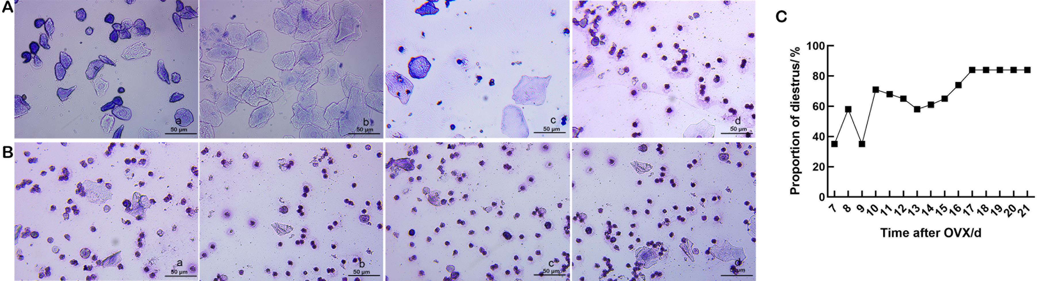
Figure 1 Vaginal smears after ovariectomy (OVX) of rats in each group (Wright's staining, ×40) (A and B)and the proportion of the diestrus period (C)

Figure 2 Body weight (A) and uterine wet weight/body weight (B) of rats from day 15–21 after ovariectomy (OVX)
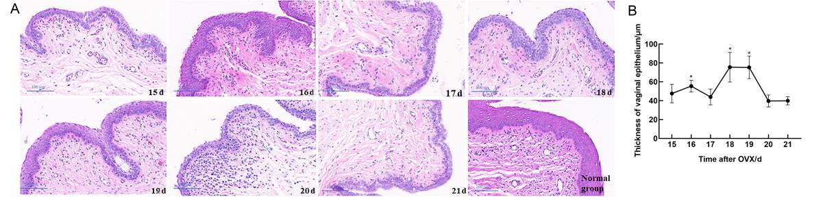
Figure 3 Histopathological changes of vaginal in rats after ovariectomy (OVX) and the thickness of vaginal epithelium
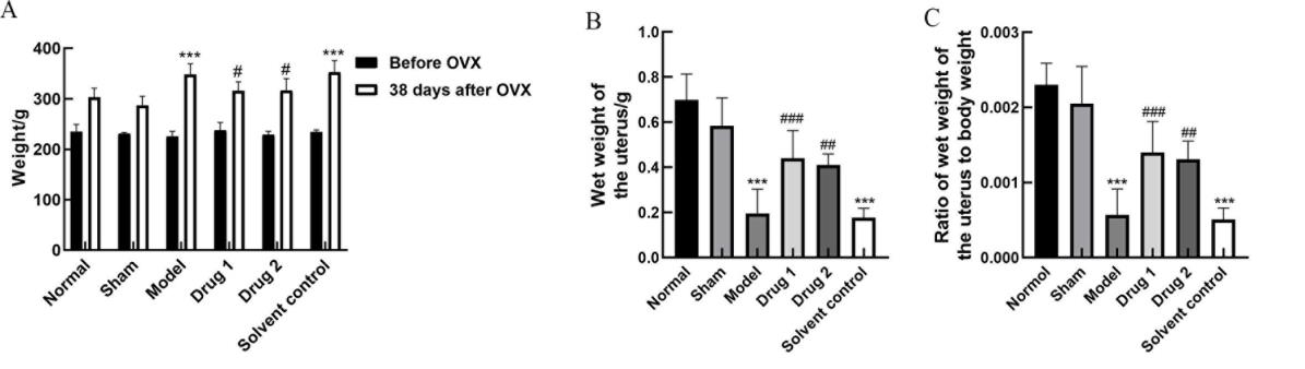
Figure 4 Body weight (A), uterine wet weight (B), and uterine wet weight/ body weight (C) of rats in each group after drug treatment
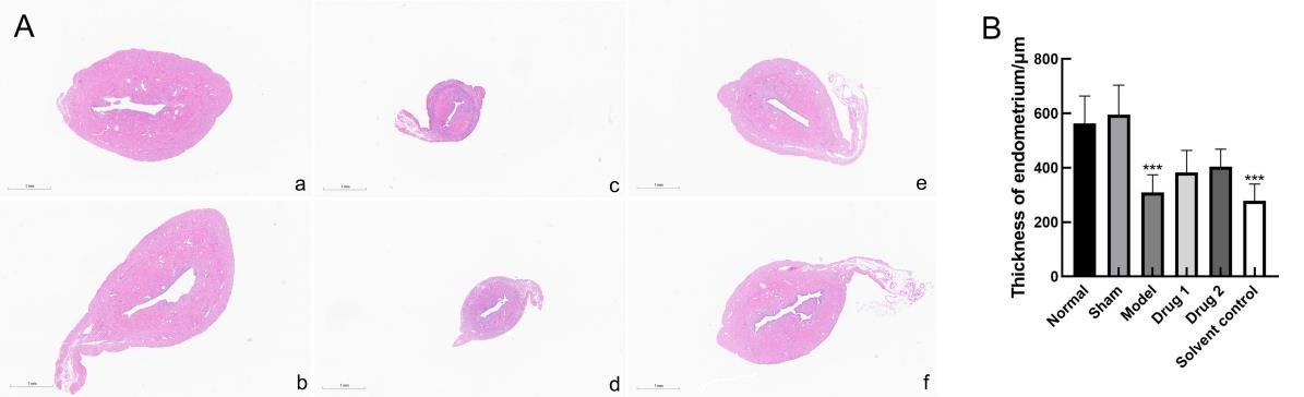
Figure 5 Histopathological changes of uterus tissue(HE, ×2)(A) and thickness of the endometrium (B)
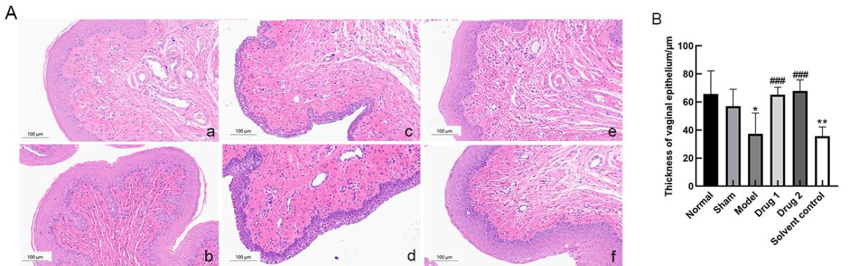
Figure 6 Histopathological changes of vaginal tissue (HE, ×20)(A) and thickness of the vaginal epithelium (B)
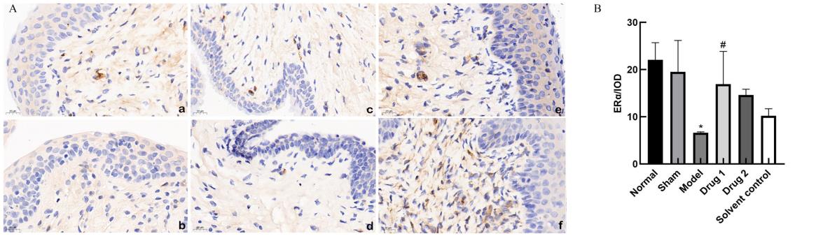
Figure 7 Immunohistochemistry (DAB, ×20)(A) and integral optical density (B) of ERα protein expression in rat vagina
| 1 | NAPPI R E, MARTINI E, CUCINELLA L, et al. Addressing vulvovaginal atrophy (VVA)/genitourinary syndrome of menopause (GSM) for healthy aging in women[J]. Front Endocrinol (Lausanne), 2019, 10:561. DOI:10.3389/fendo. 2019. 00561 . |
| 2 | MALDONADO P A, MONTOYA T I, ACEVEDO J F, et al. Effects of vaginal conjugated equine estrogens and ospemifene on the rat vaginal wall and lower urinary tract[J]. Biol Reprod, 2016, 96(1):81-92. DOI:10.1095/biolreprod.116. 144428 . |
| 3 | YOU S, LIU S B, DONG X J, et al. Intravaginal administration of human type III collagen-derived biomaterial with high cell-adhesion activity to treat vaginal atrophy in rats[J]. ACS Biomater Sci Eng, 2020, 6(4):1977-1988. DOI:10.1021/acsbiomaterials.9b01649 . |
| 4 | ZANNI P C, NEGRI M, SALCI T P, et al. Animal models for the effective development of atrophic vaginitis therapies: possibilities and limitations[J]. Expert Opin Drug Discov, 2014,9(3):269-281. DOI:10.1517/17460441.2014.877883 . |
| 5 | 吴琼. 雌激素联合甲硝唑治疗萎缩性阴道炎的临床疗效及其安全性[J]. 临床合理用药杂志, 2021, 14(21):145-147. DOI:10.15887/j.cnki.13-1389/r.2021.21.053 . |
| WU Q. Clinical efficacy and safety of estrogen combined with metronidazole in the treatment of atrophic vaginitis[J]. Chin J Clin Ration Drug Use, 2021, 14(21):145-147. DOI:10.15887/j.cnki.13-1389/r.2021.21.053 . | |
| 6 | DONDERS G G G, RUBAN K, BELLEN G,et al. Pharmaco-therapy for the treatment of vaginal atrophy[J]. Expert Opin Pharmacother, 2019, 20(7):821-835. DOI:10.1080/14656566. 2019. 1574752 . |
| 7 | MAYER L P, DEVINE P J, DYER C A, et al. The follicle-deplete mouse ovary produces androgen[J]. Biol Reprod, 2004, 71(1):130-138. DOI:10.1095/biolreprod.103.016113 . |
| 8 | ACOSTA J I, MAYER L, TALBOOM J S, et al. Transitional versus surgical menopause in a rodent model: etiology of ovarian hormone loss impacts memory and the acetylcholine system[J]. Endocrinology, 2009, 150(9):4248-4259. DOI:10.1210/en.2008-1802 . |
| 9 | SOCIETY N A M. The role of local vaginal estrogen for treatment of vaginal atrophy in postmenopausal women: 2007 position statement of The North American Menopause Society[J]. Menopause, 2007, 14(3):370-371. DOI:10.1097/gme.0b013e3180533b2a . |
| 10 | LÓPEZ-BELMONTE J, NIETO C, ESTEVEZ J, et al. Comparative uterine effects on ovariectomized rats after repeated treatment with different vaginal estrogen formulations[J]. Maturitas, 2012, 72(4):353-358. DOI:10.1016/j.maturitas.2012.05.007 . |
| 11 | PUNNONEN R, VILSKA S, GRÖNROOS M, et al. The vaginal absorption of oestrogens in post-menopausal women[J]. Maturitas, 1980, 2(4):321-326. DOI:10.1016/0378-5122(80)90034-1 . |
| 12 | CECCARELLI S, D'AMICI S, VESCARELLI E, et al. Topical KGF treatment as a therapeutic strategy for vaginal atrophy in a model of ovariectomized mice[J]. J Cell Mol Med, 2014, 18(9):1895-1907. DOI:10.1111/jcmm.12334 . |
| 13 | VAILATI S, MELLONI E, RISCASSI E, et al. Evaluation of the effects of a new intravaginal gel, containing purified bovine colostrum, on vaginal blood flow and vaginal atrophy in ovariectomized rat[J]. Sex Med, 2013, 1(2):35-43. DOI:10.1002/sm2.8 . |
| 14 | WOLFF J P, CACHELOU R, GUÉRITÉE N. Absence of systemic hormonal effects in an oestradiol diether topically active on the vaginal mucosa[J]. Maturitas, 1982, 4(4):239-246. DOI:10.1016/0378-5122(82)90054-8 . |
| 15 | PUP L D. Management of vaginal dryness and dyspareunia in estrogen sensitive cancer patients[J]. Gynecol Endocrinol, 2012, 28(9):740-745. DOI:10.3109/09513590.2011.652717 . |
| 16 | 闫丽, 温和, 唐桂毅, 等. 大鼠阴道细胞涂片不同染色方法在动情周期判定中的价值[J]. 药物评价研究, 2020, 43(1):72-76. DOI:10.7501/j.issn.1674-6376.2020.01.012 . |
| YAN L, WEN H, TANG G Y, et al. Diagnostic value of three staining methods for rat vaginal cell smears in determination of estrous cycle[J]. Drug Eval Res, 2020, 43(1):72-76. DOI:10.7501/j.issn.1674-6376.2020.01.012 . | |
| 17 | 刘翠萍, 张颖, 李宁莉. 干扰素联合红外线局部照射治疗人乳头瘤病毒及相关阴道炎和宫颈炎大鼠的作用及相关机制研究[J]. 临床和实验医学杂志, 2021, 20(13):1353-1357. DOI:10.3969/j.issn.1671-4695.2021.13.003 . |
| LIU C P, ZHANG Y, LI N L. Effect and mechanism of interferon combined with infrared local radiation in the treatment of rats with HPV related vaginitis and cervicitis[J]. J Clin Exp Med, 2021, 20(13):1353-1357. DOI:10.3969/j.issn.1671-4695. 2021. 13.003 . | |
| 18 | LI T, MA Y Y, ZHANG H, et al. Differential regulation of morphology and estrogen receptor-alpha expression in the vagina of ovariectomized adult virgin rats by estrogen replacement: a histological study[J]. Int J Endocrinol, 2016, 2016:1093512. DOI:10.1155/2016/1093512 . |
| 19 | 游爽. Ⅲ型胶原蛋白对大鼠阴道萎缩的作用及机制研究[D]. 重庆: 重庆医科大学, 2020. |
| YOU S. The effect and mechanism of type Ⅲ collagen on vaginal atrophy in rats [D].Chongqing: Chongqing Medical University, 2020. | |
| 20 | 李淑桢, 王琦, 李沁园, 等. 基于下丘脑-垂体-卵巢轴探讨藏药二十五味鬼臼丸对绝经后骨质疏松症大鼠的干预作用[J]. 中草药, 2021, 52(20):6282-6290. DOI:10.7501/j.issn.0253-2670.2021.20.019 . |
| LI S Z, WANG Q, LI Q Y, et al. Study on intervention effect of Tibetan medicine Ershiwuwei Guijiu Pill on PMOP rats based on HPOA[J]. Chin Tradit Herb Drugs, 2021, 52(20):6282-6290. DOI:10.7501/j.issn.0253-2670.2021.20.019 . | |
| 21 | YANG Q N, WANG C Y, JIN Y, et al. Disocin prevents postmenopausal atherosclerosis in ovariectomized LDLR-/- mice through a PGC-1α/ERα pathway leading to promotion of autophagy and inhibition of oxidative stress, inflammation and apoptosis[J]. Pharmacol Res, 2019, 148:104414. DOI:10.1016/j.phrs.2019.104414 . |
| 22 | 谢冰颖, 黄景文, 陈赛楠, 等. 补肾法对围绝经期综合征大鼠性激素及卵巢雌激素受体的影响[J]. 福建中医药, 2021, 52(10):11-13, 22. DOI:10.13260/j.cnki.jfjtcm.012341 . |
| XIE B Y, HUANG J W, CHEN S N, et al. Effect of kidney-invigorating method on the levels of sex hormone and ovarian estrogen receptor in perimenopausal syndrome rats[J]. Fujian J Tradit Chin Med, 2021, 52(10):11-13, 22. DOI:10.13260/j.cnki.jfjtcm.012341 . |
| [1] | WEN Fuli, XU Yongjun, FU Yunfeng, XUE Laien, CUI Linlin, WANG Junzhu, LU Jun, ZHENG Heping. Effectiveness Evaluation of Pre-Service Training for Laboratory Animal Practitioners Based on the "Three-Segmentation" Model [J]. Laboratory Animal and Comparative Medicine, 2026, (): 1-8. |
| [2] | LI Longxue, WAN Chongfan, ZHANG Qi, LEI Ruting, WANG Xiaoyue, CHENG Leyan, LAI Qi, LIU Ronghua, LIU Xuan, XU Tielong. Molecular Mechanisms of Qingfei Paidu Decoction in the Prevention and Treatment of Acute Lung Injury in Mice Based on miRNA Sequencing [J]. Laboratory Animal and Comparative Medicine, 2026, 46(3): 311-320. |
| [3] | PAN Linqin, DENG Xiangliang, LUO Yunxia. Advances in Integrative Translational Research on Animal Models of Ischemic Stroke in Traditional Chinese and Western Medicine [J]. Laboratory Animal and Comparative Medicine, 2026, 46(3): 344-356. |
| [4] | WANG Juan, XU Jiahui, TIAN Yunyuan, ZHANG Mengmeng, LI Min, WANG Siwang, LI Yao. Comparison and Behavioral Observation of Two Female Mice Models of Ulcerative Colitis [J]. Laboratory Animal and Comparative Medicine, 2026, 46(3): 332-343. |
| [5] | BU Yu, HOU Jinting, LI Yuanyuan, SHA Jingtao, XIE Chenlu, WANG Wengang, SUN Xingwei. A Review and Evaluation of Integrated Disease and Syndrome Animal Models for Hemorrhoids in Traditional Chinese and Western Medicine [J]. Laboratory Animal and Comparative Medicine, 2026, 46(3): 357-366. |
| [6] | WANG Xiuran, LI Hao, CHEN Zhengtao, YU Yang, ZHANG Suying, TAO Ru, WANG Kezhou. Innovation and Practice in the Construction of "Three-in-one" Talent Training Systems for Laboratory Animal Professionals in Medical Colleges [J]. Laboratory Animal and Comparative Medicine, 2026, 46(3): 446-455. |
| [7] | LI Jiafei, ZHANG Zhenhao, WANG Shuo, TIAN Ge, WEN Shuang, YAN Yuxue, CUI Ran, YE Zhen, CUI Yongchun. Effects of Autonomic Neuromodulators on Atrial Electrical Remodeling and Histopathological Changes in a Rat Model of Atrial Fibrillation [J]. Laboratory Animal and Comparative Medicine, 2026, 46(3): 321-331. |
| [8] | Committee of Experts on Medical Animal Experiments, Chinese Research Hospital Association , Committee of Regenerative Medicine Branch, Chinese Medicinal Biotech Association , HAN Fabin, CHEN Lin, CHEN Zhiguo, LU Ming, LI Yingjun. Guidelines for Selecting Preclinical Animal Models for Drugs and Stem Cell Therapies for Parkinson Disease (2026 Edition) [J]. Laboratory Animal and Comparative Medicine, 2026, 46(2): 153-177. |
| [9] | LI Hui. Advances in Animal Models for Biolinguistic Research [J]. Laboratory Animal and Comparative Medicine, 2026, 46(2): 297-305. |
| [10] | RONG Wenshuang, NIU Yuanfei, LIU Meiting, YANG Mengyuan, CUI Shuang, MA Lina, FU Yao, WANG Lianmei, CAO Junling. Influence of Antigen Type on the Establishment of an Induced Sjögren Syndrome Mouse Model [J]. Laboratory Animal and Comparative Medicine, 2026, 46(2): 178-190. |
| [11] | LIU Sai, FU Bin, LI Sidi, CHEN Zhida, ZHANG Yue, GUO Zhongkun, WANG Yongan, WANG Kezhou. Adra2a Regulates LPS-Induced Inflammation in Hepatocytes of Lbp-/- Mice via the MAPK Signaling Pathway [J]. Laboratory Animal and Comparative Medicine, 2026, 46(2): 212-221. |
| [12] | LIU Yishu, CAI Liping. Analysis of Current Practices and Exploration of Alternative Technologies in Use of Laboratory Animals for Minimally Invasive Surgery Education and Training [J]. Laboratory Animal and Comparative Medicine, 2026, 46(2): 279-287. |
| [13] | TANG Xiaohang, GU Yingmin, LÜ Yangyang, HUANG Mingshu, TIAN Xuesong. Evaluation of the Histological Staining Performance of Rat Eyeball Sections Prepared Using a Self-Developed Fixative [J]. Laboratory Animal and Comparative Medicine, 2026, 46(2): 261-270. |
| [14] | SONG Jing, YANG Zongtong, LI Xiaojing, LI Zifa, SU Fengyun, XU Dongchuan, SUI Zaiyun. Effects of Xiebai San on the Morphological Structures of Lung and Intestinal Tissues and Expression Levels of PI3K and Akt in Rats with Allergic Asthma [J]. Laboratory Animal and Comparative Medicine, 2026, 46(2): 191-204. |
| [15] | JIANG Haitao, YUAN Hantao, HUANG Wenting, YANG Rongrong, CHEN Xiaochun, YU Baoqing, LI Sibo. Regulation of Rat Intervertebral Disc Annulus Fibrosus Cell Proliferation and Apoptosis by Yaoshu Zhuyu Fang via miR-17-5P/MDM2/p53 Pathway [J]. Laboratory Animal and Comparative Medicine, 2026, 46(1): 55-65. |
| Viewed | ||||||
|
Full text |
|
|||||
|
Abstract |
|
|||||