



Laboratory Animal and Comparative Medicine ›› 2026, Vol. 46 ›› Issue (5): 668-676.DOI: 10.12300/j.issn.1674-5817.2026.001
• Animal Models of Human Diseases • Previous Articles Next Articles
ZHANG Panpan1,2, XU Jinsen2, CHENG Youmin3, WU Huasong3, LIN Jingyu1,2( )(
)( )
)
Received:2026-01-03
Revised:2026-06-11
Online:2026-10-25
Published:2026-10-16
Correspondence to:
LIN Jingyu
CLC Number:
ZHANG Panpan,XU Jinsen,CHENG Youmin,et al. Establishment and Evaluation of a Rat Model of Chronic Obstructive Pulmonary Disease with Phlegm-Dampness Syndrome[J]. Laboratory Animal and Comparative Medicine, 2026, 46(5): 668-676. DOI: 10.12300/j.issn.1674-5817.2026.001.
Add to citation manager EndNote|Ris|BibTeX
URL: https://www.slarc.org.cn/dwyx/EN/10.12300/j.issn.1674-5817.2026.001
Traditional Chinese medicine syndrome scoring scale for rats with phlegm-dampness syndrome in chronic obstructive pulmonary disease models
| 评分 Score | 大鼠行为 Rat behavior | 精神状态 Mental state | 毛发、耳、尾 Hair, ear, tail | 舌苔 Tongue coating | 饮食、体重 Diet and body weight |
|---|---|---|---|---|---|
| 0 | 正常 | 正常 | 正常 | 正常 | 正常 |
| 1 | 咳嗽、扎堆、倦怠、弓背、轻度动作迟缓 | 萎靡、嗜睡 | 毛发稀疏,耳、尾血色淡 | 舌苔淡红,有薄苔 | 进食减少、饮水增加 |
| 2 | 喘息、重度动作迟缓 | 反应迟钝 | 毛色干枯无光泽、毛发蓬松 | 舌苔白滑 | 体重减轻或增长缓慢 |
Table 1
Traditional Chinese medicine syndrome scoring scale for rats with phlegm-dampness syndrome in chronic obstructive pulmonary disease models
| 评分 Score | 大鼠行为 Rat behavior | 精神状态 Mental state | 毛发、耳、尾 Hair, ear, tail | 舌苔 Tongue coating | 饮食、体重 Diet and body weight |
|---|---|---|---|---|---|
| 0 | 正常 | 正常 | 正常 | 正常 | 正常 |
| 1 | 咳嗽、扎堆、倦怠、弓背、轻度动作迟缓 | 萎靡、嗜睡 | 毛发稀疏,耳、尾血色淡 | 舌苔淡红,有薄苔 | 进食减少、饮水增加 |
| 2 | 喘息、重度动作迟缓 | 反应迟钝 | 毛色干枯无光泽、毛发蓬松 | 舌苔白滑 | 体重减轻或增长缓慢 |
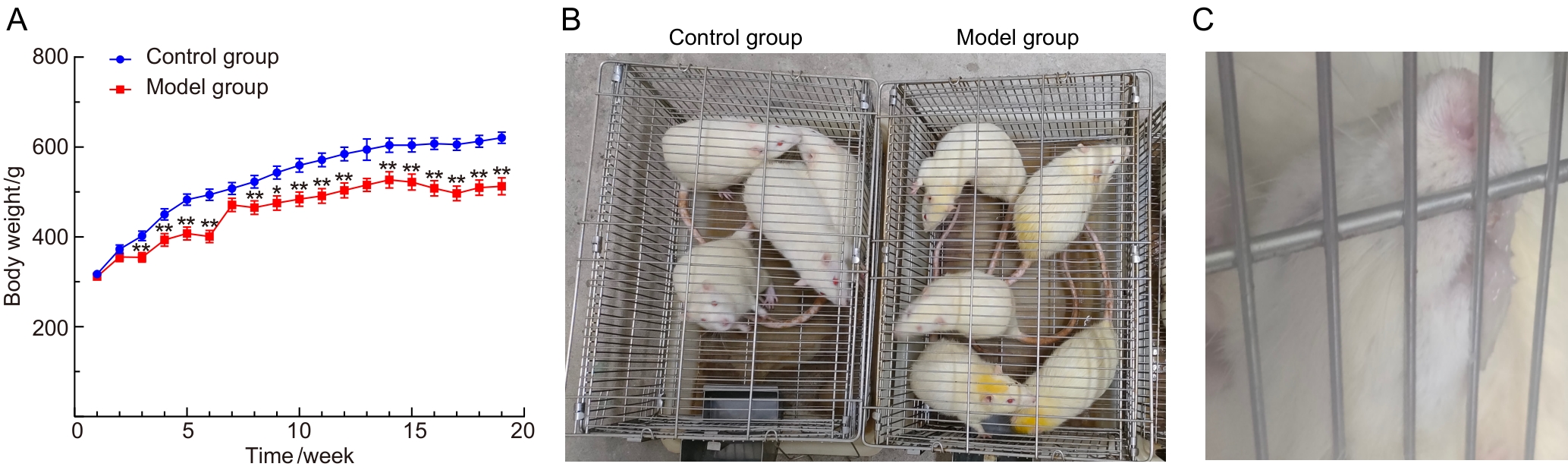
Figure 1
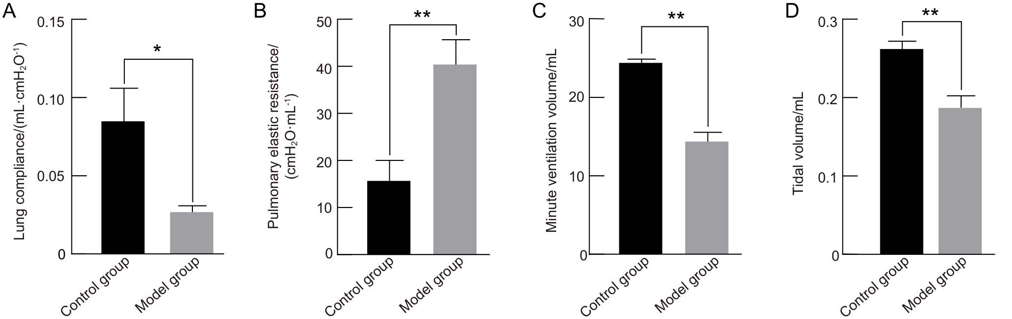
Figure 2
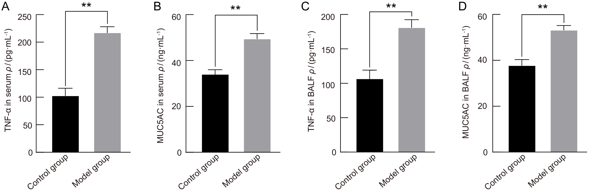
Figure 3
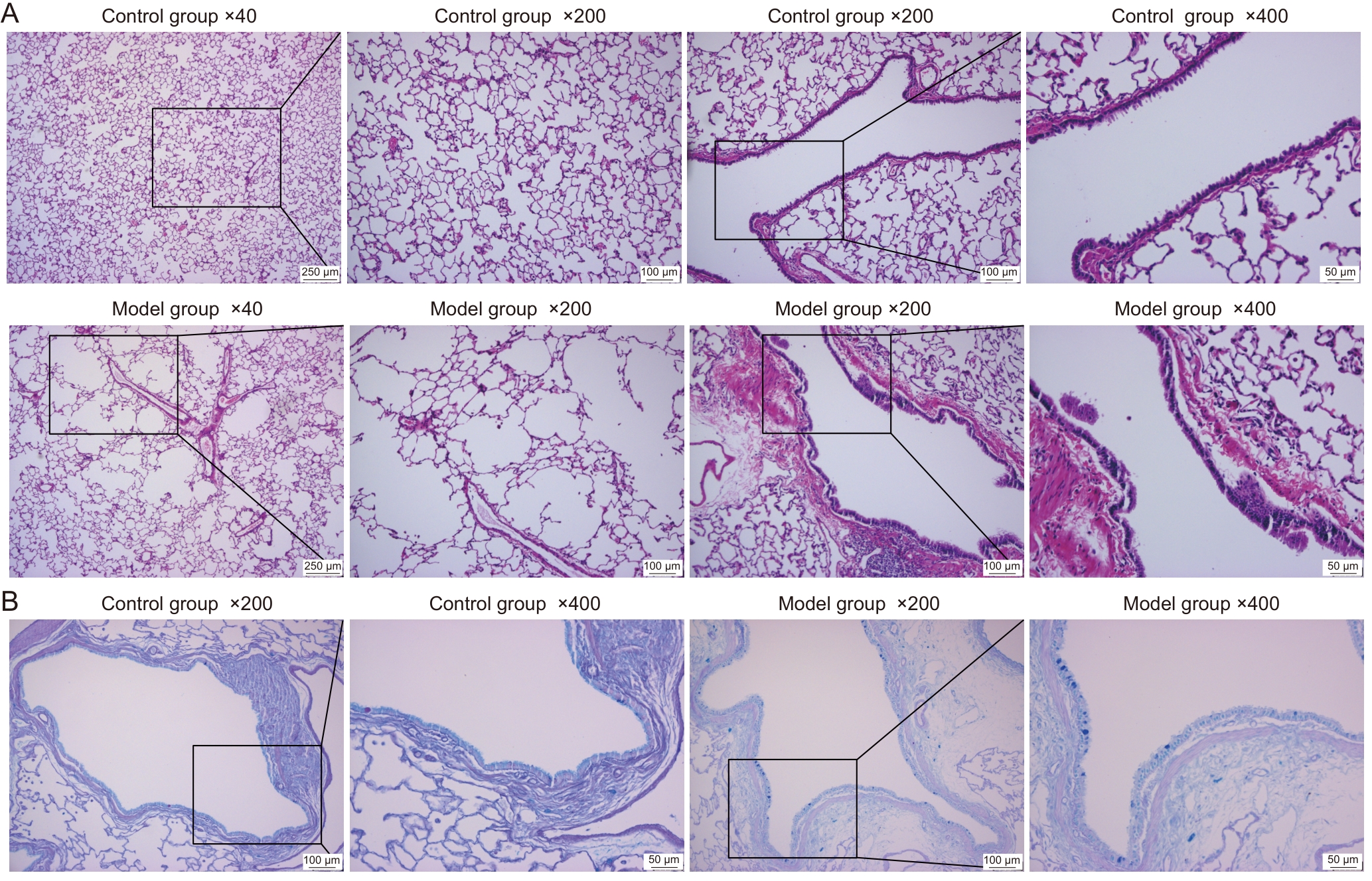
Figure 4
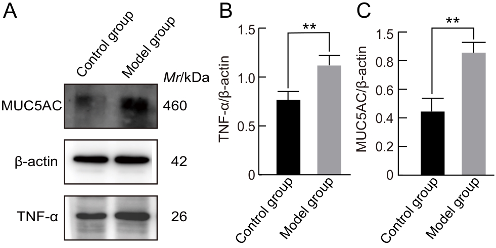
Figure 5
| [1] |
BOERS E, ALLEN A, BARRETT M, et al. Forecasting the global economic and health burden of COPD from 2025 through 2050[J]. Chest, 2025, 168(4): 880-889. DOI: 10.1016/j.chest. 2025.03.029.
|
| [2] |
AGUSTÍ A, CELLI B R, CRINER G J, et al. Global initiative for chronic obstructive lung disease 2023 report: gold executive summary[J]. Eur Respir J, 2023, 61(4): 2300239. DOI: 10.1183/13993003.00239-2023.
|
| [3] |
李建生. 国际中医临床实践指南 慢性阻塞性肺疾病[J]. 世界中医药, 2020, 15(7): 1084-1092. DOI: 10.3969/j.issn.1673-7202.2020.07.026.
|
|
LI J S. International clinical practice guideline of Chinese medicine chronic obstructive pulmonary disease[J]. World Chin Med, 2020, 15(7): 1084-1092. DOI: 10.3969/j.issn.1673-7202.2020.07.026.
|
|
| [4] |
王笑铭, 孟晨晨, 范鹿, 等. 中医证候类动物模型构建方法概述[J]. 实验动物与比较医学, 2025, 45(5): 596-610. DOI: 10.12300/j.issn.1674-5817.2025.038.
|
|
WANG X M, MENG C C, FAN L, et al. An overview of strategies for constructing animal models of traditional Chinese medicine syndromes[J]. Lab Anim Comp Med, 2025, 45(5): 596-610. DOI: 10.12300/j.issn.1674-5817.2025.038.
|
|
| [5] |
冯硕, 胡晶, 张会娜, 等. 中药新药研发中人用经验数据的评价方法探索及思考[J]. 中国中药杂志, 2022, 47(6): 1700-1704. DOI: 10.19540/j.cnki.cjcmm.20220105.501.
|
|
FENG S, HU J, ZHANG H N, et al. Exploration and reflection on evaluation methodology of human use experience for new Chinese drug development[J]. China J Chin Mater Med, 2022, 47(6): 1700-1704. DOI: 10.19540/j.cnki.cjcmm.20220105.501.
|
|
| [6] |
黄锦榕, 洪敏俐, 李灿东, 等. 基于聚类分析的慢阻肺稳定期中医证型分布特征及其与临床综合指标相关性研究[J]. 亚太传统医药, 2018, 14(5): 107-111. DOI: 10.11954/ytctyy.201805038.
|
|
HUANG J R, HONG M L, LI C D, et al. Study of distribution characteristics of TCM syndromes in the stable COPD based on the cluster analysis and its correlation with clinical comprehensive indexes[J]. Asia Pac Tradit Med, 2018, 14(5): 107-111. DOI: 10.11954/ytctyy.201805038.
|
|
| [7] |
邵雪洁, 徐可心, 廖一羲, 等. 呼吸系统疾病病证结合动物模型的研究进展[J]. 中华中医药杂志, 2024, 39(6): 2982-2986.
|
|
SHAO X J, XU K X, LIAO Y X, et al. Research progress of animal models of combination of disease and syndrome in respiratory diseases[J]. China J Tradit Chin Med Pharm, 2024, 39(6): 2982-2986.
|
|
| [8] |
黄培炜, 林静瑜, 周凡, 等. 尺泽穴位注射对慢阻肺大鼠气道组织形态学和氧化水平的影响[J]. 临床肺科杂志, 2020, 25(11): 1667-1671. DOI: 10.3969/j.issn.1009-6663.2020.11.010.
|
|
HUANG P W, LIN J Y, ZHOU F, et al. Effect of LU5 point injection on airway histomorphology and oxidative stress in rats with COPD[J]. J Clin Pulm Med, 2020, 25(11): 1667-1671. DOI: 10.3969/j.issn.1009-6663.2020.11.010.
|
|
| [9] |
刘思美, 强喆, 吴思澜, 等. 痰湿阻肺型COPD大鼠模型的构建及代谢组学分析[J]. 中国实验动物学报, 2023, 31(1): 51-63. DOI: 10.3969/j.issn.1005-4847.2023.01.007.
|
|
LIU S M, QIANG Z, WU S L, et al. Establishment and metabolomics analysis of a rat model of chronic obstructive pulmonary disease with phlegm-dampness syndrome[J]. Acta Lab Anim Sci Sin, 2023, 31(1): 51-63. DOI: 10.3969/j.issn.1005-4847.2023.01.007.
|
|
| [10] |
春柳, 李建生, 马锦地, 等. 基于现代名老中医经验的痰饮常见证候及其特征的研究[J]. 辽宁中医杂志, 2018, 45(3): 496-498. DOI: 10.13192/j.issn.1000-1719.2018.03.013.
|
|
CHUN L, LI J S, MA J D, et al. Study on the common syndromes and characteristics of phlegm and drink based on the experience of modern famous and old Chinese medicine practitioners[J]. Liaoning J Tradit Chin Med, 2018, 45(3): 496-498. DOI: 10.13192/j.issn.1000-1719.2018.03.013.
|
|
| [11] |
罗齐军, 陈媛丽, 夏燕华, 等. 三子六君汤联合穴位贴敷治疗慢阻肺急性加重期临床研究[J]. 亚太传统医药, 2017, 13(14): 142-144. DOI: 10.11954/ytctyy.201714066.
|
|
LUO Q J, CHEN Y L, XIA Y H, et al. Clinical observation of AECOPD treated by San zi Liu Jun decoction combined with acupoint application of Chinese herbal medicine[J]. Asia Pac Tradit Med, 2017, 13(14): 142-144. DOI: 10.11954/ytctyy.201714066.
|
|
| [12] |
李会革, 王迪浔. 实验动物呼吸力学指标的测定[J]. 同济医科大学学报, 1995, 24(2): 91-93.
|
|
LI H G, WANG D X. Measurement of respiratory mechanics in experimental animals[J]. Acta Univ Med Tongji, 1995, 24(2): 91-93.
|
|
| [13] |
陈美凤, 加慧, 夏书月. 不同COPD动物模型特点及应用的研究进展[J]. 中国实验动物学报, 2020, 28(4): 563-569. DOI: 10.3969/j.issn.1005-4847.2020.04.019.
|
|
CHEN M F, JIA H, XIA S Y. Research progress on the characteristics of animal models of chronic obstructive pulmonary disease and related clinical applications[J]. Acta Lab Anim Sci Sin, 2020, 28(4): 563-569. DOI: 10.3969/j.issn.1005-4847.2020.04.019.
|
|
| [14] |
刘迪, 张洪春. 慢性阻塞性肺疾病基因工程动物模型研究进展[J]. 中国生物工程杂志, 2020, 40(4): 59-68. DOI: 10.13523/j.cb.1909072.
|
|
LIU D, ZHANG H C. Advances in genetically engineered animal models of chronic obstructive pulmonary disease[J]. China Biotechnol, 2020, 40(4): 59-68. DOI: 10.13523/j.cb.1909072.
|
|
| [15] |
张泽宇, 梅晓峰, 陶柳颖, 等. 香烟烟雾联合肺炎克雷伯杆菌诱导慢阻肺模型小鼠气道重塑机制[J]. 中国实验动物学报, 2025, 33(5): 644-655. DOI: 10.3969/j.issn.1005-4847.2025.05.003.
|
|
ZHANG Z Y, MEI X F, TAO L Y, et al. Mechanism of airway remolding in a mouse model of chronic obstructive pulmonary disease induced by cigarette smoke combined with Klebsiella pneumoniae[J]. Acta Lab Anim Sci Sin, 2025, 33(5): 644-655. DOI: 10.3969/j.issn.1005-4847.2025.05.003.
|
|
| [16] |
李武, 刘雲华, 石拓, 等. LPS+CS诱导12周建立符合临床标准的COPD小鼠模型[J]. 陆军军医大学学报, 2023, 45(9): 982-988. DOI: 10.16016/j.2097-0927.202301084.
|
|
LI W, LIU Y H, SHI T, et al. Establish of a mouse model of COPD that meets clinical criteria by LPS+ cigarette smoke exposure for 12 weeks[J]. J Army Med Univ, 2023, 45(9): 982-988. DOI: 10.16016/j.2097-0927.202301084.
|
|
| [17] |
邓铁涛. 中医诊断学[M]. 3版. 上海: 上海科学技术出版社, 2013.
|
|
DENG T T. Diagnostics of traditional Chinese medicine[M]. 3rd ed. Shanghai: Shanghai Scientific and Technical Publishers, 2013.
|
|
| [18] |
YUK J M, KIM J K, KIM I S, et al. TNF in human tuberculosis: a double-edged sword[J]. Immune Netw, 2024, 24(1): e4. DOI: 10.4110/in.2024.24.e4.
|
| [19] |
SONG K S, LEE W J, CHUNG K C, et al. Interleukin-1β and tumor necrosis factor-α induce MUC5AC overexpression through a mechanism involving ERK/p38 mitogen-activated protein kinases-MSK1-CREB activation in human airway epithelial cells[J]. J Biol Chem, 2003, 278(26): 23243-23250. DOI: 10.1074/jbc.M300096200.
|
| [20] |
LORA J M, ZHANG D M, LIAO S M, et al. Tumor necrosis factor-α triggers mucus production in airway epithelium through an IκB kinase β-dependent mechanism[J]. J Biol Chem, 2005, 280(43): 36510-36517. DOI: 10.1074/jbc.M507977200.
|
| [21] |
FU H T, ZHANG Y, ZHANG P, et al. Tumor necrosis factor-α promotes airway mucus hypersecretion by repressing miR-146a-5p and miR-134-5p levels in human airway epithelial cells[J]. Transl Cancer Res, 2021, 10(9): 4047-4056. DOI: 10.21037/tcr-20-3375.
|
| [22] |
YAO Y, ZHOU J, DIAO X, et al. Association between tumor necrosis factor-α and chronic obstructive pulmonary disease: a systematic review and meta-analysis[J]. Ther Adv Respir Dis, 2019, 13: 1753466619866096. DOI: 10.1177/1753466619866096.
|
| [23] |
杜利莉, 章丹宁, 薛梅, 等. 中医证候动物模型建立及评价方法研究[J]. 南京中医药大学学报, 2025, 41(1): 11-18. DOI: 10.14148/j.issn.1672-0482.2025.0011.
|
|
DU L L, ZHANG D N, XUE M, et al. Research on establishment and evaluation methods of animal model of TCM syndromes[J]. J Nanjing Univ Tradit Chin Med, 2025, 41(1): 11-18. DOI: 10.14148/j.issn.1672-0482.2025.0011.
|
|
| [24] |
林浩. 慢性阻塞性肺病寒饮蕴肺证大鼠模型血清细胞因子表达的动态研究[D]. 济南: 山东中医药大学, 2014.
|
|
LIN H. Dynamic study on serum cytokine expressions in rat models of chronic obstructive pulmonary disease with cold‑fluid accumulating in lung syndrome[D]. Jinan: Shandong University of Traditional Chinese Medicine, 2014.
|
|
| [25] |
孙广仁, 王洪武, 高博, 等. COPD寒饮蕴肺证大鼠病证结合模型的建立[J]. 山东中医药大学学报, 2007, 31(3): 242-244. DOI: 10.3969/j.issn.1007-659X.2007.03.025.
|
|
SUN G R, WANG H W, GAO B, et al. Establishment of a disease-syndrome combined rat model of COPD with cold fluid retention in lung syndrome[J]. J Shandong Univ Tradit Chin Med, 2007, 31(3): 242-244. DOI: 10.3969/j.issn.1007-659X.2007.03.025.
|
|
| [26] |
高旅, 刘丽娜, 史正刚. 从中医角度看气道黏液高分泌[J]. 中国中西医结合儿科学, 2019, 11(2): 133-135. DOI: 10.3969/j.issn.1674-3865.2019.02.011.
|
|
GAO L, LIU L N, SHI Z G. Airway mucus hypersecretion from the perspective of traditional Chinese medicine[J]. Chin Pediatr Integr Tradit West Med, 2019, 11(2): 133-135. DOI: 10.3969/j.issn.1674-3865.2019.02.011.
|
|
| [27] |
RADICIONI G, CEPPE A, FORD A A, et al. Airway mucin MUC5AC and MUC5B concentrations and the initiation and progression of chronic obstructive pulmonary disease: an analysis of the SPIROMICS cohort[J]. Lancet Respir Med, 2021, 9(11): 1241-1254. DOI: 10.1016/S2213-2600(21)00079-5.
|
| [28] |
LI J Y, YE Z G. The potential role and regulatory mechanisms of MUC5AC in chronic obstructive pulmonary disease[J]. Molecules, 2020, 25(19): 4437. DOI: 10.3390/molecules25194437.
|
| [1] | ZHANG Haitao, WEI Yalan, FAN Xiaojing, CHEN Lu, ZHUANG Xudong, WANG Xinrui. Anti-Inflammatory Effects of the Triterpenoid Saponin-Enriched Fraction from She Medicine Clematis florida and Exploration of Its Mechanisms in a Rat Model of Cervicitis [J]. Laboratory Animal and Comparative Medicine, 2026, 46(5): 616-628. |
| [2] | TIAN Yan, LAI Dong, LAI Fei, JI Changfu. Study on the Relationship Between Changes in Heparin-Binding Protein Levels and Coagulation Disorders and Vascular Endothelial Injury in a Rat Model of Sepsis [J]. Laboratory Animal and Comparative Medicine, 2026, 46(5): 700-706. |
| [3] | LI Yangjin, WU Zhenhong, HUANG Zongxiu. 28-Day Oral Toxicity Study of Ganoderma lucidum Spore Oil in Rats [J]. Laboratory Animal and Comparative Medicine, 2026, 46(5): 660-667. |
| [4] | LI Yanping, HUANG Shan, YAO Jengyuan, SHI Yimin, SUI Huaxiu, HUANG Houtan, YU Yanping, MI Yan, LIN Tahui. Protective Effect and Mechanism of Artemitin on Ozone-Induced Pulmonary Inflammation in Mice [J]. Laboratory Animal and Comparative Medicine, 2026, 46(5): 640-649. |
| [5] | CHEN Wanlin, DONG Qingrui, LI Mengtao, LIN Nan, ZHAO Hu, ZHANG Zaizhong, ZENG Lingjun, LI Pei, YANG Jin, WANG Yu, HUANG Aiwen. Application of Carbon Nanoparticle-Manganese Dioxide Injection in Gastrointestinal Staining-Based Marking and Localization in Rats and Pigs [J]. Laboratory Animal and Comparative Medicine, 2026, 46(5): 727-739. |
| [6] | LIAN Ronggui, LIAO Naishun. In Vivo Tracking of Adipose-Derived Mesenchymal Stem Cell Therapy for Subacute Liver Injury Using Rare-Earth Down-Conversion Nanoparticles [J]. Laboratory Animal and Comparative Medicine, 2026, 46(5): 716-726. |
| [7] | AI Xiufeng, ZHANG Lizong, FANG Mingsun, LÜ Dongying, CHEN Chu, CAI Zhaowei, WANG Dejun. Analysis of Differences in the Intestinal Flora of Rats and Mice after Drinking Chlorinated Water Based on 16S rRNA Sequencing [J]. Laboratory Animal and Comparative Medicine, 2026, 46(3): 437-445. |
| [8] | HE Jiaqi, ZHOU Yuanyuan, NIE Yongqiang, WANG Zhaoxia, XU Wangjie. Establishment and Multidimensional Pathological Evaluations of a Cigarette Smoke Exposure-Induced Chronic Obstructive Pulmonary Disease Mouse Model [J]. Laboratory Animal and Comparative Medicine, 2026, 46(1): 11-19. |
| [9] | QIN Chao, LI Shuangxing, ZHAO Tingting, JIANG Chenchen, ZHAO Jing, YANG Yanwei, LIN Zhi, WANG Sanlong, WEN Hairuo. Study on the 90-day Feeding Experimental Background Data of SD Rats for Drug Safety Evaluation [J]. Laboratory Animal and Comparative Medicine, 2025, 45(4): 439-448. |
| [10] | PAN Yicong, JIANG Wenhong, HU Ming, QIN Xiao. Optimization of Surgical Procedure and Efficacy Evaluation of Aortic Calcification Model in Rats with Chronic Kidney Disease [J]. Laboratory Animal and Comparative Medicine, 2025, 45(3): 279-289. |
| [11] | Jian GE, Jingfen SUN, Yongjie WU. Taurine Has no Protective Effect on Rat Corneal Endothelial Cells Injured by Benzalkonium Chloride [J]. Laboratory Animal and Comparative Medicine, 2023, 43(1): 39-43. |
| [12] | Qin XU, Yan NI, Wenhui SHI, Jianying LI, Jiangwei LIU, Hongqiong ZHAO, Xinming XU. Analysis on Ileum and Colon Microflora of SPF Male SD Rats based on High-throughput Sequencing [J]. Laboratory Animal and Comparative Medicine, 2023, 43(1): 53-60. |
| [13] | Xinpeng LU, Rong LIU, Wenbo Huang, Jin ZHAO, Hongtao LI. A Comparative Study of Chronic Obstructive Pulmonary Disease Rat Models Established by Different Methods [J]. Laboratory Animal and Comparative Medicine, 2022, 42(3): 201-206. |
| [14] | ZUO Ximeng, SHI Xiaoguang, LIU Jieli, YANG Zhenrui, GAO Xiang, LAI Rui, ZHAO Ze, WANG Tangshun. Establishment of a Rat Model of Granulomatous Lobular Mastitis [J]. Laboratory Animal and Comparative Medicine, 2021, 41(4): 299-304. |
| [15] | XU Qin, WANG Wei, DONG Xiang, LI Jianying, SHI Wenhui, LIU Xiaolu, LIU Aizhong, MA Na, SONG Laiyang, LIU Jiangwei. Chronic Toxicity Study on Deinococcus Radiodurans R1 in Rats [J]. Laboratory Animal and Comparative Medicine, 2021, 41(4): 333-342. |
| Viewed | ||||||
|
Full text |
|
|||||
|
Abstract |
|
|||||
