
Laboratory Animal and Comparative Medicine ›› 2022, Vol. 42 ›› Issue (3): 177-186.DOI: 10.12300/j.issn.1674-5817.2022.001
• Animal Models of Human Diseases • Previous Articles Next Articles
Jiani LIU1,2,3( ), Jiangang LIU1(
 ), Yun WEI1()(), Hao LI4, Zenggang LUO1, Yi WANG3, Kun LI5
Received:2022-01-04
Revised:2022-03-25
Online:2022-06-25
Published:2022-07-01
Contact:
Jiangang LIU, Yun WEI
About author:LIU Jiangang E-mail: liujiangang2002@sina.comCLC Number:
Jiani LIU, Jiangang LIU, Yun WEI, Hao LI, Zenggang LUO, Yi WANG, Kun LI. Pathological and Synaptic Morphological Changes of the Olfactory Bulb in APP/PS1 Model Mice at Different Ages and the Intervention Effect of Memantine[J]. Laboratory Animal and Comparative Medicine, 2022, 42(3): 177-186.
Add to citation manager EndNote|Ris|BibTeX
URL: https://www.slarc.org.cn/dwyx/EN/10.12300/j.issn.1674-5817.2022.001
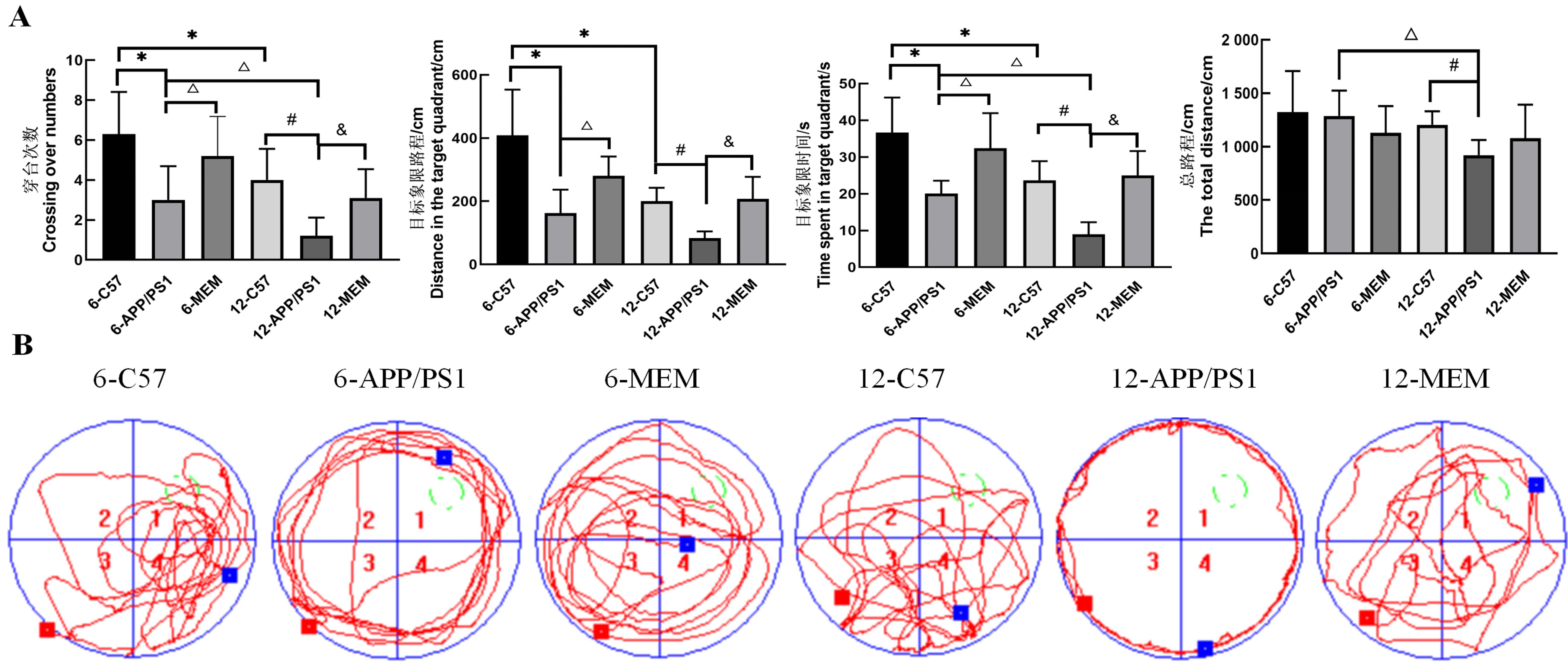
Figure 1 Morris water maze test to evaluate the behavioral changes of mice in each groupNote: Fig. A shows the crossing numbers, distance in the target quadrant, time spent in the target quadrant, and the total distance of mice in each group. Fig. B shows the swimming track of mice in each group in the water maze (the green circle represents the position of the original escape platform, the red dot represents the entry point, and the blue dot represents the 90 s cut-off point). 6-C57 refers to 6-month-old C57BL/6 mice in the blank control group. 6-APP/PS1 refers to 6-month-old APP/PS1 double transgenic mice in the model group; 6-MEM refers to APP/PSI double transgenic mice treated with 2.6 mg/kg memantine hydrochloride from 3- to 6-month-old; 12-C57 refers to 12-month-old C57BL/6 mice; 12-APP/PS1 refers to 12-month-old APP/PS1 double transgenic mice; 12-MEM refers to APP/PS1 double transgenic mice treated with 2.6 mg/kg memantine hydrochloride from 9- to 12-month-old. Each group contained 10 mice. Compared with 6-C57, *P < 0.05; compared with 12-C57, #P < 0.05; compared with 6-APP/PS1, △P < 0.05; compared with 12-APP/PS1, &P < 0.05.
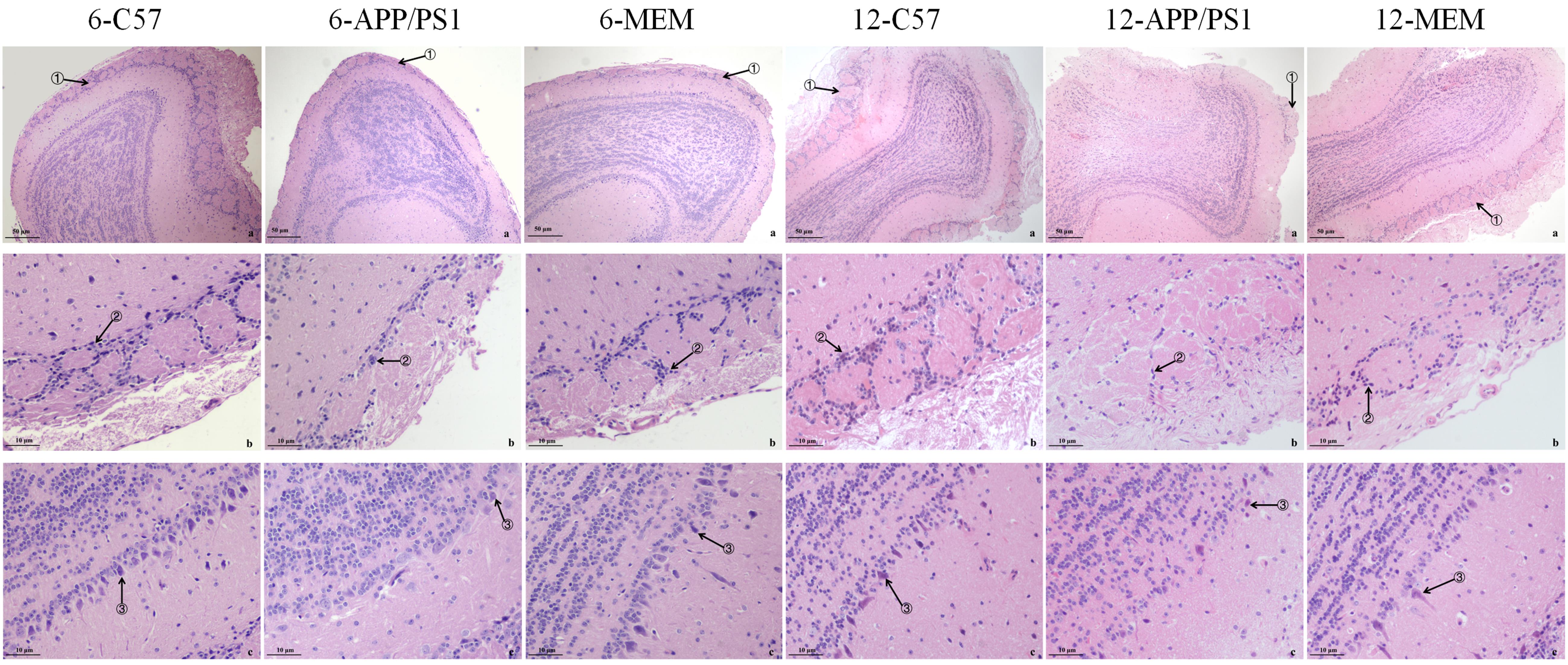
Figure 2 Pathological changes of the olfactory bulb in mice in each group observed by optical microscopy after hematoxylin-eosin stainingNote: Fig. a is the olfactory bulb (×40); Fig. b is the glomerular layer (×200); Fig. c is the mitral cell layer (×200). Arrows ①, ②, and ③ show glomeruli, periglomerular, and mitral cells, respectively. 6-C57 refers to 6-month-old C57BL/6 mice in the blank control group; 6-APP/PS1 refers to 6-month-old APP/PS1 double transgenic mice in the model group; 6-MEM refers to APP/PSI double transgenic mice treated with 2.6 mg/kg memantine hydrochloride from 3- to 6-month-old; 12-C57 refers to 12-month-old C57BL/6 mice; 12-APP/PS1 refers to 12-month-old APP/PS1 double transgenic mice; 12-MEM refers to APP/PS1 double transgenic mice treated with 2.6 mg/kg memantine hydrochloride from 9- to 12-month-old. Each group contained five mice.
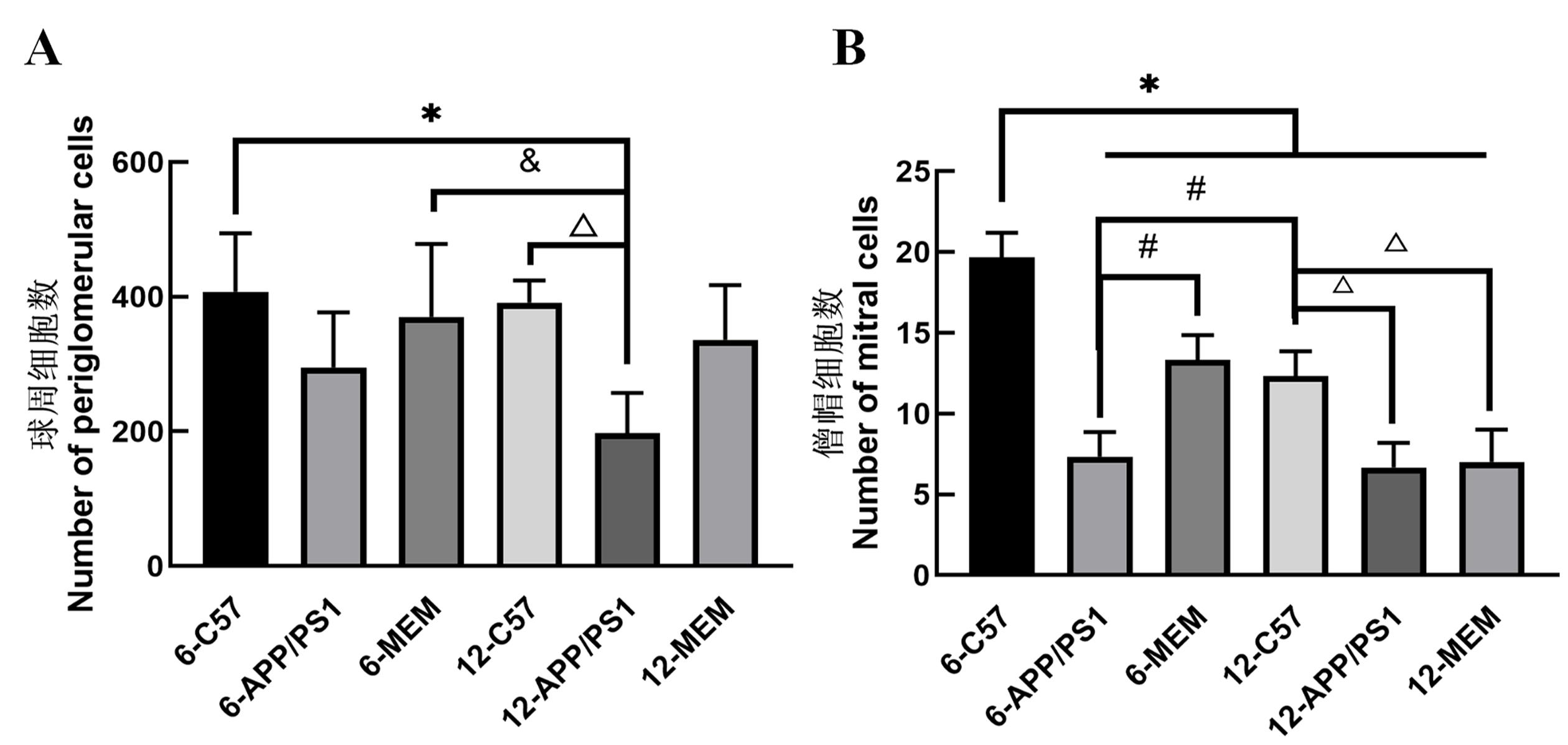
Figure 3 Effects of memantine on periglomerular cells (A) and mitral cells (B)Note: 6-C57 refers to 6-month-old C57BL/6 mice in the blank control group; 6-APP/PS1 refers to 6-month-old APP/PS1 double transgenic mice in model group; 6-MEM refers to APP/PSI double transgenic mice treated with 2.6 mg/kg memantine hydrochloride from 3- to 6-month-old; 12-C57 refers to 12-month-old C57BL/6 mice; 12-APP/PS1 refers to 12-month-old APP/PS1 double trans-genic mice; 12-MEM refers to APP/PS1 double transgenic mice treated with 2.6 mg/kg memantine hydrochloride from 9- to 12-month-old. Each group contained five mice. Compared with 6-C57, *P < 0.05; compared with 12-C57, #P < 0.05; compared with 6-APP/PS1, △P < 0.05; compared with 12-APP/PS1, &P < 0.05.
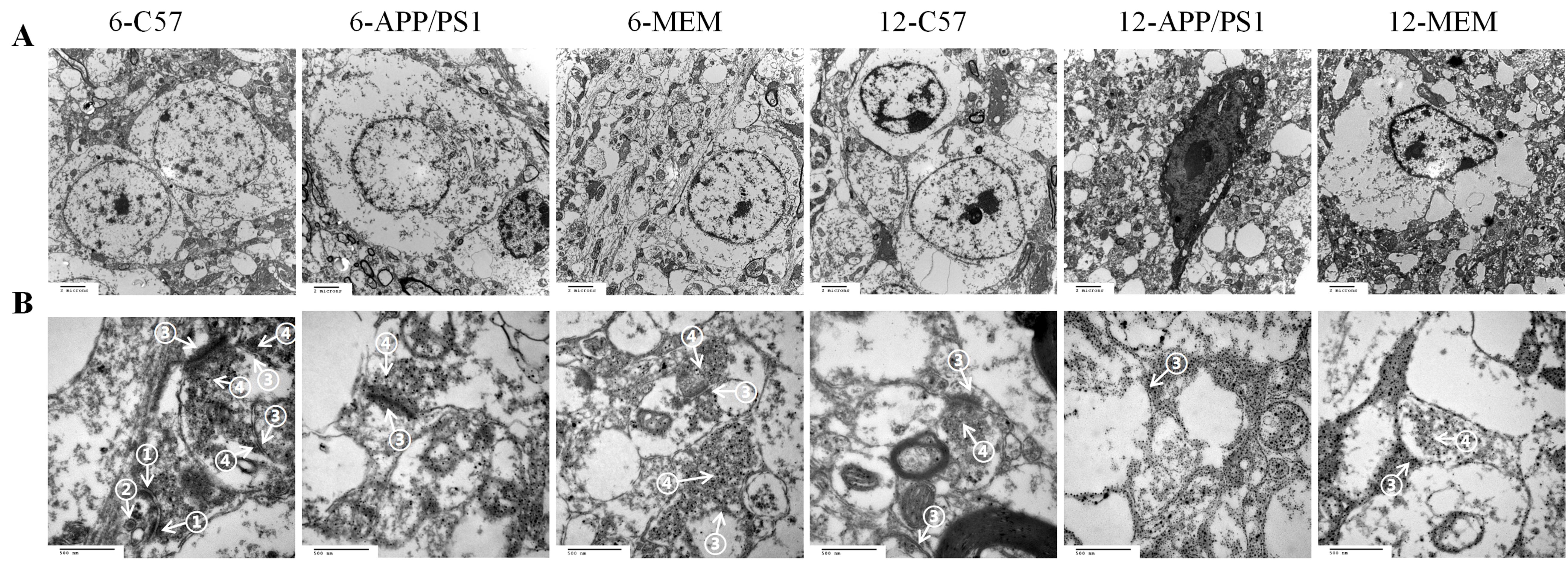
Figure 4 Ultrastructure of the olfactory bulb nerve cells (A, ×7 000) and synapses (B, ×60 000) in each group observed by transmission electron microscope after ultrathin section stainingNote: 6-C57 refers to 6-month-old C57BL/6 mice in the blank control group; 6-APP/PS1 refers to 6-month-old APP/PS1 double transgenic mice in model group; 6-MEM refers to APP/PSI double transgenic mice treated with 2.6 mg/kg memantine hydrochloride from 3- to 6-month-old; 12-C57 refers to 12-month-old C57BL/6 mice; 12-APP/PS1 refers to 12-month-old APP/PS1 double transgenic mice; 12-MEM refers to APP/PS1 double transgenic mice treated with 2.6 mg/kg memantine hydrochloride from 9- to 12-month-old. Each group contained five mice. Arrows ① show symmetric synapses, arrows ② show synaptic vesicles (containing inhibitory neurotransmitters), arrows ③ show asymmetric synapses, and arrows ④ show synaptic vesicles (containing excitatory neurotransmitters).
| 1 | DIBATTISTA M, PIFFERI S, MENINI A, et al. Alzheimer's disease: what can we learn from the peripheral olfactory system?[J]. Front Neurosci, 2020, 14:440. DOI:10.3389/fnins. 2020.00440 . |
| 2 | CHEN M, CHEN Y N, HUO Q W, et al. Enhancing GABAergic signaling ameliorates aberrant gamma oscillations of olfactory bulb in AD mouse models[J]. Mol Neurodegener, 2021, 16(1):14. DOI:10.1186/s13024-021-00434-7 . |
| 3 | XU Z P, YANG S L, ZHAO S, et al. Biomarkers for early diagnostic of mild cognitive impairment in type-2 diabetes patients: a multicentre, retrospective, nested case-control study[J]. EBioMedicine, 2016, 5:105-113. DOI:10.1016/j.ebiom.2016.02.014 . |
| 4 | SAIZ-SANCHEZ D, FLORES-CUADRADO A, UBEDA-BAÑON I, et al. Interneurons in the human olfactory system in Alzheimer's disease[J]. Exp Neurol, 2016, 276:13-21. DOI:10. 1016/j.expneurol.2015.11.009 . |
| 5 | DOTY R L. Olfactory dysfunction in neurodegenerative diseases: is there a common pathological substrate?[J]. Lancet Neurol, 2017, 16(6):478-488. DOI:10.1016/S1474-4422(17)30123-0 . |
| 6 | DAMMALLI M, DEY G, MADUGUNDU A K, et al. Proteomic analysis of the human olfactory bulb[J]. OMICS, 2017, 21(8):440-453. DOI:10.1089/omi.2017.0084 . |
| 7 | MURPHY C. Olfactory and other sensory impairments in Alzheimer disease[J]. Nat Rev Neurol, 2019, 15(1):11-24. DOI:10.1038/s41582-018-0097-5 . |
| 8 | ROUBY C, THOMAS-DANGUIN T, VIGOUROUX M, et al. The Lyon clinical olfactory test: validation and measurement of hyposmia and anosmia in healthy and diseased populations[J]. Int J Otolaryngol, 2011, 2011:203805. DOI:10.1155/2011/203805 . |
| 9 | BALLEZA-TAPIA H, HUANOSTA-GUTIÉRREZ A, MÁRQUEZ-RAMOS A, et al. Amyloid β oligomers decrease hippocampal spontaneous network activity in an age-dependent manner[J]. Curr Alzheimer Res, 2010, 7(5):453-462. DOI:10.2174/156720510791383859 . |
| 10 | DANDO S J, MACKAY-SIM A, NORTON R, et al. Pathogens penetrating the central nervous system: infection pathways and the cellular and molecular mechanisms of invasion[J]. Clin Microbiol Rev, 2014, 27(4):691-726. DOI:10.1128/cmr. 00118-13 . |
| 11 | MCSHANE R, WESTBY M J, ROBERTS E, et al. Memantine for dementia[J]. Cochrane Database Syst Rev, 2019, 3(3): CD003154. DOI:10.1002/14651858.CD003154.pub6 . |
| 12 | QIAO O, ZHANG X Y, ZHANG Y, et al. Cerebralcare Granule® enhances memantine hydrochloride efficacy in APP/PS1 mice by ameliorating amyloid pathology and cognitive functions[J]. Chin Med, 2021, 16(1):47. DOI:10.1186/s13020-021-00456-9 . |
| 13 | LIU M Y, WANG S, YAO W F, et al. Memantine improves spatial learning and memory impairments by regulating NGF signaling in APP/PS1 transgenic mice[J]. Neuroscience, 2014, 273:141-151. DOI:10.1016/j.neuroscience.2014.05.011 . |
| 14 | 金昊. APP/PS1小鼠不同时期内嗅皮质、海马体积变化特征及对学习记忆能力的影响[D]. 福州: 福建中医药大学, 2018. |
| JIN H. Changes of volume of entorhinal cortex and hippocampus in APP/PS1 mice at different stages and their effects on learning and memory ability[D]. Fuzhou: Fujian University of Chinese Medicine, 2018. | |
| 15 | YUAN Z, ZHOU H Y, ZHOU N, et al. Dynamic evaluation indices in spatial learning and memory of rat vascular dementia in the Morris water maze[J]. Sci Rep, 2019, 9:7224. DOI:10.1038/s41598-019-43738-x . |
| 16 | ZHANG X M, ZHAO F, WANG C F, et al. AVP(4-8) improves cognitive behaviors and hippocampal synaptic plasticity in the APP/PS1 mouse model of Alzheimer's disease[J]. Neurosci Bull, 2020, 36(3):254-262. DOI:10.1007/s12264-019-00434-0 . |
| 17 | KARUNAKARAN S. Unraveling early signs of navigational impairment in APPswe/PS1dE9 mice using Morris water maze[J]. Front Neurosci, 2020, 14:568200. DOI:10.3389/fnins. 2020. 568200 . |
| 18 | COLES M, WATT G, KREILAUS F, et al. Medium-dose chronic cannabidiol treatment reverses object recognition memory deficits of APP Swe /PS1ΔE9 transgenic female mice[J]. Front Pharmacol, 2020, 11:587604. DOI:10.3389/fphar.2020.587604 . |
| 19 | GIMÉNEZ-LLORT L, MARIN-PARDO D, MARAZUELA P, et al. Survival bias and crosstalk between chronological and behavioral age: age- and genotype-sensitivity tests define behavioral signatures in middle-aged, old, and long-lived mice with normal and AD-associated aging[J]. Biomedicines, 2021, 9(6):636. DOI:10.3390/biomedicines9060636 . |
| 20 | LI M Q, SU S X, CAI W H, et al. Differentially expressed genes in the brain of aging mice with cognitive alteration and depression- and anxiety-like behaviors[J]. Front Cell Dev Biol, 2020, 8:814. DOI:10.3389/fcell.2020.00814 . |
| 21 | LI P, XU J, GU H H, et al. Memantine ameliorates cognitive deficit in AD mice via enhancement of entorhinal-CA1 projection[J]. BMC Neurosci, 2021, 22(1):41. DOI:10.1186/s12868-021-00647-y . |
| 22 | DOTY R L. Olfactory dysfunction in neurodegenerative diseases: is there a common pathological substrate? [J]. Lancet Neurol, 2017, 16(6):478-488. DOI:10.1016/S1474-4422(17)30123-0 . |
| 23 | UBEDA-BAÑON I, SAIZ-SANCHEZ D, FLORES-CUADRADO A, et al. The human olfactory system in two proteinopathies: Alzheimer's and Parkinson's diseases[J]. Transl Neurodegener, 2020, 9(1):22. DOI:10.1186/s40035-020-00200-7 . |
| 24 | MORIGUCHI S, INAGAKI R, FUKUNAGA K. Memantine improves cognitive deficits via KATP channel inhibition in olfactory bulbectomized mice[J]. Mol Cell Neurosci, 2021, 117:103680. DOI:10.1016/j.mcn.2021.103680 . |
| 25 | LI W Y, LI S S, SHEN L H, et al. Impairment of dendrodendritic inhibition in the olfactory bulb of APP/PS1 mice[J]. Front Aging Neurosci, 2019, 11:2. DOI:10.3389/fnagi.2019.00002 . |
| 26 | ZOU Y M, LU D, LIU L P, et al. Olfactory dysfunction in Alzheimer's disease[J]. Neuropsychiatr Dis Treat, 2016, 12:869-875. DOI:10.2147/NDT.S104886 . |
| 27 | DONG H W, ENNIS M. Activation of group II metabotropic glutamate receptors suppresses excitability of mouse main olfactory bulb external tufted and mitral cells[J]. Front Cell Neurosci, 2018, 11:436. DOI:10.3389/fncel.2017.00436 . |
| 28 | RIBAK C E, VAUGHN J E, SAITO K, et al. Glutamate decarboxylase localization in neurons of the olfactory bulb[J]. Brain Res, 1977, 126(1):1-18. DOI:10.1016/0006-8993(77)90211-6 . |
| 29 | WANG R, REDDY P H. Role of glutamate and NMDA receptors in Alzheimer's disease[J]. J Alzheimers Dis, 2017, 57(4):1041-1048. DOI:10.3233/JAD-160763 . |
| 30 | PARSONS C G, STÖFFLER A, DANYSZ W. Memantine: a NMDA receptor antagonist that improves memory by restoration of homeostasis in the glutamatergic system - too little activation is bad, too much is even worse[J]. Neuropharmacology, 2007, 53(6):699-723. DOI:10.1016/j.neuropharm.2007.07.013 . |
| 31 | ALBERDI E, SÁNCHEZ-GÓMEZ M V, CAVALIERE F, et al. Amyloid β oligomers induce Ca2+ dysregulation and neuronal death through activation of ionotropic glutamate receptors[J]. Cell Calcium, 2010, 47(3):264-272. DOI:10.1016/j.ceca. 2009.12.010 . |
| 32 | TERAVSKIS P J, ASHE K H, LIAO D Z. The accumulation of tau in postsynaptic structures: a common feature in multiple neurodegenerative diseases? [J]. Neuroscientist, 2020, 26(5-6):503-520. DOI:10.1177/1073858420916696 . |
| 33 | LIMA CALDEIRA G, PEÇA J, CARVALHO A L. New insights on synaptic dysfunction in neuropsychiatric disorders[J]. Curr Opin Neurobiol, 2019, 57:62-70. DOI:10.1016/j.conb.2019.01.004 . |
| 34 | GARAD M, EDELMANN E, LEßMANN V. Impairment of spike-timing-dependent plasticity at schaffer collateral-CA1 synapses in adult APP/PS1 mice depends on proximity of aβ plaques[J]. Int J Mol Sci, 2021, 22(3):1378. DOI:10.3390/ijms22031378 . |
| 35 | LI W Y, LI S S, SHEN L H, et al. Impairment of dendrodendritic inhibition in the olfactory bulb of APP/PS1 mice[J]. Front Aging Neurosci, 2019, 11:2. DOI:10.3389/fnagi.2019.00002 . |
| 36 | PALOP J J, MUCKE L. Amyloid-β-induced neuronal dys-function in Alzheimer's disease: from synapses toward neural networks[J]. Nat Neurosci, 2010, 13(7):812-818. DOI:10.1038/nn.2583 . |
| 37 | WESSON D W, BORKOWSKI A H, LANDRETH G E, et al. Sensory network dysfunction, behavioral impairments, and their reversibility in an Alzheimer's β-amyloidosis mouse model[J]. J Neurosci, 2011, 31(44):15962-15971. DOI:10.1523/JNEUROSCI.2085-11.2011 . |
| 38 | BOYLE P A, YANG J Y, YU L, et al. Varied effects of age-related neuropathologies on the trajectory of late life cognitive decline[J]. Brain, 2017, 140(3):804-812. DOI:10.1093/brain/aww341 . |
| 39 | SCHUBERT C R, FISCHER M E, PINTO A A, et al. Sensory impairments and risk of mortality in older adults[J]. J Gerontol A Biol Sci Med Sci, 2016, 72(5):710-715. DOI:10.1093/gerona/glw036 . |
| 40 | EKSTRÖM I, SJÖLUND S, NORDIN S, et al. Smell loss predicts mortality risk regardless of dementia conversion[J]. J Am Geriatr Soc, 2017, 65(6):1238-1243. DOI:10.1111/jgs.14770 . |
| 41 | FRIESEN M, ZIEGLER-WALDKIRCH S, EGENOLF M, et al. Distinct Aβ pathology in the olfactory bulb and olfactory deficits in a mouse model of Aβ and α-syn co-pathology[J]. Brain Pathol, 2022, 32(3): e13032. DOI:10.1111/bpa.13032 . |
| 42 | WESSON D W, BORKOWSKI A H, LANDRETH G E, et al. Sensory network dysfunction, behavioral impairments, and their reversibility in an Alzheimer's β-amyloidosis mouse model[J]. J Neurosci, 2011, 31(44):15962-15971. DOI:10.1523/JNEUROSCI.2085-11.2011 . |
| [1] | LI Na, WANG Wei, LI Chen-jing, ZHANG Ying-yan, ZHANG Sheng, ZHANG Hai-yan, ZHANG Zhou. Analysis on Reproductive Performance and Growth of APPswe/PS1dE9 Transgenic Mice Model for Alzheimer's Disease [J]. Laboratory Animal and Comparative Medicine, 2012, 32(3): 222-227. |
| [2] | RONG Zhou-hui1, QU Jie-ming2, SHEN Ai-di1,YANG You-ming3, ZHOU Guang-xin3, HE Li-xian2. Observation of Inflammatory Cells in Bronchoalveolar Lavage Fluid and Pathological Morphology of the Airway in Passive Smoking Rats [J]. , 2001, 21(1): 33-35. |
| Viewed | ||||||
|
Full text |
|
|||||
|
Abstract |
|
|||||