Laboratory Animal and Comparative Medicine ›› 2020, Vol. 40 ›› Issue (5): 426-.DOI: 10.3969/j.issn.1674-5817.2020.05.011
Previous Articles Next Articles
FAN Shuqiong1,2, ZHENG Li1
Received:
Online:
Published:
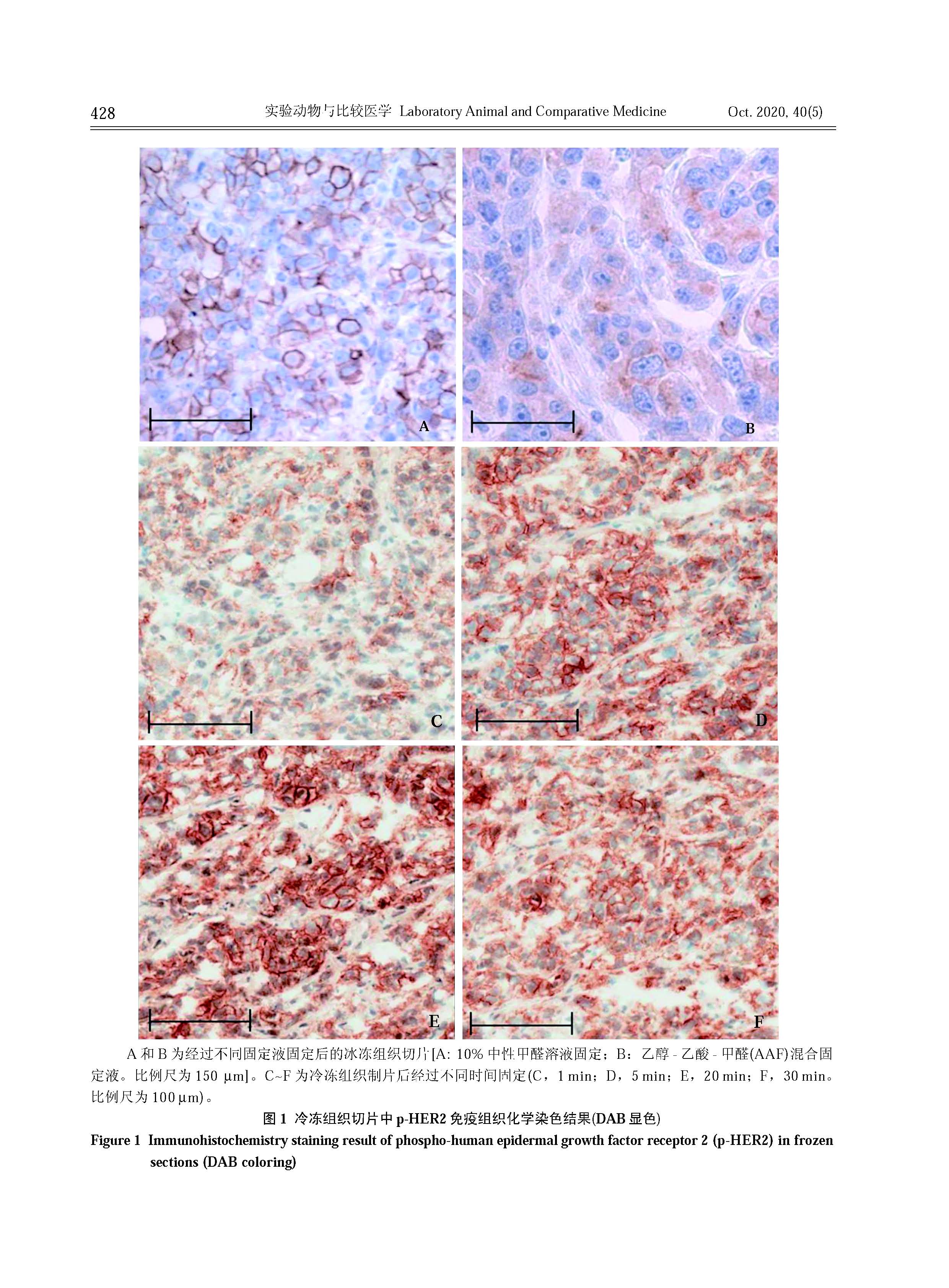
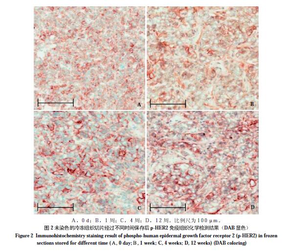
Abstract: Objective To investigate the optimized fixation and storage conditions for the immunohistochemistry detection of phospho-human epidermal growth factor receptor 2 (HER2) on frozen sections. Methods Xenograft model was established by subcutaneously injecting human breast cancer cell BT474 into the abdomen of BALB/c-nu/nu mice. The mice were sacrificed when the tumor volume up to 300 mm3. The tumor samples were quickly frozen, sectioned, fixed in different reagents and stored for different time period. Phospho-HER2 immunohistochemistry was performed to evaluate the change of protein expression under these conditions. Results Good cell morphology was kept by the fixation of frozen section in 50% formalin for 5 minutes. The expression of phospho-HER2 protein was stable within 12 weeks after fixation when the tumor tissues were stored in -20 ℃. Conclusion The immunohistochemistry staining on frozen tissues takes less time and can be a supplement or replacement of traditional staining on formalin-fixed and paraffin-embedded sections.
Key words: Frozen section, Human epidermal growth factor receptor 2, Immunohistochemistry; BALB/c-nu/nu mice
CLC Number:
Q95-33
FAN Shuqiong, ZHENG Li. Exploration of Phospho-HER2 Immunohistochemistry Staining on Frozen Tumor Tissues[J]. Laboratory Animal and Comparative Medicine, 2020, 40(5): 426-.
0 / / Recommend
Add to citation manager EndNote|Ris|BibTeX
URL: https://www.slarc.org.cn/dwyx/EN/10.3969/j.issn.1674-5817.2020.05.011
https://www.slarc.org.cn/dwyx/EN/Y2020/V40/I5/426